


Changing the Strategy: Introduction of the 120-Pulse Shockwave C2+ Catheter at St. Francis Heart Center
© 2023 HMP Global. All Rights Reserved.
Any views and opinions expressed are those of the author(s) and/or participants and do not necessarily reflect the views, policy, or position of EP Lab Digest or HMP Global, their employees, and affiliates.
St. Francis Heart Center was the first to introduce the Shockwave C2+ catheter (Shockwave Medical) in the United States. Can you share your experience?
 Since August 2023’s limited market release when we first started using the C2+, we have done 30+ cases over the past few months. The concept behind the development of the C2+ was that if more pulses were available to the operators, it would facilitate the management of more complex, calcified lesions.
Almost all lesions can be managed in 120 pulses. In fact, in many cases multiple lesions or vessels could be treated with 120 pulses. To put the value of the C2+ in perspective, in all the intravascular lithotripsy (IVL) studies to date, the mean number of pulses used was around 70 with a standard deviation of about 40 pulses.1,2 This means that 95% of all cases could be performed with the 120 pulses available in the C2+ catheter.
Have the additional 40 pulses with the Shockwave C2+ catheter changed how you develop or deploy a “pulse management” strategy?
Yes. Most importantly, we believe that focusing the IVL only on the calcium in the tightest segment of the artery is a mistake. While most focal stenotic calcification is only 5 to 7 millimeters (mm) long, the vessel calcium often actually extends 30 or 40 mm around the lesion. Operators have generally focused their attention on this 5 mm of very, very dense calcium, but then end up putting in a much longer 38 mm stent. The areas that are remote to these very tight segments still have significant calcium, many time eccentric, and aren’t being modified. We changed our approach so that any location that will be covered with stent is pulsed. This is where the 120 pulses of the C2+ come with a real advantage. Often eccentric pieces of calcium that are remote from the tightest segment limit the stent expansion.
Many studies have shown that following drug-eluting stent (DES) placement, the minimal stent area is usually outside of the baseline maximum calcified area. This teaches us that we are good at treating and modifying the tight segment, but we are not so good at treating the not-so-tight segments. This is an advantage of IVL over other modalities. When atherectomy is performed in a larger part of the vessel, the atherectomy burr doesn’t touch the calcium in the wall; it just floats right through the lumen. IVL scaffolds the wall, so it can still modify calcium in the larger parts of the artery, not just in the tighter segments.
In which lesion types are the 40 additional pulses of the Shockwave C2+ catheter most valuable?
In the DISRUPT CAD series of trials, it became clear that IVL was highly effective in severely calcified lesions identified by angiography. However, there are various different subtypes of calcification, including nodular calcification and eccentric calcification, that cannot be differentiated by angiography. This year, we had two important publications2,3 that showed the following. First, IVL is highly effective in nodular calcification.3 Clinical studies to date show that intervening on nodular calcification with atherectomy leads a threefold higher hazard compared to non-nodular lesions. This isn’t the case with IVL, where at two years, the event rate is similar. Moreover, what we found is acutely when you perform IVL, the minimal stent area and minimal stent expansion are the same whether or not a nodule is present. The IVL is working on the nodule and breaking it into smaller pieces, allowing it to be modified. A good analogy is to think about a nodule like a mountain. Atherectomy cuts off the very top of the mountain. You feel like you have done a good job by performing atherectomy, but the only thing that has been done is that the top of the mountain has been cut off. IVL pulverizes the entire mountain into smaller pieces of rock. As the mountain crumbles, you can push it out of the way more easily with a stent. Albeit, there haven’t been any head-to-head studies. We don’t have data looking at the minimal stent expansion and minimal stent area in nodular calcification when using atherectomy. When using IVL, we know that whether or not there is a nodule, IVL gives you the same stent expansion in the same stent area.
Next, consider eccentric calcium. The more calcium you have, the more IVL will break it, and the less calcium you have, the less IVL will break it. IVL is effective in proportion to the amount of calcium present in the wall. For example, for a 90-degree arc of calcium, you are fourfold less likely to get fractures in that calcium, because there is fourfold less calcium. For a 180-degree arc of calcium, fractures are 50% less likely, and for a 270-degree arc of calcium, fractures are 25% less likely. Remember, the IVL balloon doesn’t know where the calcium is located. You inflate the balloon and the energy will hit the calcium wherever it is located, which means that the greater the substrate of calcium, the more fractures will occur. The less the substrate of calcium, the less IVL will fracture. Eccentric calcium is a very interesting phenomenon that truly identifies the mechanism of action of IVL, because there is no way to break eccentric calcium with a noncompliant balloon. It is physically impossible. Take the example of a wishbone. You couldn’t break a wishbone using a balloon from the inside if it is already broken in half. You can only break the wishbone if the whole wishbone is intact, and once you break it, you can’t keep breaking it. It is the same phenomenon with IVL. If you have a 180-degree or greater arc of calcium and use a balloon inside, you can break it with a balloon. Half of that impact or some proportion of it will be related to the balloon itself. But when you have an already broken wishbone and you get another break in it, that is the result of IVL.
How do you think the Shockwave C2+ catheter will change calcium modification utilization in the lab?
There has been a sort of dogma that in very long lesions it is better to do atherectomy, but with the C2+, that thinking should no longer be relevant, because we now have 120 pulses to treat the whole vessel. Those of us who are experienced with IVL learned how to ration the original 80 pulses, so when we first started using the Shockwave C2+ catheter with 120 pulses, we had to figure out how to use the extra pulses in a meaningful way. So we changed our strategy. Rather than lesion preparation, we instead do vessel preparation. We go distally to the limit of the calcified segment where we are planning to place the distal end of the stent, and start pulsing from there and pull back. We will save 80 pulses (which previously was our original entire amount) for the most calcified segment. If you divide up 120 pulses and use 80 pulses in the max calcium site, it means we have 40 ‘free’ pulses to spend. We use those 40 pulses all over the remainder of the calcium within the lesion. What is the advantage of dividing the 120 pulses in this way? We are able to pulse all parts of the artery that will be stented. We also know that the more pulses you deliver, the more calcium fractures you will get. In the most calcified segments, this strategy allows us to achieve even better lesion preparation. In our pulse management strategy, we have nicknamed the pulses into the max calcified segment as the “power surge”. A power surge is where we use 50%-60% of the IVL energy in the most calcified segment, and then take the rest of that energy and distribute it along the length of the lesion.
The Shockwave C2+ catheter has encouraged a move from focal lesion preparation to entire vessel preparation using IVL.
The maximum calcified lesion length by angiographic core laboratory analysis is 5-7 mm. However, the mean stent length in the studies is 35 mm. That math doesn’t make sense. It is telling us that calcium that previously we were leaving alone is surrounding the maximum calcified segment. As I mentioned, in the PREPARE-CALC study4 and other studies, leaving the calcified areas remote from the max calcium site untreated means the minimal stent area frequently ends up outside of a maximized calcified segment from the baseline. Now the event-defining cross-section has become the minimal stent area in an unprepped area. That is one reason that head-to-head trials of lesion modification that use calcium modification devices against balloon angioplasty alone have failed. It’s simple. When you spend all your time fixing the alternator, but there is also a problem in the radiator, then you are going to end up in the same scenario with a failing engine.
Is there a maximum lesion length that you take into consideration for one-catheter Shockwave C2+ cases?
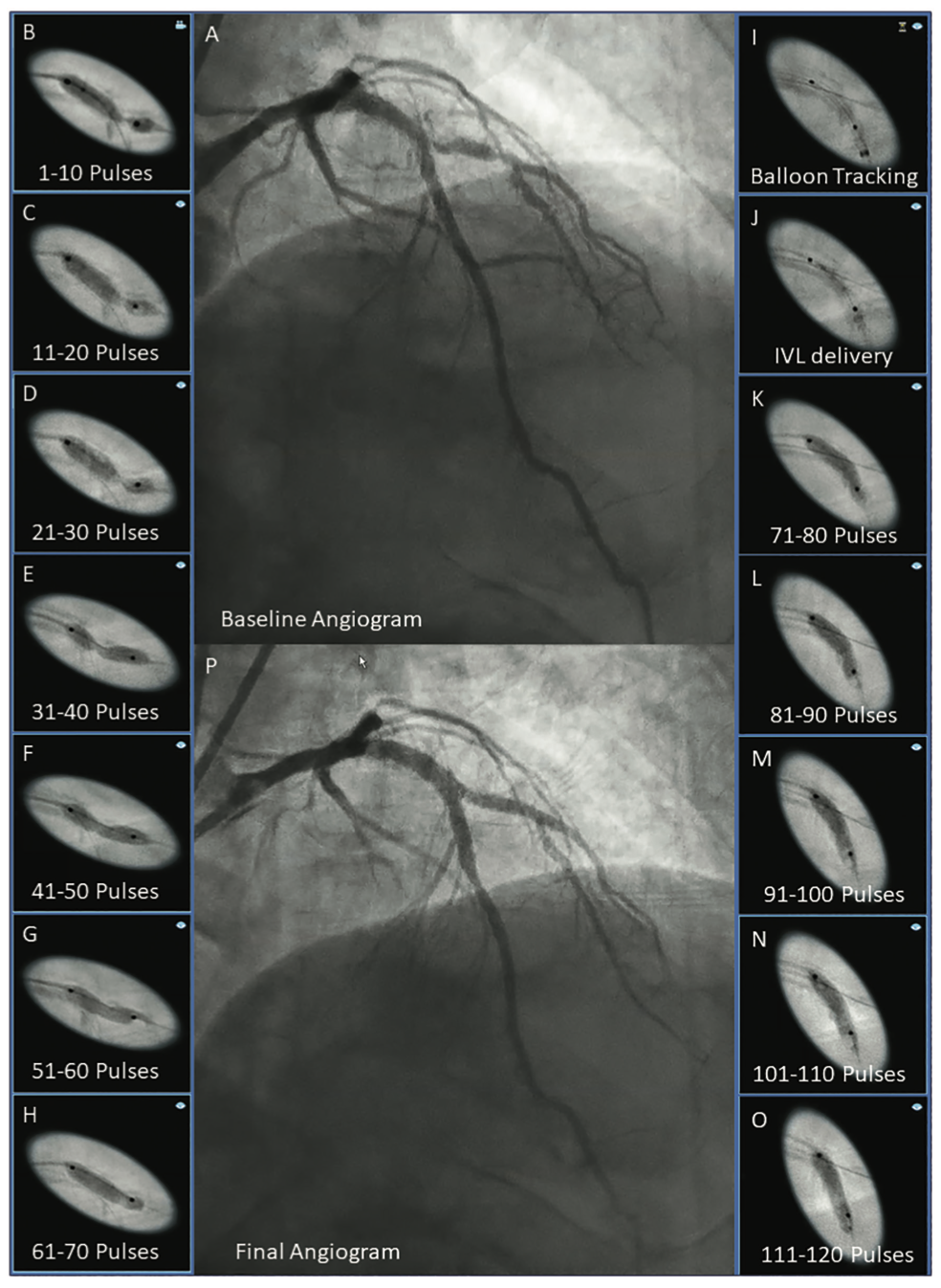
No, the ability to use 120 pulses has essentially eliminated that. In fact, we will often use one C2+ in two different vessels. Yesterday we had a severely calcified left anterior descending (LAD) and diagonal bifurcation (Figure). We delivered the C2+ to the diagonal and delivered 70 pulses into a very resistant lesion that yielded. We then removed the used balloon, and advanced a guide extension catheter into the LAD lesion using balloon-assisted tracking and delivered the remaining 50 pulses. This allowed two-vessel preparation with one catheter. Of course, the same technique could be used in two completely separate vessels like a LAD and circumflex. The key to getting the use of the residual pulses in a used balloon is delivery through a guide extension catheter.
Do you have any additional suggestions for the use of the Shockwave C2+ catheter?
The safety of IVL is unparalleled. It is also faster and there is no special workflow. You can do IVL at any hospital, anywhere, with or without surgical backup. You don’t need to buy any special additional equipment. The use of IVL is simple and straightforward. I think it is great that people adopted IVL so quickly because of its simplicity, but at the same time, it is really not that simple to maximize its effect. Meaning when you get into more complicated lesions, you have to understand that adjuvant tools are necessary, on top of just the balloon, to get your work done. For example, if you have a very tight lesion, you might want to use a guide extension catheter and a small balloon in order to get your guide extension across the lesion and get your IVL balloon across the lesion before trying atherectomy. Many of the lesions that are considered balloon uncrossable actually aren’t; you just have to make yourself a little space to deliver the IVL. We had wrongly thought of it as an all-or-none phenomenon — that the IVL balloon is not going to cross that lesion or atherectomy is the only choice for that lesion. It is not cheating to first try to get a balloon across using conventional techniques. In our cath lab’s experience, we use a guide extension catheter in 90% of cases. The guide extension catheter allows us to do two things. First, it allows us to use the 120 pulses throughout the lesion much more easily, because the IVL catheter is not as deliverable as a conventional balloon. It is bulkier, so it is harder to move around. Second, using a guide extension catheter also allows you a unique opportunity, especially with the C2+, to treat multiple lesions or vessels. Imagine you have a lesion in the LAD and a calcified lesion in the circumflex. Traditionally, you would either use two IVL balloons or use 40 pulses in each artery, which isn’t enough pulses. Using the C2+ and guide extension catheter, you can now split up your 120 pulses into both arteries. You do 60 pulses in the LAD and 60 in the circumflex. The timing for this use of IVL is perfect, as the reimbursement rules are changing and we are generally moving towards using one balloon rather than more. A guide extension catheter will be a fraction of the cost of another IVL balloon.
Any final thoughts?
The two places where we have changed our strategy are in long lesions and in treating two separate vessels with a single balloon. With C2+, I use the same amount of pulses as before (80) in the maximum calcified segment but now use all the ‘bonus’ pulses for the calcification surrounding the maximized lesion. We take one IVL balloon, start distally, and as we come all the way up, treat the whole lesion. At that point, if there are pulses left over, we go back to the maximum calcium segment and deliver the remaining pulses. The Shockwave C2+ catheter has afforded us the opportunity to not only treat the maximum calcified segment in the lesion, but to do entire lesion preparation for the whole segment that will be stented.
This interview is sponsored by Shockwave Medical. Dr. Ali is a paid consultant of Shockwave Medical.
References
1. Ali ZA, Nef H, Escaned J, et al. Safety and effectiveness of coronary intravascular lithotripsy for treatment of severely calcified coronary stenoses: the Disrupt CAD II study. Circ Cardiovasc Interv. 2019 Oct; 12(10): e008434. doi:10.1161/CIRCINTERVENTIONS.119.008434
2. Ali ZA, Kereiakes DJ, Hill JM, et al. Impact of calcium eccentricity on the safety and effectiveness of coronary intravascular lithotripsy: pooled analysis from the Disrupt CAD studies. Circ Cardiovasc Interv. 2023 Oct;16(10):e012898. doi: 10.1161/CIRCINTERVENTIONS.123.012898
3. Ali ZA, Kereiakes D, Hill J, et al. Safety and effectiveness of coronary intravascular lithotripsy for treatment of calcified nodules. JACC Cardiovasc Interv. 2023 May 8; 16(9): 1122-1124. doi:10.1016/j.jcin.2023.02.015
4. Abdel-Wahab M, Toelg R, Byrne RA, et al. High-speed rotational atherectomy versus modified balloons prior to drug-eluting stent implantation in severely calcified coronary lesions. Circ Cardiovasc Interv. 2018 Oct; 11(10): e007415. doi:10.1161/CIRCINTERVENTIONS.118.007415
Keep reading about how to treat coronary calcium:
Go to Cath Lab Digest's current issue page
SPL-70449


