How to Diagnose and Treat INOCA in 2022
Every cath lab encounters patients with chest pain (assumed to be ischemic in origin) who have normal or nonobstructed coronary arteries, a syndrome called INOCA (ischemia but no coronary artery disease). The ischemia is often thought to be due to microvascular dysfunction (small vessels in the myocardium not dilating appropriately, resulting in reduced coronary flow reserve). Until recently, many labs, including ours, had no way to quantitate coronary flow reserve.
Recently, we received new software from Abbott (Coroventis CoroFlow) that permits us to measure both fractional flow reserve as well as coronary flow reserve at the same time by a thermodilution method. I have always been a fan of measuring coronary flow reserve since the development of intracoronary Doppler in 1990. But our fellows and lab staff have often asked “what’s the point?” since we have few available treatments to help a patient with impaired coronary flow reserve. Since INOCA patients comprise up to 30% of patients in most cardiac cath labs,1,2 I thought this would be a good time to review INOCA and how we should be thinking about it for treatment.
Ischemia, Myocardial Oxygen Supply-Demand Imbalance, and Coronary Microvascular Disease
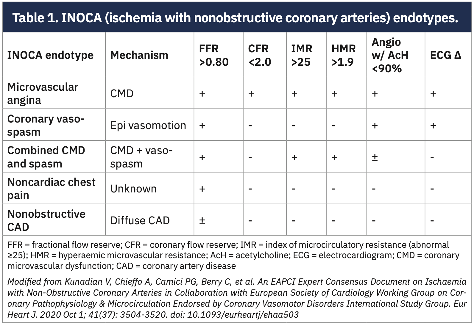
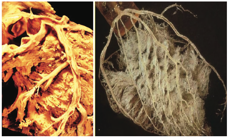
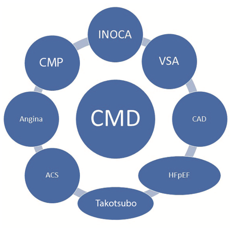
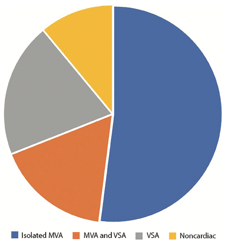
Ischemia is caused by an imbalance in the myocardial oxygen supply-demand balance. The inability to increase coronary blood flow (oxygen supply) enough to meet increased myocardial oxygen demand (eg, during exercise) results in ischemia, manifesting as left ventricular dysfunction, electrocardiographic or perfusion abnormalities, and angina. Ischemia is most commonly due to atherosclerotic obstructions in the epicardial arteries, >400 microns in size: those we can see on angiography (Figure 1). However, ischemia can also be caused by a reduced ability to increase blood flow due to an impaired myocardial microcirculation (vessels <400 microns, which we cannot see on angiography), manifesting as a reduced coronary flow reserve (CFR, maximal flow/resting flow). There are several types of patients who present with chest pain and no coronary artery obstructions (Table 1 and Figure 2), the most common being coronary microvascular dysfunction or vasospastic angina (VSA, eg, Prinzmetal’s angina). The prevalence of the INOCA endotypes is shown on Figure 3.
If the Patient’s Chest Pain is Not Due to Coronary Artery Obstructions, What Then?
ACS = acute coronary syndrome; CAD = coronary artery disease; CMD = coronary microvascular dysfunction; HF = heart failure; HFpEF = HF with preserved ejection fraction; INOCA = ischemia with nonobstructive coronary arteries; MINOCA = myocardial infarction with nonobstructive coronary arteries; PCI = percutaneous coronary intervention
Modified from Del Buono MG, Montone RA, Camilli M, et al. Coronary microvascular dysfunction across the spectrum of cardiovascular diseases: JACC state-of-the-art review. J Am Coll Cardiol. 2021 Sep 28; 78(13): 1352-1371. doi: 10.1016/j.jacc.2021.07.042
Although this sounds like an easy question, angiography does not always provide an answer. Chest pain can occur either in a typical cardiac fashion with exertion, relieved with rest or nitroglycerin (classic angina pectoris), or it can occur under different conditions, such as at rest, or have a constant nature, features of noncardiac pain that were previously termed atypical angina. When patients are suspected of having obstructive coronary artery disease and atherosclerosis, coronary angiography is usually performed after stress testing for myocardial ischemia and initiating treatment. Often the angiogram will show mild or even nonobstructive coronary artery disease, which requires assessment for an accurate diagnosis. Epicardial ischemia in the lab is detected by fractional flow reserve (FFR) or non-hyperemic pressure ratios (NHPR) like instantaneous wave-free ratio (iFR).
Modified from Ford TJ, Yii E, Sidik N, et al. Ischemia and no obstructive coronary artery disease: prevalence and correlates of coronary vasomotion disorders. Circ Cardiovasc Interv. 2019 Dec; 12(12): e008126. doi: 10.1161/CIRCINTERVENTIONS.119.008126
If epicardial arteries are not physiologically obstructed, then the mechanisms of chest pain in INOCA patients are often attributed to coronary microvascular dysfunction or abnormal epicardial vasomotion (ie, vasospasm), or both. By measuring CFR (and the index of myocardial resistance [IMR]), a tailored therapeutic approach can be applied to reduce chest pain, improve quality of life, and stop repeated and perhaps unnecessary interventions. The therapeutic approach to patients with INOCA depends on the type and clinical presentation with which the syndrome is associated. For example, INOCA in some patients may be due to hypertrophic cardiomyopathy, myocarditis, aortic stenosis, and infiltrative cardiomyopathies, wherein treatments vary widely and therapeutic decisions become particularly challenging. Other patients with INOCA may have systemic inflammatory syndromes, autoimmune disorders like lupus or rheumatoid arthritis, or may have primary metabolic disorders, again posing conundrums for the team taking care of these patients. Because of the uncertainty about INOCA mechanisms, many clinicians now believe it is important to understand not only epicardial patency, but also the status of the microvascular circulation. To do this, we need to make measurements beyond the angiogram alone. This is where intracoronary physiology adds value. Figure 3 shows the predominant mechanisms of INOCA.
How are CFR Thermodilution Measurements Made?
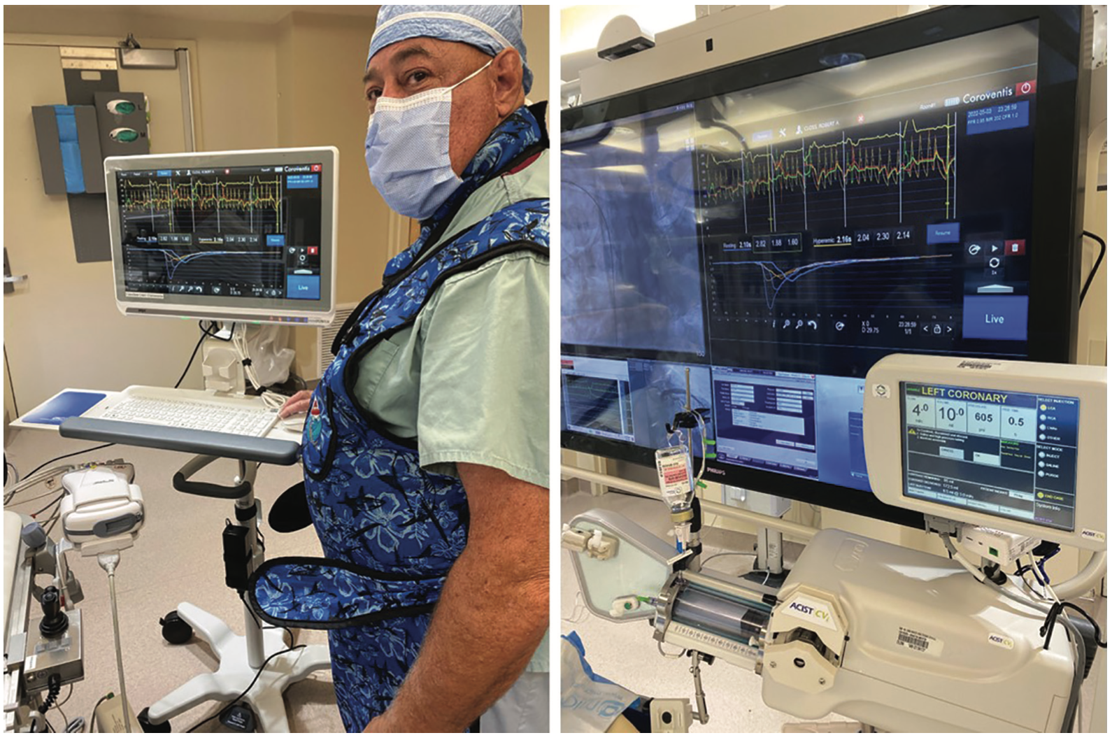
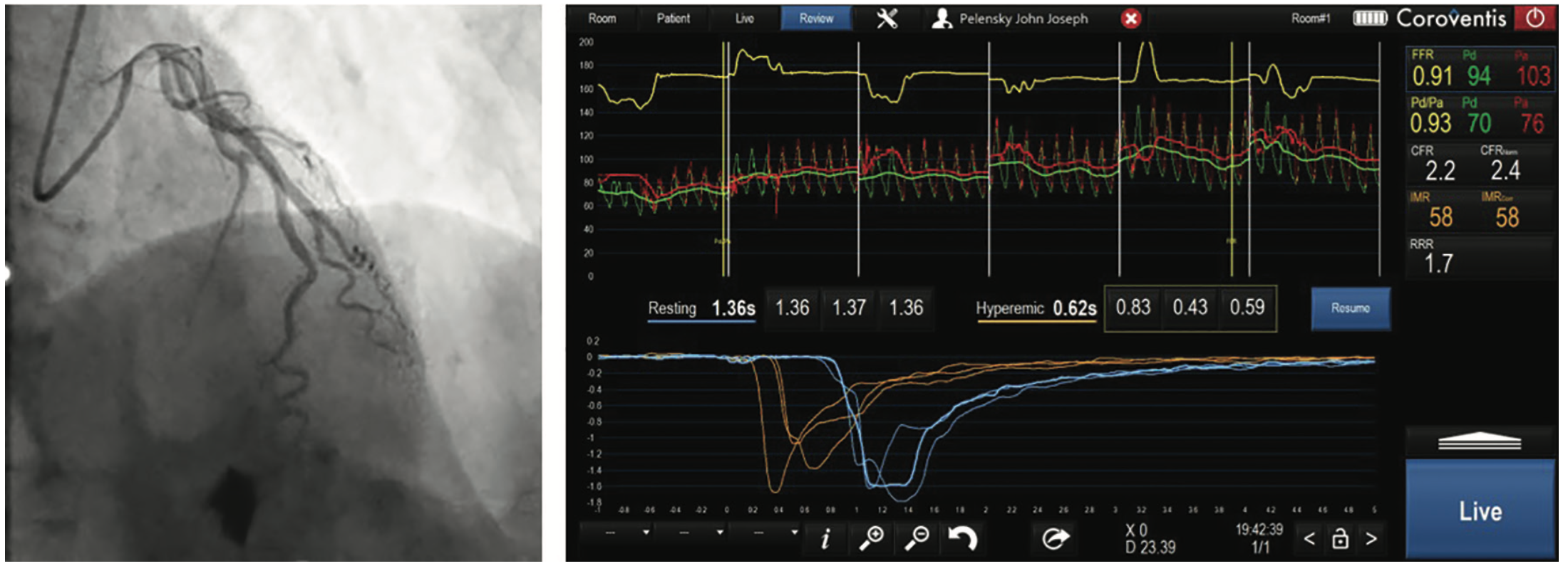
Let’s review a quick example. A 72-year-old man had angina with features of both cardiac and noncardiac causes (previously known as typical or atypical angina). He had a stress test with an equivocal result and presented to the cath lab, where we find unobstructed and near-normal coronaries on angiography. To better understand this patient’s chest pain, we measured intracoronary physiology with a pressure wire (PressureWire X [Abbott Vascular]) with the CorFlow system (Figure 4A) to provide all of the information about the coronary circulation.
The technique to measure CFR is the same used as for routine FFR measurements. To obtain a CFR value, a PressureWire X guidewire is used to measure the transit time of flow by thermodilution (Tmn) at rest and again during intraveous (IV) adenosine-induced hyperemia. Thermodilution-derived CFR is the ratio between resting and hyperemic temperature arrival time, Tmn.3
As with any routine FFR procedure, it begins after heparinization (5000u IV, as used for percutaneous coronary intervention). Next, position the guide catheter, and zero both guide and guidewire pressures. Advance the guide wire distally (about two-thirds of the artery. The sensor should be 6-10 cm from the guide tip). Flush the guide with saline. Following the computer prompts, vigorously inject 3 mLs of saline and close the injection port to monitor pressure. Repeat the bolus 3 times at rest and 3 times at max hyperemia during adenosine IV infusion (not intracoronary boluses). The software will prompt you for the actions needed and interpret the signals to give you CFR (as well as resting distal coronary pressure to aortic pressure ratio [Pd/Pa], resting full-cycle ratio [RFR], FFR, and IMR [index of microvascular resistance]) (Figure 4B). CFR indicates the maximum increase in coronary artery flow above the normal resting flow. CFR represents flow across both epicardial and microvascular territories, making it less specific than the IMR, which measures myocardial resistance, excluding the contribution of epicardial narrowings.
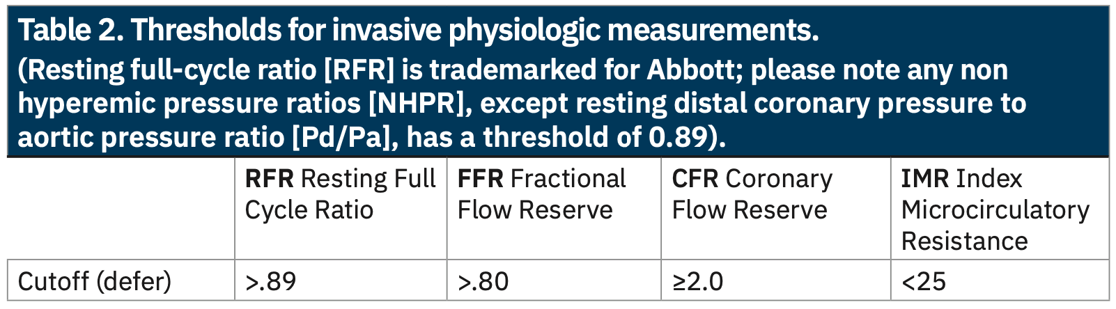
(Resting full-cycle ratio [RFR] is trademarked for Abbott; please note any non hyperemic pressure ratios [NHPR], except resting distal coronary pressure to aortic pressure ratio [Pd/Pa], has a threshold of 0.89).
Experimental models in both animals and humans have shown a reasonable level of agreement between bolus thermodilution-based CFR and intracoronary Doppler-based CFR.3 Using CFRthermo has proved to be much more feasible technically than using Doppler, as the latter is very sensitive to loading conditions and wire position in the artery. The interpretation of CFR depends on understanding the true resting and hyperemic responses. For example, a low CFR can be due to either high resting flow with normal hyperemia or normal resting flow with low hyperemia. For this reason, IMR contributes to our knowledge of microvascular resistance.
Returning to our patient example (Figure 4B), the FFR is negative for ischemia, but the CFR is low, 2.2, and the IMR high, 58, demonstrating coronary microvascular dysfunction, which establishes a diagnosis of microvascular disease. Table 2 lists normal physiologic values.
The Index of Microvascular Resistance (IMR)
From the same measurements of bolus thermodilution-derived Tmn, Fearon et al4 introduced the index of microvascular resistance (IMR), the first to specifically assess the microcirculation independent of the epicardial resistance. IMR indicates the level of microcirculatory resistance in the target artery territory. IMR is computed as follows:
IMR = distal pressure (Pd) at hyperemia x Tmn
CFR = coronary flow reserve; CMD = coronary microvascular dysfunction; CP = chest pain; Dia = diameter; ECG = electrocardiogram; FFR = fractional flow reserve; IMR = index of microvascular resistance; LVEDP = left ventricular end-diastolic pressure; VSA = vasospastic angina. (A) Negative non-invasive or invasive testing for epicardial ischemia. (B) Combo wire is an alternative option to measure FFR, CFR, and IMR.
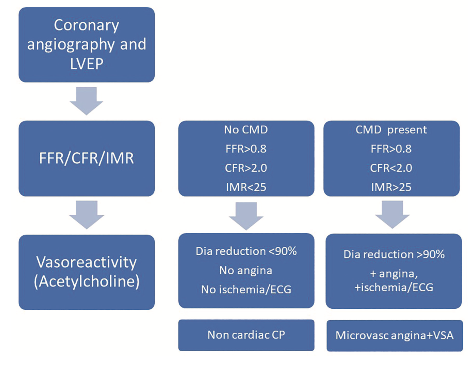
Normal IMR values (<25) indicate no coronary microvascular dysfunction. IMR values (≥25) indicate coronary microvascular dysfunction. Although the guidewire setup and serial bolus saline injections may take 2-3 minutes more than a routine FFR, the additional information of the CFR and IMR (and RFR pullback) are worth the time for a complete understanding of the patient’s status. Figure 5 illustrates the comprehensive evaluation of INOCA. The classification of INOCA endotypes begins with diagnostic angiography, followed by FFR, CFR, and IMR, and concludes with a provocation study of vasospasm (VSA).5,6
What is Vasoactive or Vasospastic Coronary Artery Disease (VSA)?
Vasospastic vascular disorders are caused by spasm of the large epicardial coronary arteries. While the exact mechanism is not well understood, hypersensitive vascular smooth muscle cells surrounding the large artery and endothelial dysfunction are identified to spasm in response to acetylcholine. Frequent or persistent VSA (eg, Prinzmetal’s angina) can result in myocardial infarction or death. The prevalence of VSA varies with ethnic origins: 24% in Japan, 19% in Taiwan, and 5% in the USA.7 VSA is more prevalent among men than women, more common between the ages of 40 and 70 years old, and decreases with age >70 years.7 Smoking is a risk factor for vasospastic angina, while diabetes and hypertension are not. For patients experiencing vasospastic angina, calcium channel blockers followed by nitrate therapy are recommended.
Treatment for INOCA
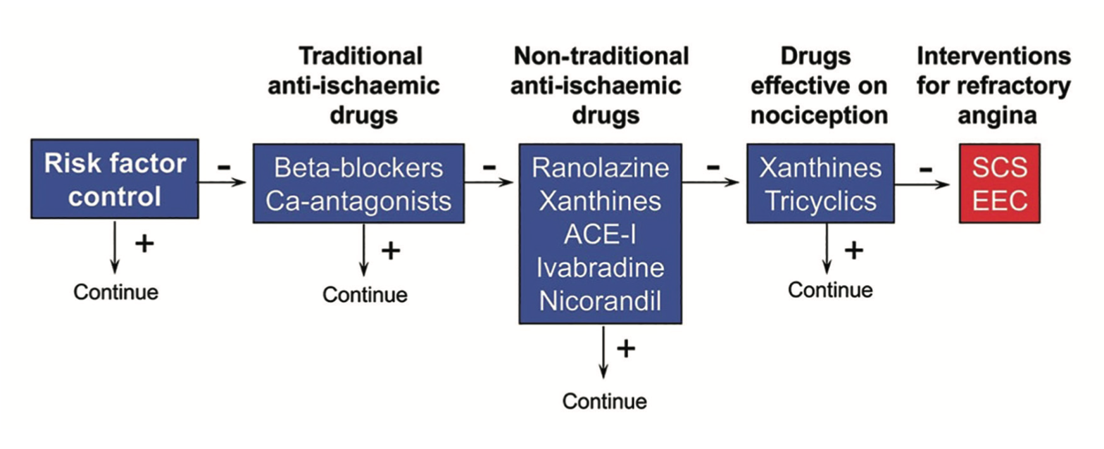
SCS = spinal cord stimulation; EEC = enhanced external counterpulsation
Reprinted with permission from Crea F, Camici PG, Bairey Merz CN. Coronary microvascular dysfunction: an update. Eur Heart J. 2014 May; 35(17): 1101-1111. doi: 10.1093/eurheartj/eht513
INOCA has specific therapies associated with improving long- and short-term prognosis.8,9 These include lifestyle factor modification and coronary artery disease (CAD) risk factor management (Figure 6). For patients in whom diagnostic microvascular angina has been associated with microvascular spasm, initial treatment with calcium channel blockers followed by ranolazine, ivabradine, or enhanced external counterpulsation (EECP) can be considered. For VSA, calcium channel blockers, nitrates, and nicorandil have been useful. Low-dose tricyclic antidepressants such as imipramine and xanthine derivatives may also be helpful. CAD risk factor treatment should continue (antihypertensive agents, statins, and diabetic medications). Lifestyle modification involving weight control, exercise, nutrition, stress, and smoking reduction should be employed as well.
The Bottom Line
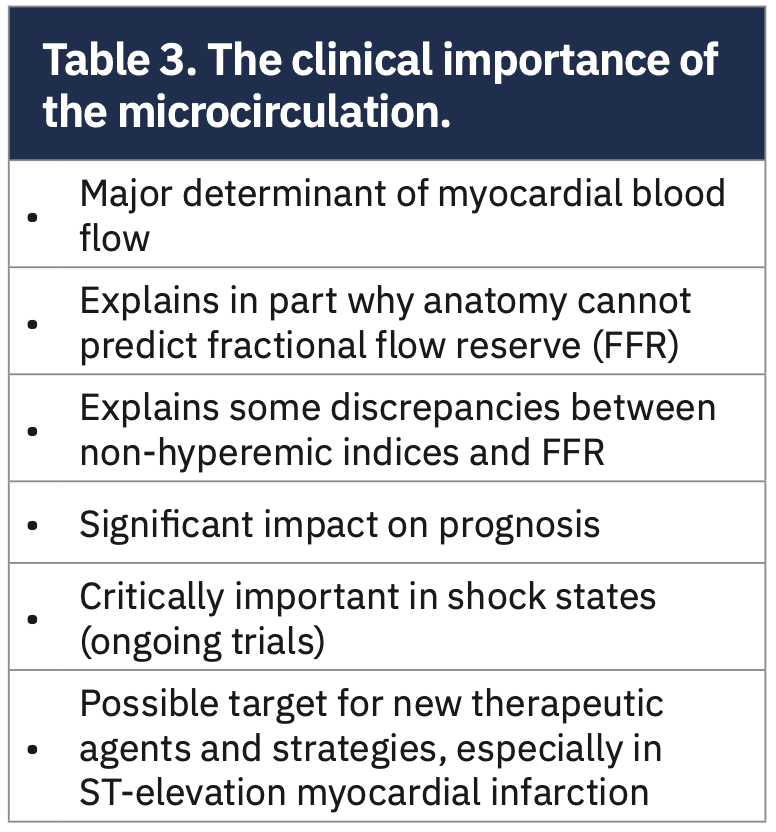
For our chest pain patients, INOCA should be recognized as clinically important in daily practice (Table 3). A systematic approach to diagnosing and treating INOCA patients can be implemented for completeness of the patient’s future care. As a field, we await targeted treatments to provide evidence-based management of patients with INOCA. It is my hope that creating awareness of INOCA and coronary microvascular dysfunction may improve our care for this common and important patient group.
Disclosures: Dr. Morton Kern reports he is a consultant for Abiomed, Abbott Vascular, Philips Volcano, ACIST Medical, and Opsens Inc.
Dr. Kern can be contacted at mortonkern2007@gmail.com
On Twitter @MortonKern
Read Part II:
Vasoreactivity Testing and Chest Pain Guidelines: Is the IIa Recommendation Warranted? INOCA Part II
References
1. Kaski JC, Crea F, Gersh BJ, Camici PG. Reappraisal of ischemic heart disease. Circulation. 2018 Oct 2; 138(14): 1463-1480. doi: 10.1161/CIRCULATIONAHA.118.031373
2. Kunadian V, Chieffo A, Camici PG, Berry C, et al. An EAPCI expert consensus document on ischaemia with non-obstructive coronary arteries in collaboration with European Society of Cardiology working group on coronary pathophysiology & microcirculation. Endorsed by Coronary Vasomotor Disorders International Study Group. Eur Heart J. 2020 Oct 1; 41(37): 3504-3520. doi: 10.1093/eurheartj/ehaa503
3. Candreva A, Gallinoro E, van ‘t Veer M, et al. Basics of coronary thermodilution. JACC Cardiovasc Interv. 2021 Mar 22; 14(6): 595-605. doi: 10.1016/j.jcin.2020.12.037
4. Fearon WF, Balsam LB, Farouque HM, et al. Novel index for invasively assessing the coronary microcirculation. Circulation. 2003 Jul 1; 107(25): 3129-3132. doi: 10.1161/01.CIR.0000080700.98607.D1. Erratum in: Circulation. 2003 Dec 23; 108(25): 3165.
5. Del Buono MG, Montone RA, Camilli M, et al. Coronary microvascular dysfunction across the spectrum of cardiovascular diseases: JACC state-of-the-art review. J Am Coll Cardiol. 2021 Sep 28; 78(13): 1352-1371. doi: 10.1016/j.jacc.2021.07.042
6. Ford TJ, Yii E, Sidik N, et al. Ischemia and no obstructive coronary artery disease: prevalence and correlates of coronary vasomotion disorders. Circ Cardiovasc Interv. 2019 Dec; 12(12): e008126. doi: 10.1161/CIRCINTERVENTIONS.119.008126
7. S Hung MJ, Hu P, Hung MY. Coronary artery spasm: review and update. Int J Med Sci. 2014; 11(11): 1161-1171. doi:10.7150/ijms.9623
8. Crea F, Camici PG, Bairey Merz CN. Coronary microvascular dysfunction: an update. Eur Heart J. 2014 May; 35(17): 1101-1111. doi: 10.1093/eurheartj/eht513
9. Ford TJ, Stanley B, Sidik N, et al. 1-year outcomes of angina management guided by invasive coronary function testing (CorMicA). JACC Cardiovasc Interv. 2020 Jan 13; 13(1): 33-45. doi: 10.1016/j.jcin.2019.11.001