


Upsizing a Radial Sheath? Methods and Cautionary Notes
© 2023 HMP Global. All Rights Reserved.
Any views and opinions expressed are those of the author(s) and/or participants and do not necessarily reflect the views, policy, or position of EP Lab Digest or HMP Global, their employees, and affiliates.
 Listen to Morton J. Kern, MD, and Arnold Seto, MD, MPA, Long Beach, California, in a discussion on this topic with Douglas E. Drachman, MD, Boston, Massachusetts.
Adopting radial artery access for cardiac catheterization was and still is a game changer. Since we began our radial-first program in 2009, the uptake of radial procedures in the United States now exceeds 50%.1 As a training program with a VA hospital, university hospital, and private large community program, we see how physician habits and patterns of adoption of new techniques influences the application of new procedures, in particular radial access and most recently, distal radial access. I, like many others, believe the radial-first approach with ultrasound-guided access is now the default method for cardiac catheterization and coronary angiography for most routine cases.
As with all procedures, radial access and angiography continue to have some challenging problems (eg, vasospasm, radial loops, subclavian tortuosity) with room for better techniques to improve success (defined as reduction in crossover to femoral artery access or reduction in arm complications). The most recent addition to the radial methodology undergoing examination and limited adoption is the use of distal radial access.
Given the different techniques and variable success rates, it’s always worthwhile discussing how others do their procedures to provide an easier and better result. With this background in mind, I received a question from Dr. Anthony D. Pisaniello, Interventional Cardiologist, Clinical Senior Lecturer, at the University of Adelaide, Australia, who asked, “I always love listening to tips and tricks for coronary angiography. Last week, I had a 6 French [F] radial sheath in situ, and wanted to perform a 7F intervention, but didn’t want to convert to femoral [access]. I thought about exchanging for a 7F Glidesheath Slender [Terumo] over a coronary wire, eg, a Grand Slam wire [Asahi Intecc]. What methods have you used to upsize radial sheaths?”
Mort Kern, Long Beach, California: Thanks for the question. In our low-volume lab, it turns out I rarely upsize my radial sheaths, but my approach is a routine exchange method: insert the new wire through the 6F sheath, remove it over the wire with compression to reduce bleeding, and advance the 7F over the wire and into the artery. This is the simplest method for the least complicated angiography patient. Caution is needed when upsizing in a patient with a calcific artery (eg, Mönckeberg’s medial sclerosis), but if using a suitably matched tapered-tip catheter and well-fitting guidewire, it should be a relatively smooth exchange. If you need to keep access in the coronary artery and need to use a bigger sheath for atheroablation, then Dr. Drachman has some good advice (see below).
 Douglas Drachman, Boston, Massachusetts: We sometimes start cases with a 5F Slender sheath and then upsize to 6F Slender sheath [as the case dictates], which is analogous to what you are asking here.
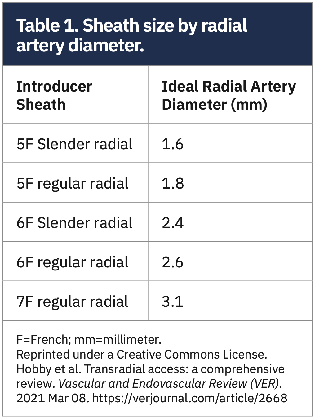
Rather than “giving up” the wire access to the ascending aorta, I typically leave an .035-inch exchange length wire extending from the existing sheath to the ascending aorta. Then I take an .035-inch compatible dilator from a standard 6F Terumo sheath kit (not the “Slender” kit) and insert that dilator into the corresponding 6F “Slender” sheath. I then introduce the Slender sheath over the .035-inch wire into the radial artery. Once you have done that, you are ready to advance your guide to the coronary and get started with the intervention. This works very well. I think you could do the same thing going from a 6F to 7F Slender. Table 1 is a sizing chart for radial artery sheaths.
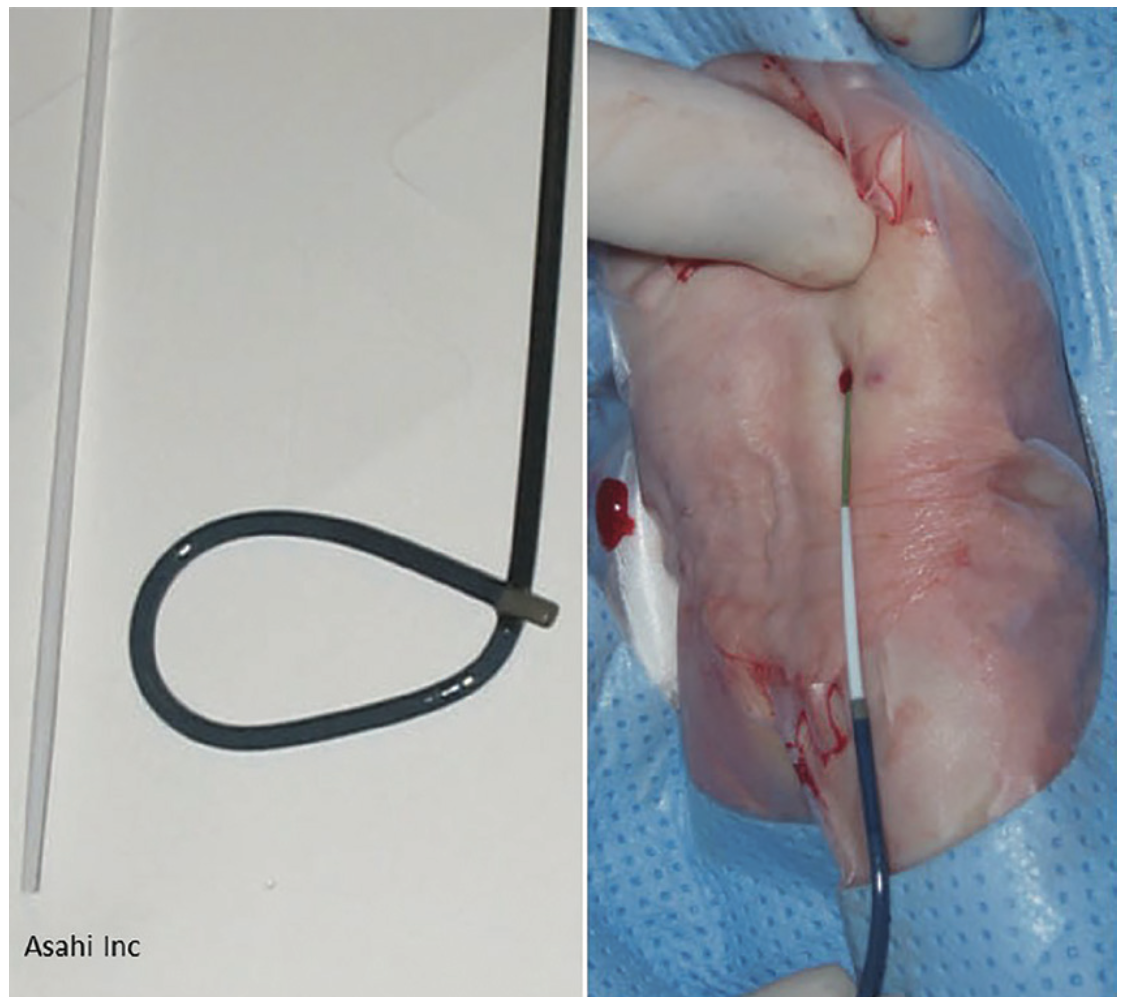
If you are already doing the intervention with a 6F guide in position and an .014-inch coronary guidewire is down the coronary, but you decide you want to upsize to 7F, it is a bit trickier, but can be done. First, back out the 6F guide so it is not engaging the coronary artery and is in the proximal ascending aorta, leaving your .014-inch (exchange length, or extension-docked) coronary guidewire in position down the coronary. Then, carefully advance an .025-inch Amplatz J-tipped wire (Boston Scientific) through the guide into the ascending aorta (but not into the coronary ostium!). Then, back the guide out of the body over the two wires, monitoring their position in the aorta/coronary to ensure they don’t move. Exchange the 6F sheath over both the .025-inch wire and the .014-inch wire for the 7F Slender sheath with an .035-inch dilator (see Figure 1). Advance a 7F guide over both wires to re-engage the coronary, and continue with your intervention after removing the .025-inch wire. Double-check that the .025-inch and .014-inch wires can both fit through the lumen of your dilator on the back table before you commit to taking everything out!
 Dmitriy N. Feldman, New York, New York: Doug, I use a standard .035-inch dilator in a Slender sheath for sheath exchanges. It works well to exchange to 5/6F Slender or to 6/7F Slender, and you don’t need to give up .035-inch long wire access. Then, with 7F Slender sheaths, use a 7F guide with mother-daughter technique, particularly for small radials.
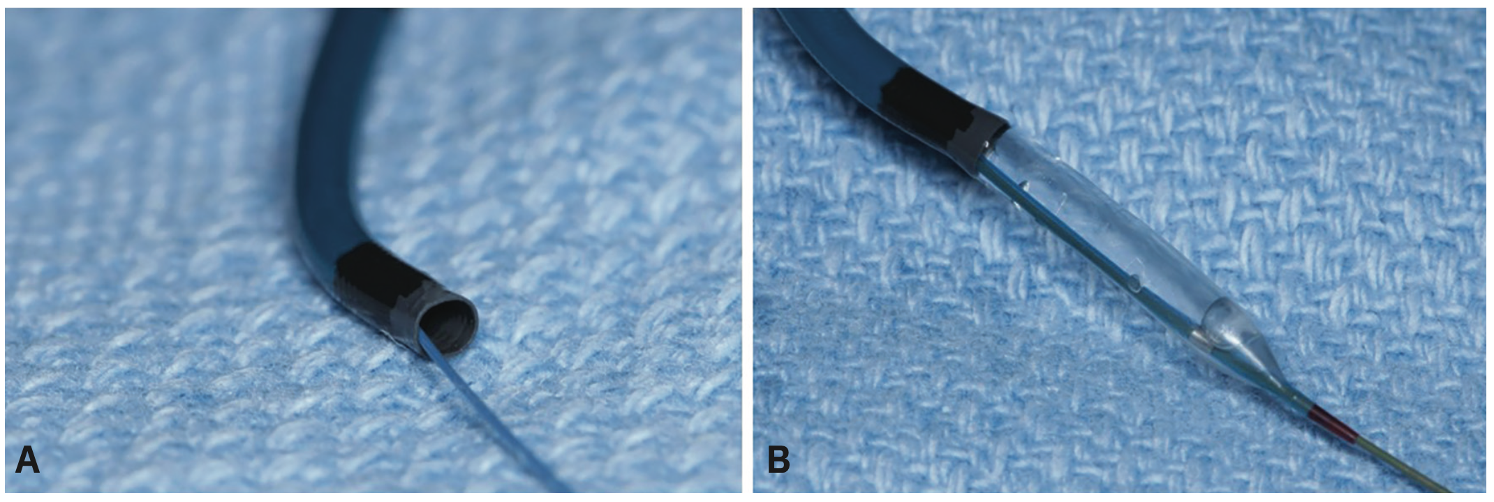
 J. Dawn Abbott, Providence, Rhode Island: I often use the option of exchanging to a sheathless guide (Eaucath Guiding Catheter [Asahi Intecc]), but also use the .035-inch compatible dilator in 7F Slender radial as well (Figure 2A-B).
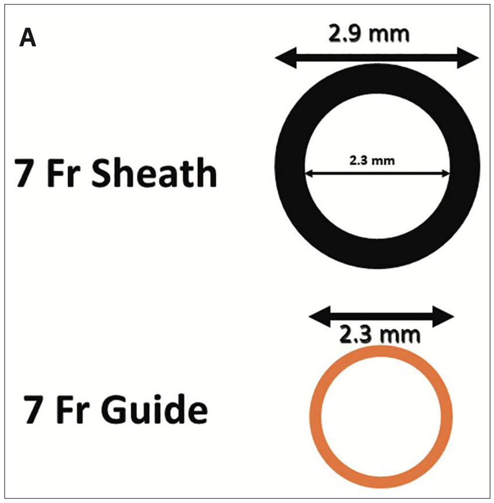
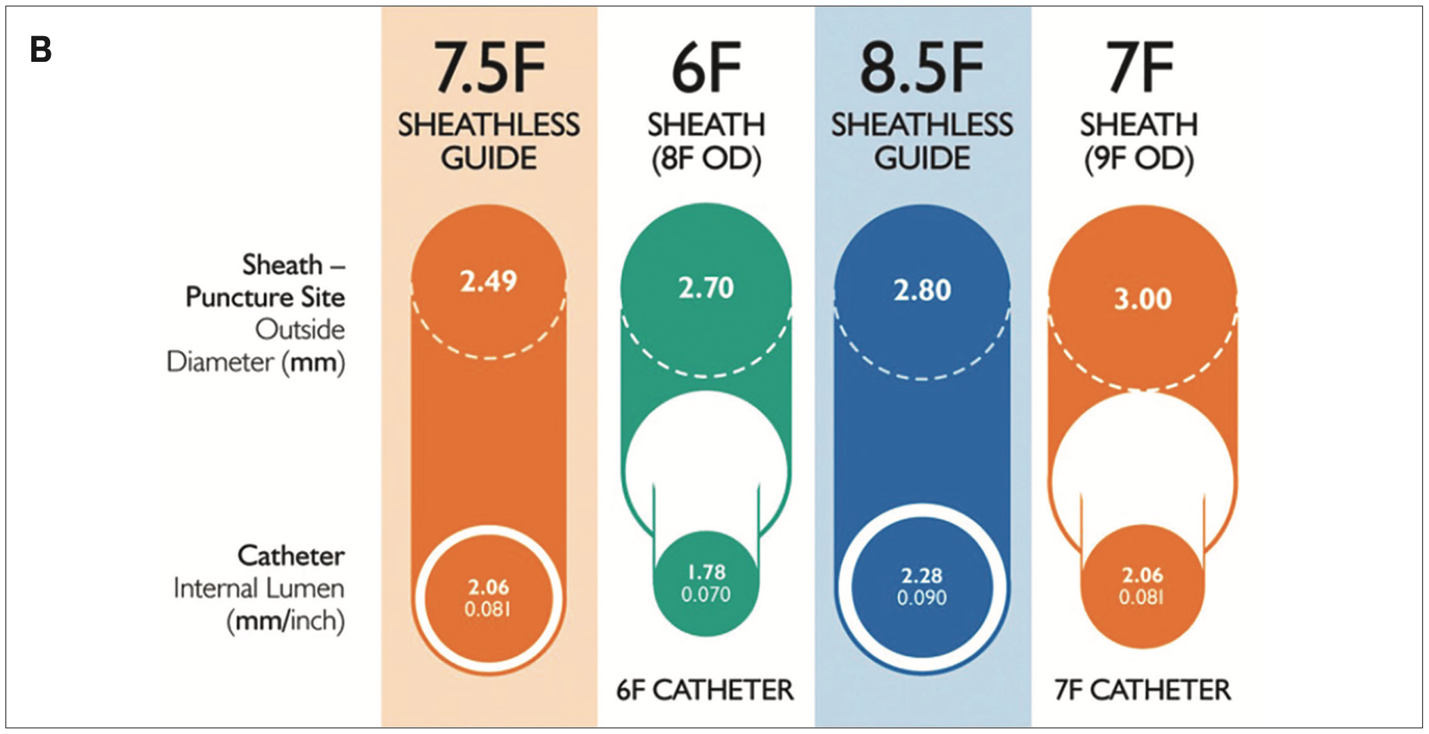
With permission from Wassef A, Cheema AN. Essential equipment for radial access problem solving: a synopsis of the techniques and technology available to assist with and improve the benefits of radial access procedures. Cardiac Interv Today. 2017 May/June; 11(3):42. https://citoday.com/articles/2017-may-june/essential-equipment-for-radial-access-problem-solving
Advancing Sheaths or Guide Catheter Without Dilators
We may not always consider the potential problem of advancing sheaths or guide catheter without dilators, or advancing a catheter when there is a mismatch between the lumen and its guiding wire (eg, an 0.14-inch wire through an .035-inch dilator lumen), which leaves an edge contacting the endothelium with the potential to disrupt the surface or even perforate it. In an earlier CLD Clinical Editor’s column2, we reviewed the balloon-assisted tracking method to help pass guide catheters up the arm over .014-inch guidewires. One of the best review articles was that of Obaid et al3 describing the balloon assisted tracking technique (BAT). Balloon-assisted tracking is a particularly useful technique for negotiating arterial loops and extreme tortuosity. BAT puts a smooth, tapered tip (the balloon catheter) in front of the diagnostic or guide catheter to ease passage through a tortuous or vasospastic arm artery (Figure 3).
The Steps for BAT
After the guide catheter is just outside the radial sheath, (1) insert an .014-inch regular guidewire through the guide, past the difficult arterial segment. (2) Advance a small percutaneous coronary intervention (PCI) balloon (<2 mm diameter) into the guide, positioning it at the end of the guide catheter and partially protruding from the distal end of the diagnostic or guiding catheter. (3) Inflate the balloon at low pressure (3 to 4 atmospheres). (4) Advance the catheter-balloon assembly over the guidewire, thus eliminating a sharp edge of the guide catheter, and facilitating a smooth and atraumatic catheter movement through the difficult vascular anatomy.
James C. Blankenship commented not only on BAT performance but also highlighted additional aspects of the procedure to become a ‘complete radialist’.4 In view of the advice about wire and sheath exchanges, I thought it would be good to paraphrase some of Dr. Blankenship’s recommendations:
1. Ultrasound imaging increases radial artery puncture success.5 It should be part of the routine.
2. Trouble with the radial artery? Look at the ulnar artery before crossing to femoral access. A recent CLD Clinical Editor’s Corner6 describes when the ulnar artery should be considered before abandoning transradial access.
3. Considering the contralateral radial access before switching to the femoral artery makes good sense. It will still have lower bleeding risk than femoral artery access.
4. When a problem of catheter advancement through the arm occurs, early angiographic imaging of the brachial artery is key. Look for a radial loop or a small, recurrent radial artery branch as the source of trouble. To reduce friction and facilitate catheter placement, one can use hydrophilic sheaths and mother-in-daughter 5F catheters with a 6F guide catheter.
5. Consider sheathless 7F guides for PCI when larger equipment or bifurcation stenting is planned.
6. Coronary catheter seating and manipulation is improved when operators have experience with several different catheters.
7. A right heart catheterization from the arm should be straightforward. Using ultrasound imaging, a brachial vein or branch can be easily cannulated. A 16-gauge Angiocath (BD) placed in the vein before the procedure will reduce access time. Using a micropuncture guidewire, we exchange the Angiocath for a 5F sheath in the brachial vein. A 5F pediatric balloon-tipped pulmonary artery catheter can then be easily positioned for right heart hemodynamics and estimated cardiac output from oxygen saturation samples. We can use an .014-inch wire to help with navigating the 5F pulmonary artery catheter. Femoral vein access is rarely necessary.
The Bottom Line
Procedure experience is gained not only by doing the procedures in various patients with challenging problems, but also by discussing the problems and methods to overcome them with expert colleagues. I hope that over the years this editor’s page has provided just such a forum for proceduralists and their cath lab teams to address problems for improved patient care.
Disclosures: Dr. Morton Kern reports he is a consultant for Abiomed, Abbott Vascular, Philips Volcano, ACIST Medical, and Opsens Inc.
Dr. Kern can be contacted at mortonkern2007@gmail.com
On Twitter @MortonKern
References
1. Doll JA, Beaver K, Naranjo D, et al. Trends in arterial access site selection and bleeding outcomes following coronary procedures, 2011-2018. Circ Cardiovasc Qual Outcomes. 2022 May; 15(5): e008359. doi:10.1161/CIRCOUTCOMES.121.008359
2. Kern M. Time to review your radial practice for best results. Cath Lab Digest 2017 Sept; 25(9): 6-10. https://www.hmpgloballearningnetwork.com/site/cathlab/article/time-review-your-radial-practice-best-results
3. Obaid D, Hailan A, Chase A, et al. Balloon-assisted tracking use reduces radial artery access failure in an experienced radial center and is feasible during primary PCI for STEMI. J Invasive Cardiol. 2017 Jul; 29(7): 219-224.
4. Blankenship JC. The compleat radialist. J Invasive Cardiol. 2017 Jul; 29(7): 225-226.
5. Seto AH, Roberts JS, Abu-Fadel MS, et al. Real-time ultrasound guidance facilitates transradial access: RAUST (Radial Artery access with Ultrasound Trial). JACC Cardiovasc Interv. 2015 Feb; 8(2): 283-291. doi:10.1016/j.jcin.2014.05.036
6. Kern MJ, Seto AH. Radial access failure: when to go ulnar? Cath Lab Digest. 2016 Nov; 24(11) 4-8. https://www.hmpgloballearningnetwork.com/site/cathlab/article/radial-access-failure-when-should-we-go-ulnar
Find More:
Grand Rounds With Morton Kern, MD
The Latest Clinical & Industry News
Podcasts: Cath Lab Conversations
Cardiovascular Ambulatory Surgery Centers (ASCs) Topic Center


