The HawkOne™ Directional Atherectomy System Puts Control in the Hands of the Operator
© 2024 HMP Global. All Rights Reserved.
Any views and opinions expressed are those of the author(s) and/or participants and do not necessarily reflect the views, policy, or position of Cath Lab Digest or HMP Global, their employees, and affiliates.
Note to readers: Cases #1 and #2 begin at the end of the interview.
Can you tell us about your practice?
 Michael Paisley, MD: We do a lot of peripheral work that includes the full spectrum of open, hybrid and endovascular peripheral vascular interventions. All of our cases are done in a hospital-based setting. Outpatient cases are run through the cath lab or a dedicated hybrid operating room.
 Brant Ullery, MD: A large percentage of our cases are multilevel and/or hybrid cases. In such cases, we have a low threshold to consider alternative arterial access sites, including both transradial (antegrade) and transpedal (retrograde) routes. In our 5-surgeon group, probably 50% of our overall volume centers around the treatment of peripheral arterial disease (PAD). All of us have embraced an endo-first approach for the overwhelming majority of our patients, particularly in the subgroup of patients with chronic limb-threatening ischemia (CLTI).
What is your treatment goal for your patients?
Brant Ullery, MD: When considering treatment options, it is important to stratify by indication given that the pathology, life expectancy, and comorbid status are different in those with claudication compared to those with rest pain or tissue loss. For claudicants, the endpoint of our intervention centers around durability — objectively measured by primary patency — and is usually reflected clinically by resolution or improvement in claudication symptoms. Patients with limb-threatening ischemia, on the other hand, represent a more vulnerable cohort as noted by the fact that nearly half of these patients die within four years of diagnosis.1 The therapeutic endpoints of peripheral intervention in such cases are, therefore, markedly different. Indeed, amputation-free survival, wound healing, and quality of life tend to dominant our goals for intervention, rather than simply primary patency.
Michael Paisley, MD: In a CLTI case, as long as the patient’s wounds heal and they are not having symptoms keeping them in the healthcare system, then it is not as crucial if the anterior tibial artery that I opened up loses patency a few months later. These aren’t focal femoral lesions that we are treating. We are seeing multilevel, tandem lesions and diffuse disease in diabetic, end stage renal disease patients, patients that are really, truly end-stage PAD, and these patients make up a majority of our practice. When it comes to treating claudicants, we are conservative. It is only when these patients fail medical therapy or have limb-threatening events or progression of disease that we offer a procedure, whether endovascular or surgical. Overall, direct in-line flow to the foot is the way to heal a wound and is achievable with both endovascular and open options. We are at least 85% endovascular in the fempop segment, but I still like a good distal vein bypass. It can be a durable option and sometimes it is the best thing to do, but not in some patients with multiple medical comorbidities and poor life expectancy.
How does the HawkOne directional atherectomy system (Medtronic) help you in your endovascular procedures?
Brant Ullery, MD: The HawkOne has long served as our dominant form of atherectomy. We have excellent case support from our local Medtronic personnel and I cannot overstate the advantage of the simplicity of use around the HawkOne. Over the last several years, the makeup of our surgical teams has changed due to the impacts from COVID and many of the trends affecting healthcare on a national level (eg, increased staff turnover, higher percentage of temporary workers). For example, a team member might include a traveling nurse who has never worked with atherectomy before. HawkOne offers an intuitive, highly reproducible procedure that is easy to use. The majority of our interventions are performed thru a 6-French access site, which nicely affords the opportunity for both radial and pedal access sites.
Michael Paisley, MD: Directional atherectomy allows us to create a controlled intimal disruption and plaque modification that can then be ballooned and expanded with reduced risk of bailout stenting.2 The HawkOne is plug and play. The learning curve is not steep and having sampled many other devices, I still think the HawkOne is the simplest to use. The HawkOne makes sense for plaque modification of eccentric calcification because it can treat a specific area versus just running a device through the vessel without a particular target.
Brant Ullery, MD: You can also take advantage of the directionality inherent in the HawkOne by specifically targeting those bulky, coral reef-like lesions that protrude into the lumen. We frequently use drug-coated balloons, but I maintain a low threshold for provisional or primary stenting in cases of prohibitive calcium or subintimal recanalization. If I have any concerns regarding inadequate stent expansion, even on circumferential, concentric plaque, I will use the HawkOne to improve luminal gain and enhance compliance of the blood vessel.2
What advice related to endovascular procedures would you give to a new physician starting their practice and using a new device for the first time?
Michael Paisley, MD: Follow the fundamentals and make sure you are not doing things that you have never heard of — that is how I keep the patient safe. As far as adopting a new device, if you can learn from the experience of the people around you, that is invaluable. If a partner has used that device, go do a case with them. If not, courses are available through industry partners. Leveraging the relationships you have with your local teams, whether it be with Medtronic or whatever device company you are working with, is extremely important. Finally, pick patients that have a good indication for the device you want to try and have a good reason to try it. Especially in the endovascular space, when you don’t know exactly what you are going to do every time as you go into a case.
On the HawkOne device in particular, there are good visual cues on the device. You can see exactly where the cutting blade is oriented when you are cutting. Then you can see when the nosecone is getting full, and you know the device is actually removing the plaque. The other good thing is that it is a rapid exchange model, so it is not necessary to do long wire transfers for long tibial lesions, which improves wire hygiene and decreases time for exchanges.
Brant Ullery, MD: We continue to deal with the repercussions of COVID, where we have to operate with new and inexperienced team members and work in less than ideal venues because of operating room capacity constraints. There can be many obstacles, some of which are modifiable and some of which are not. Taking a treatment modality like the HawkOne that puts all the control in your hands as the operator can help eliminate some of the unexpected challenges and unreliability of a varied team and operating room venue. Some atherectomy devices require a second pair of hands at the back of the wire, and that second person may or may not be trained on that specific device. There may be organizational and operational challenges regarding equipment, inventory, pharmacy, or consoles that are stored in different locations. Maximizing the control of the user experience at the level of the operator helps to ensure a reliable solution for treatment, regardless of procedural setting and team dynamic. Medtronic is unique in that it has treatment modalities to hit all categories, from plain balloon angioplasty to self-expanding stent, to drug-coated balloon, to directional atherectomy. Having the full armamentarium available allows us to better troubleshoot and tailor therapy to the given lesion in the given patient.
This article is supported by Medtronic.
References
1. Mustapha JA, Katzen BT, Neville RF, Lookstein RA, Zeller T, Miller LE, Jaff MR. Determinants of long-term outcomes and costs in the management of critical limb ischemia: a population-based cohort study. J Am Heart Assoc. 2018 Aug 21; 7(16): e009724. doi:10.1161/JAHA.118.009724
2. Babaev A, Halista M, Bakirova Z, et al. Directional versus orbital atherectomy of femoropopliteal artery lesions: angiographic and intravascular ultrasound outcomes. Catheter Cardiovasc Interv. October 2022; 100(4); 687–695. doi:10.1002/ccd.30339
Case #1: Treatment of Multi-Level Disease for Claudication With Rest Pain
Brant W. Ullery, MD, MBA, FACS, FSVS
In this case, a woman in her 70s was referred to my office with several months of progressive, disabling, right leg claudication and nocturnal rest pain. She was a former smoker with history of atrial fibrillation, chronic obstructive pulmonary disease, diabetes mellitus, and sick sinus syndrome. A right lower extremity arterial duplex noted diffuse moderate-to-severe arterial occlusive disease of the distal right femoropopliteal system and chronic occlusion of the proximal right anterior tibial artery and posterior tibial artery. The patient’s pre-intervention ankle-brachial indices (ABI) were 0.54 on the right and 0.76 on the left, with monophasic waveforms noted in the right leg distal to the mid superficial femoral artery (SFA).
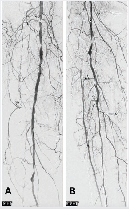
Ultrasound-guided access of the left common femoral artery was obtained, and right lower extremity angiography confirmed the presence of multiple tandem lesions in the right distal SFA and distal popliteal artery in excess of 75% stenosis (Figure 1A), as well as chronic occlusions of the proximal anterior tibial and posterior tibial arteries (Figure 1B). Her dominant infrapopliteal runoff was via a relatively disease-free peroneal artery.
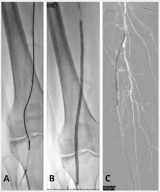
Next, a 6 Fr 65 cm Pinnacle Destination sheath (Terumo) was advanced over the aortic bifurcation to the level of the mid right SFA. Successful wire catheterization of the right anterior tibial artery was achieved with aid of an .018-inch 135 cm TrailBlazer support catheter (Medtronic) and .014-inch Glide Advantage wire (Terumo). A SpiderFX embolic protection device (Medtronic) was placed to the level of the distal anterior tibial artery. A total of four passes of the HawkOne M directional atherectomy system (Medtronic) was performed in the distal femoropopliteal and proximal anterior tibial arteries to serve as vessel preparation (Figure 2A).
Balloon angioplasty of the right distal SFA and popliteal arteries was subsequently performed using a 6 mm x 250 mm IN.PACT Admiral drug-coated PTA balloon catheter (Medtronic) (Figure 2B). Additional balloon angioplasty of the proximal anterior tibial artery was then completed using a 3.0 mm x 120 mm Chocolate PTA balloon catheter (Medtronic) (Figure 2C).
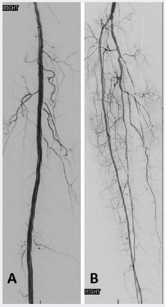
Completion angiography demonstrated a widely patent right distal femoropopliteal segment (Figure 3A) and restoration of 2-vessel infrapopliteal runoff, which included recanalization of the occluded proximal anterior tibial artery (Figure 3B). One-month follow-up duplex noted interval improvement in the right ABI to 0.93, including biphasic waveforms at the ankle in the peroneal and anterior tibial artery distributions. She reported full resolution of her disabling right lower extremity symptoms.
Case #2: Multi-Level Disease Treatment in CLTI
Michael J. Paisley, MD
A male in his late 70s with comorbidities including non-insulin dependent type 2 diabetes mellitus, smoking, hypertension, and chronic kidney disease presented to the emergency department with a history of worsening right foot pain and dry gangrene of the right 3rd toe. A physical exam demonstrated palpable bilateral femoral pulses, and non-palpable popliteal and distal pulses. A right lower extremity arterial duplex showed diffuse calcification with >75% stenosis of the distal superficial femoral artery, 50-74% stenosis of the mid popliteal artery, and occlusion of the peroneal artery with monophasic waveforms in the posterior and anterior tibial arteries with non-compressible ankle brachial index.
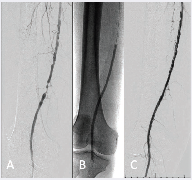
Ultrasound-guided access of the left common femoral artery was obtained and a right lower extremity angiogram was performed, demonstrating tandem distal superficial femoral artery (SFA) stenosis greater than 75% and 50% stenosis of the popliteal artery (Figure 1A). Below the knee there were tandem chronic total occlusions (CTO) of the mid and distal peroneal artery with dominant posterior tibial runoff, a distal occlusion at the level of the malleolus, and a distal anterior tibial artery occlusion without reconstitution of the dorsalis pedis artery (Figure 2A-B).
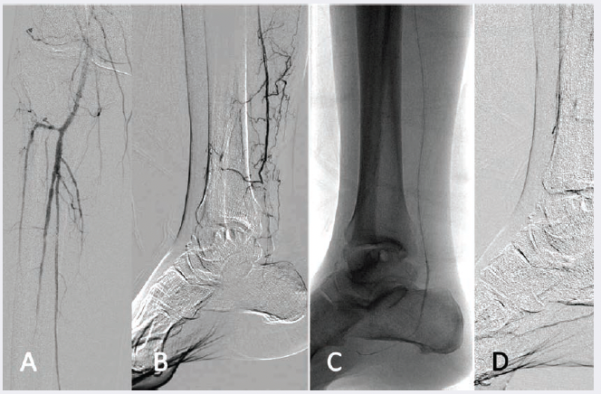
Selective angiogram of the distal posterior tibial artery with distal occlusion (B). 2 mm balloon
angioplasty of the distal posterior tibial and medial tarsal artery (C) with recanalization (D).
A 6 French 45 cm Pinnacle Destination sheath (Terumo) was seated in the proximal right SFA. An .035-inch Glidewire advantage (Terumo) and TrailBlazer support catheter (Medtronic) system traversed the SFA stenoses and a 5.0 mm SpiderFX Embolic Protection Device (Medtronic) was seated in the below-knee popliteal artery. A HawkOne M directional atherectomy system (Medtronic) was passed a total of 6 times in the distal superficial femoral and popliteal arteries for plaque remodeling and vessel preparation. Plain balloon angioplasty was performed in the popliteal and superficial femoral arteries. After confirmation of no dissections and resolution of stenosis, the segment was treated with drug-coated balloon angioplasty using the IN.PACT Admiral drug-coated balloon. The distal embolic protection device was removed.
An .014 wire was used to cross the posterior tibial artery CTO into the medial plantar artery. Angioplasty was performed using a 2.0 mm balloon with recanalization to the plantar artery (Figure 2C). Completion angiography demonstrated a widely patent SFA, popliteal artery (Figure 1C), and posterior tibial artery with inline flow to the medial tarsal artery (Figure 2D).
Follow-up arterial duplex ultrasound at 6 weeks showed biphasic waveforms throughout the SFA, popliteal, anterior tibial and posterior tibial arteries, and previously demonstrated peroneal artery occlusion. There was no hemodynamically significant stenosis visualized in the treated segments and non-compressible ABIs. The patient subsequently underwent a right 2nd and 3rd toe amputation for osteomyelitis, and on follow-up there was a granulating wound bed without residual infection.
For US Audiences Only
HawkOne™ Directional Atherectomy System
Indications for Use: The HawkOne™ directional atherectomy system is intended for use in atherectomy of the peripheral vasculature. The HawkOne catheter is indicated for use in conjunction with the SpiderFX embolic protection device in the treatment of severely calcified lesions. The HawkOne catheter is NOT intended for use in the coronary, carotid, iliac, or renal vasculature.
Contraindications: Do not use in the coronary arteries, carotid artery, or in the iliac, or renal vasculature. Do not use for in-stent restenosis at the peripheral vascular site.
Potential Adverse Effects of the Device on Health: The potential complications include, but are not limited to, amputation, aneurysm, arterial dissection, arterial perforation, arterial rupture, arterial spasm, arteriovenous fistula, bleeding complications, death, embolism or arterial thrombosis, emergency or non-emergency arterial bypass surgery, entry site complications, hypotension, infection, ischemia, restenosis of the treated segment, total occlusion of the peripheral artery, or vascular complications that could require surgical repair.
TrailBlazer™ Support Catheter
Indications for Use: TrailBlazer Support Catheter are percutaneous, single lumen catheters designed for use in the peripheral vascular system. TrailBlazer Support Catheters are intended to guide and support a guide wire during access of the vasculature, allow for wire exchanges and provide a conduit for the delivery of saline solutions or diagnostic contrast agents.
Chocolate™* PTA Balloon Catheter
Indications for Use: The Chocolate™* PTA balloon catheter is intended for balloon dilatation of lesions in the peripheral vasculature, including the iliac, femoral, ilio-femoral, popliteal, infra-popliteal, and renal arteries.
SpiderFX™ Embolic Protection Device
Lower Extremity Indications for Use: The SpiderFX Embolic Protection Device is indicated for use as a guidewire and embolic protection system to contain and remove embolic material in conjunction with the TurboHawk™ Peripheral Plaque Excision System, either during standalone procedures or together with PTA and/or stenting, in the treatment of severely calcified lesions in arteries of the lower extremities. The vessel diameter at the filter basket placement site should be between 3.0 mm and 6.0 mm.
IN.PACT™ Admiral™ Drug Coated PTA Balloon Catheter
Indications for Use: The IN.PACT™ Admiral™ Paclitaxel-coated PTA Balloon Catheter is indicated for percutaneous transluminal angioplasty, after appropriate vessel preparation, of de novo, restenotic, or in-stent restenotic lesions with lengths up to 360 mm in superficial femoral or popliteal arteries with reference vessel diameters of 4-7 mm.
Contraindications
The IN.PACT Admiral DCB is contraindicated for use in:
• Coronary arteries, renal arteries, and supra-aortic/cerebrovascular arteries
• Patients who cannot receive recommended antiplatelet and/or anticoagulant therapy
• Patients judged to have a lesion that prevents complete inflation of an angioplasty balloon or proper placement of the delivery system
• Patients with known allergies or sensitivities to paclitaxel
• Women who are breastfeeding, pregnant or are intending to become pregnant or men intending to father children. It is unknown whether paclitaxel will be excreted in human milk and whether there is a potential for adverse reaction in nursing infants from paclitaxel exposure.
Warnings
• Use the product prior to the Use-by Date specified on the package.
• Contents are supplied sterile. Do not use the product if the inner packaging is damaged or opened.
• Do not use air or any gaseous medium to inflate the balloon. Use only the recommended inflation medium (equal parts contrast medium and saline solution).
• Do not move the guidewire during inflation of the IN.PACT Admiral DCB.
• Do not exceed the rated burst pressure (RBP). The RBP is 14 atm (1419 kPa) for all balloons except the 200 and 250 mm balloons. For the 200 and 250 mm balloons the RBP is 11 atm (1115 kPa). The RBP is based on the results of in vitro testing. Use of pressures higher than RBP may result in a ruptured balloon with possible intimal damage and dissection.
• The safety and effectiveness of using multiple IN.PACT Admiral DCBs with a total drug dosage exceeding 34,854 µg of paclitaxel in a patient has not been clinically evaluated.
Precautions
• This product should only be used by physicians trained in percutaneous transluminal angioplasty (PTA).
• This product is designed for single patient use only. Do not reuse, reprocess, or resterilize this product. Reuse, reprocessing, or resterilization may compromise the structural integrity of the device and/or create a risk of contamination of the device, which could result in patient injury, illness, or death.
• Assess risks and benefits before treating patients with a history of severe reaction to contrast agents.
• The safety and effectiveness of the IN.PACT Admiral DCB used in conjunction with other drug-eluting stents or drug-coated balloons in the same procedure or following treatment failure has not been evaluated.
• The extent of the patient’s exposure to the drug coating is directly related to the number of balloons used. Refer to the Instructions for Use (IFU) for details regarding the use of multiple balloons and paclitaxel content.
• The use of this product carries the risks associated with percutaneous transluminal angioplasty, including thrombosis, vascular complications, and/or bleeding events
• Vessel preparation using only pre-dilatation was studied in the clinical study. Other methods of vessel preparation, such as atherectomy, have not been studied clinically with IN.PACT Admiral DCB.
• This product is not intended for the expansion or delivery of a stent.
Potential Adverse Effects
The potential adverse effects (e.g. complications) associated with the use of the device are: abrupt vessel closure; access site pain; allergic reaction to contrast medium, antiplatelet therapy, or catheter system components (materials, drugs, and excipients); amputation/loss of limb; arrhythmias; arterial aneurysm; arterial thrombosis; arteriovenous (AV) fistula; death; dissection; embolization; fever; hematoma; hemorrhage; hypotension/hypertension; inflammation; ischemia or infarction of tissue/organ; local infection at access site; local or distal embolic events; perforation or rupture of the artery; pseudoaneurysm; renal insufficiency or failure; restenosis of the dilated artery; sepsis or systemic infection; shock; stroke; systemic embolization; vessel spasms or recoil; vessel trauma which requires surgical repair.
Potential complications of peripheral balloon catheterization include, but are not limited to the following: balloon rupture; detachment of a component of the balloon and/or catheter system; failure of the balloon to perform as intended; failure to cross the lesion.
Although systemic effects are not anticipated, potential adverse events that may be unique to the paclitaxel drug coating include, but are not limited to: allergic/immunologic reaction; alopecia; anemia; gastrointestinal symptoms; hematologic dyscrasia (including leucopenia, neutropenia, thrombocytopenia); hepatic enzyme changes; histologic changes in vessel wall, including inflammation, cellular damage, or necrosis; myalgia/arthralgia; myelosuppression; peripheral neuropathy.
Refer to the Physician’s Desk Reference for more information on the potential adverse effects observed with paclitaxel. There may be other potential adverse effects that are unforeseen at this time.
Please reference appropriate product Instructions for Use for a detailed list of indications, warnings, precautions and potential adverse effects. This content is available electronically at www.manuals.medtronic.com.
UC202408114EN ©2024 Medtronic. All brands are trademarks of their respective owners.