Carbon Monoxide Poisoning: Incidence, Diagnosis & Treatment
Your ambulance is dispatched to a 42-year-old man following a syncopal episode. Over the weekend, he has had a headache, chills, nausea and dizziness -common flu-like symptoms. The patient is treated and transported to a local emergency department, where he is given intravenous hydration and is discharged neurologically intact with normal vital signs. Two days later, you are dispatched to the same address, where you find the patient unconscious on his living room floor. Following aggressive airway management, oxygenation and ventilation, the patient is transported to the ED, where he is diagnosed with carbon monoxide (CO) poisoning. He is treated with hyperbaric oxygen, and is discharged with chronic headaches and memory problems. After returning to his home two days earlier, he had again been exposed to CO from a faulty furnace, which caused permanent neurologic deficits.
Many substances can cause dramatic poisonings, and even death, but CO-an odorless, colorless, two-molecule gas produced by burning material containing carbon-accounts for greater mortality and morbidity than all other poisonings combined. Carbon monoxide causes thousands of needless deaths each year in the United States.1,2 It is the leading cause of accidental poisoning deaths in America. Patients who survive the initial poisoning still face the prospect of delayed neurologic dysfunction, which occurs in 14% to 40% of serious cases.
Initial symptoms of CO poisoning, such as headaches, nausea and fatigue, are often mistaken for the flu, because the deadly gas goes undetected in a home. Early confirmation of CO poisoning and continuous monitoring of carboxyhemoglobin levels can now be performed in the field with new, noninvasive pulse CO-oximetry.
Carbon monoxide, an insidious byproduct of incomplete hydrocarbon combustion, is generated in toxic amounts by internal-combustion engines, fossil-fuel heating systems and fires. Carbon monoxide emissions from modern automobiles, though reduced by regulatory standards, are still highly toxic in poorly ventilated spaces. A stable gas at physiologic temperatures, CO diffuses rapidly across the alveolar capillary membrane and binds tightly to iron centers in hemoglobin.
Dangers of CO Poisoning
Anything that creates a flame, even a candle, is a potential source of CO. During incomplete combustion, carbon, hydrogen and available oxygen combine to form carbon dioxide, water, heat and the deadly CO. Any disruption of the burning process or shortage of oxygen can increase CO production and accumulation to dangerous levels.
Carbon monoxide gas enters the blood system during normal breathing. Inhaled CO combines with hemoglobin to form carboxyhemoglobin (COHb). Once this conversion occurs, the hemoglobin is no longer available for transporting oxygen to other parts of the body. As the amount of CO increases in the bloodstream, the tissues become hypoxic. The rate at which carboxyhemoglobin accumulates in the body is a factor of the concentration of gas being inhaled (parts per million or percent) and duration of exposure. Smokers have a pre-existing build-up of carboxyhemoglobin dependent upon the frequency of smoking. Aggravating the effects of exposure is the long half-life of carboxyhemoglobin in the bloodstream.
Carboxyhemoglobin decreases blood oxygen content and hinders the release of oxygen from hemoglobin to tissues.3 Carbon monoxide quickly binds with hemoglobin with an affinity 200-250 times greater than that of oxygen to form COHb. The resulting
decrease in arterial oxygen content and shift of the oxyhemoglobin dissociation curve to the left explains the acute hypoxic symptoms (primarily neurologic and cardiac) seen in patients with CO poisoning; however, the toxic effects of CO cannot be explained by this process alone.4 COHb levels do not correlate well with symptoms or outcome, and this process cannot account for the phenomenon of delayed neurologic sequelae.5 In patients with severe poisoning, carboxyhemoglobin compromises delivery of oxygen to tissue and leads to tissue hypoxia and its immediate functional implications, especially for organs with high oxygen demands, such as the brain and heart.6,7
An ambient CO level of 100 ppm produces a COHb of 16% at equilibration, which is enough to produce significant clinical symptoms. This can be increased if exposure time is increased.8 Binding of CO to hemoglobin causes an increased binding of oxygen molecules at the three other oxygen- binding sites, resulting in a leftward shift in the oxyhemoglobin dissociation curve and decreasing the availability of oxygen to the already-hypoxic tissues.
Although an elevated COHb level is the primary diagnostic indicator of CO poisoning, it does not predict the severity of clinical signs and symptoms, particularly those affecting the brain.9 This poor correlation between COHb levels and neurologic presentation, which has long been recognized, is related to unmeasured tissue uptake of CO, which increases during hypoxia because of competition between CO and oxygen at the oxygen-binding sites on hemoglobins.
Evidence that myocardial infarction and other cardiopulmonary complaints have been directly related to high exposure levels of CO in the environment is now becoming available. Any exposure level, including low levels, is especially hazardous to fetuses, infants, children, the elderly and individuals with medical problems like anemia and heart or lung disease. Toxicology experts agree that it is difficult to estimate the true number of CO incidents because the symptoms of CO poisoning resemble so many other common ailments.10,11
CO Sources
The body produces CO as a by-product of hemoglobin degradation, but the gas does not reach toxic concentrations unless it is inhaled from exogenous sources, such as the incomplete combustion of carbon-based fossil fuels. According to a 10-year review of CO-related deaths, more than half of unintentional deaths were caused by motor vehicle exhaust. Although natural gas is touted as a clean fuel, combustion from forklifts and ice-rink resurfacers in enclosed environments has resulted in poisonings, as has burning charcoal, wood, kerosene or natural gas for heating and cooking (Table I).
Carbon monoxide is formed when organic compounds burn. The most common sources are motor vehicle exhaust, fire smoke, engine fumes and nonelectric heaters. Carbon monoxide poisoning is often associated with malfunctioning or obstructed exhaust systems and with suicide attempts.
Sources of CO include:
- Gas water heaters
- Kerosene space heaters
- Charcoal and hibachi grills
- Propane stoves
- Cigarette smoke
- Propane-fueled forklifts
- Gas-powered concrete saws
- Indoor tractor pulls
- Swimming behind a motorboat or under a houseboat
- Spray paint, solvents, degreasers and paint removers.
Although most fatalities result from fires, stoves, portable heaters and automobile exhaust cause approximately one-third of deaths. Cigarette smoke is a significant source of CO and has a significant impact on myocardial oxygen delivery.
Increasing evidence implicates ambient urban CO levels in rates of angina, arrhythmias and cardiac arrest. Presuming that the evidence is quantifiable, and depending on the true extent, this implies a significant underreporting of CO-associated injuries and deaths.
Carbon monoxide can occur in the presence of other toxins, complicating both diagnosis and treatment. It is a major contributor in the thousands of smoke-inhalation deaths that occur each year. People who work with methylene chloride, a paint stripper, can be poisoned because the fumes are readily absorbed and converted to CO in the liver. In such cases, peak carboxyhemoglobin (COHb) levels may be delayed and prolonged because of ongoing production.
Symptoms of CO Poisoning
The acute symptoms of CO poisoning are reflected in the susceptibility to hypoxia of organs with high metabolic oxygen consumption-the brain and heart. Initially, patients may complain of headache, dizziness or nausea, resulting in an incorrect diagnosis of influenza. In infants, vomiting may be the only presenting symptom and may be misdiagnosed as gastroenteritis. Neurologic symptoms, such as coma or seizures, can occur in patients with prolonged CO exposure. Elderly patients, especially those with coronary artery disease, may have accompanying myocardial ischemia, which may proceed to myocardial infarction.
Although the brain and heart are sensitive to CO poisoning, other organs are also affected. Prolonged exposures, especially those resulting in coma or altered mental status, may be accompanied by retinal hemorrhages and lactic acidosis. Cherry-red skin color, a very late indicator of significantly elevated carboxyhemoglobin levels, is associated with severe CO poisoning, but is seen in only 2%-3% of symptomatic cases. Cherry-red skin color is not a reliable sign of CO poisoning.
Identifying patients with CO poisoning can be difficult, as symptoms are often vague. In acute CO poisoning, the initial flu-like symptoms can be followed by dizziness, ataxia, agitation, impairment of consciousness and respiratory failure. Cerebral edema and metabolic acidosis may develop in serious cases. An inaccurate diagnosis in the emergency department of any of these symptoms may result in the discharge of patients back into the environment that has been poisoning them.
Patient Assessment
Due to the vague and common symptoms, physical assessment is of limited value. Inhalation injury or burns should always alert the prehospital clinician to the possibility of CO exposure.
- Vital signs
- Tachycardia
- Hypertension or hypotension
- Changes in the 12-lead ECG indicative of myocardial ischemia
- Marked tachypnea (rare; severe intoxication often associated with mild or no tachypnea)
- Skin: Classic cherry red skin is rare (i.e., “When you're cherry red, you're dead”); pallor is present more often.
- Noncardiogenic pulmonary edema
- Neurologic and/or neuropsychiatric symptoms
- Patients display memory disturbance (most common), including amnesia.
- Long-term exposures or severe acute exposures frequently result in long-term neuropsychiatric deficits. Additionally, some individuals develop delayed neuropsychiatric symptoms, often after severe intoxications associated with coma.
- After recovery from the initial incident, patients may present several days to weeks later with neuropsychiatric symptoms like those just described.
Confirmation of CO Poisoning
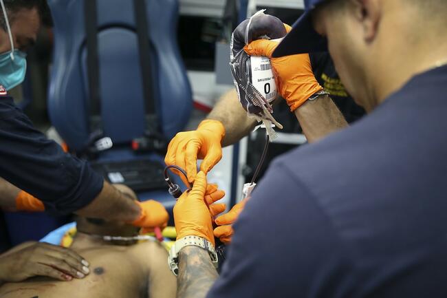
A hospital CO-oximeter can determine the percentage of CO-saturated hemoglobin in the blood. These methods for accurately measuring CO poisoning have been limited to invasive blood tests analyzed by blood gas machines with CO-oximetry measurement capability and are not available in the field.12 While many hospitals have blood gas machines with CO-oximetry, many smaller hospitals do not, which makes confirmed diagnosis of CO poisoning in these situations impossible. In EMS, there has been no easy-to-use method to measure CO poisoning, and prehospital clinicians have had to rely on a high degree of clinical suspicion, general patient condition and environmental factors to make treatment and transport decisions.
Traditional Pulse Oximetry and Carboxyhemoglobin
The traditional pulse oximeter measures functional arterial oxyhemoglobin saturation (SpO2) because it utilizes only two wavelengths of light: red and infrared. The pulse oximeter can differentiate only between deoxygenated (or reduced) hemoglobin, which has the ability to transport oxygen, and saturated hemoglobin. Carboxyhemo-globin (COHb) and methemoglobin are also absorbed at similar wavelengths; the traditional pulse oximeter is incapable of distinguishing these dyshemoglobins.13 Patients who are at risk for high concentrations of either CO (patients with smoke inhalation or CO poisoning) should be monitored using CO-oximetry. Pulse oximeters will display an oxygen saturation that is approximately equal to the percentage of hemoglobin combined with oxygen, plus the percentage of hemoglobin combined with CO. If 25% of a patient's hemoglobin is saturated with CO and has a true oxygen saturation of 70%, a traditional pulse oximeter will display an SpO2 of about 95%. Traditional pulse oximetry is of no value in patient assessment in the presence of high concentrations of COHb.14
Pulse CO-Oximetry
Pulse CO-oximetry uses advanced hardware and software that is able to collect more information than traditional pulse oximetry and allows EMS providers to accurately monitor CO and other vital parameters continuously and noninvasively.
Pulse CO-oximetry technology combines multiple wavelengths of light for analysis of physiological data. Processing of the data permits pulse CO-oximeters to accurately measure carboxyhemoglobin SpCO in the field without the need for invasive testing that is available only in some hospital environments. The rapid and noninvasive measurement of COHb SpCO with pulse CO-oximetry may improve treatment and transport decisions.
Initial Treatment
Initial treatment of patients with symptomatic CO poisoning is relatively straightforward. A nonrebreather mask supplies 80%-100% oxygen to quickly clear COHb from the blood. This therapy reduces the half-life of COHb from 4-5 hours (breathing room air) to approximately one hour. The presence of hypotension implies myocardial dysfunction or peripheral vasodilation, which can be treated with fluids and vasopressors as needed. In patients with an altered mental status, a fingerstick glucose test is essential to rule out hypoglycemia. Hyperglycemia may exacerbate central nervous system damage, thus should be treated with insulin.
Complications of CO poisoning can be treated with supportive measures. Seizures occasionally result, requiring routine administration of benzodiazepines. Patients may benefit from continuous 12-lead ECG monitoring for signs of myocardial ischemia. Continuous monitoring with pulse CO-oximetry will provide the hospital with an initial measurement during assessment and a trend with treatment.
While it may be tempting to base treatment decisions on specific COHb levels, these levels do not correlate well with symptoms and definitely do not predict sequelae. A single measurement is not representative of peak level or total tissue exposure; however, COHb levels are important in diagnosing CO exposure. In nonsmoking patients, a COHb level greater than 5% confirms exposure if 100% oxygen therapy has been administered for no more than one hour. Patients who smoke more than two packs per day can have COHb levels approaching 10%. Any patient with a high COHb level (>25%) or serious symptoms (e.g., syncope) may need more intensive treatment beyond routine oxygen therapy.
Hyperbaric Oxygen
Once a patient with acute CO poisoning has been identified and is receiving initial treatment, a transport decision must be made. The patient may benefit from hyperbaric oxygen (HBO).15 The clinical utility of hyperbaric oxygen has been best studied in the context of CO poisoning.16 The most obvious effect is enhanced clearance of COHb (half-life < 30 minutes with HBO), but this is usually clinically unimportant. In fact, because of the long delay between a patient's initial presentation and actual entry into the chamber, it is unlikely that much COHb remains;17 however, patients may still benefit from other, more important physiologic effects of hyperbaric oxygen.
COHb levels should not be used as the basis for treating CO poisoning. HBO therapy should be considered for patients who do not initially meet the criteria for such therapy but have persistent neurologic symptoms despite several hours of 100% oxygen therapy.18 This is especially true in patients with a severe headache or impaired mental function. However, the decision to use HBO therapy should be made early by the physician, since efficacy may decrease with delay, especially beyond six hours. The final considerations regarding use of hyperbaric oxygen should be stability of the patient's condition and distance to the nearest chamber or appropriate facility, based on local policies and procedures.
Pregnancy
Fetal hemoglobin has a high affinity for CO; thus, a fetus may be more susceptible to toxic effects than the mother. This may explain why pregnant patients with only moderate symptoms and no syncope have had devastating fetal outcomes.19,20 The primary concern in pregnant patients is that COHb clearance may take 4-5 times longer in the fetus than in the mother. Pregnant patients with CO poisoning do need aggressive prehospital oxygen treatment, and HBO therapy should be offered if neurologic symptoms or signs of fetal distress are present.21 Many centers use hyperbaric oxygen in any pregnant patient with a COHb level of at least 15%, regardless of symptoms.
Prevention
Being aware of the symptoms of CO poisoning can lead to early intervention and prevent needless deaths. Burn victims, especially those with evidence of smoke inhalation from an enclosed fire, should undergo rapid pulse CO-oximetry testing for COHb SpCO levels and receive appropriate treatment. CO poisoning should be suspected in patients presenting with flu-like symptoms, which they may not attribute to a faulty furnace or other source. Symptoms coinciding with the use of a combustion engine (e.g., motor vehicle, boat, forklift) in an enclosed area should also raise suspicion. EMS providers can help facilitate evaluation of an offending environment by local utilities or fire department personnel and raise patient awareness of symptoms and potential sources of CO. In addition, CO detectors can have a profound impact on home safety and are recommended by many safety organizations.22
Primary prevention of residential CO exposure can be accomplished through simple precautions. Although residential CO detectors are important for early detection, they should be considered a secondary prevention method. High oil and gas prices and power outages during winter months can contribute to consumer use of improperly vented heating sources. Public education campaigns, especially during winter months, combined with provision of battery-operated CO detectors for low-income persons, might reduce CO poisonings.
CO from a running vehicle inside an attached garage may enter the home, even with the garage door open. Normal circulation does not provide adequate fresh air to reliably prevent dangerous accumulations of CO inside the house. Check all fuel-burning household heating equipment (fireplaces, furnaces, water heaters, wood stoves, and space or portable heaters) each year at the onset of the heating season. All chimneys and chimney connectors should be evaluated for proper installation, cracks, blockages or leaks. Before enclosing central heating equipment in a smaller room, check with your fuel supplier to ensure that air is provided for proper combustion. When using a fireplace, open the flue for adequate ventilation. Always use barbecue grills capable of producing CO outside-never in the home or garage.
Conclusion
Carbon monoxide is a colorless, odorless, poisonous gas that causes vague flu-like symptoms that are often misinterpreted by both patients and physicians. Fortunately, its slow action allows time for diagnosis and appropriate intervention. Patients who present to EMS with flu-like symptoms should be questioned regarding risk factors that may warrant testing for CO poisoning. The use of pulse CO-oximetry can rapidly identify those patients exposed to CO and provide physiologic data for improved treatment and transport decisions. The mainstays of treatment are the liberal use of 100% oxygen therapy and attention to potential poisoning of housemates. The use of hyperbaric oxygen is encouraged in significant poisonings. n
Table I: Risks for Exposure to Carbon Monoxide
- Children riding in the back of enclosed pickup trucks (particularly high risk)
- Industrial workers at pulp mills, steel foundries and plants producing formaldehyde or coke
- Personnel at fire scenes
- Using heating sources during power outages
- Individuals working indoors with combustion engines or combustible gases
References
- r 1. Raub JA, Mathieu-Nolf M, Hampson NB, Thom SR. Carbon monoxide poisoning-a public health perspective. Toxicology 145(1):1-14, Apr 7, 2000.
- r 2. Cobb N, Etzel RA. Unintentional carbon monoxide-related deaths in the United States, 1979 through 1988. JAMA 266(5):659-663, 1991.
- r 3. Hardy KR, Thom SR. Pathophysiology and treatment of carbon monoxide poisoning. J Clin Toxicol 32(6):613-629, 1994.
- r 4. Piantadosi CA. Toxicity of carbon monoxide: Hemoglobin vs histotoxic mechanisms. In: Penney DG, ed. Carbon Monoxide, pp. 163-186. Boca Raton, FL: CRC Press, 1996.
- r 5. Choi IS. Delayed neurologic sequelae in carbon monoxide intoxication. Arch Neurol 40(7):433-435, 1983.
- r 6. Sokal JA, Kralkowska E. The relationship between exposure duration, carboxyhemoglobin, blood glucose, pyruvate and lactate and the severity of intoxication in 39 cases of acute carbon monoxide poisoning in man. Arch Toxicol 57(3):196-199, 1985.
- r 7. Brown SD, Piantadosi CA. Recovery of energy metabolism in rat brain after carbon monoxide hypoxia. J Clin Invest 89(2):666-672, 1992.
- r 8. Thom SR, Xu YA, Ischiropoulos H. Vascular endothelial cells generate peroxynitrite in response to carbon monoxide exposure. Chem Res Toxicol 10(9):1023-1031, 1997.
- r 9. Thom SR. Antagonism of carbon monoxide-mediated brain lipid peroxidation by hyperbaric oxygen. Toxicol Appl Pharmacol 105(2):340-344, 1990.
- r 10. Heckerling PS, Leikin JB, Maturen A. Occult carbon monoxide poisoning: validation of a prediction model. Am J Med 84(2):251-256, 1988.
- r 11. Satran D, Henry CR, Adkinson C, et al. Cardiovascular manifestations of moderate to severe carbon monoxide poisoning. J Am Coll Cardiol 45(9):1513-1516, May 3, 2005.
- r 12. Turnbull TL, Hart RG, Strange GR, et al. Emergency department screening for unsuspected carbon monoxide exposure. Ann Emerg Med 17(5):478-483, 1988.
- r 13. Buckley RG, Aks SE, Eshom JL, et al. The pulse oximetry gap in carbon monoxide intoxication. Ann Emerg Med 24(2):252-255, 1994.
- r 14. Bozeman WP, Hampson NB. Pulse oximetry in CO poisoning-additional data. Chest 117(1):295-296, Jan 2000.
- r 15. Sloan EP, Murphy DG, Hart R, et al. Complications and protocol considerations in carbon monoxide-poisoned patients who require hyperbaric oxygen therapy: Report from a ten-year experience. Ann Emerg Med 18(6):629-634, 1989.
- r 16. Tomaszewski CA, Thom SR. Use of hyperbaric oxygen in toxicology. Emerg Med Clin North Am 12(2):437-459, 1994.
- r 17. Hampson HB, Little CE. Hyperbaric treatment of patients with carbon monoxide poisoning in the United States. Undersea Hyperb Med 32(1):21-26, Jan-Feb 2005.
- r 18. Messier LD, Myers RA. A neuropsychological screening battery for emergency assessment of carbon-monoxide-poisoned patients. J Clin Psychol 47(5):675-684, 1991.
- r 19. Koren G, Sharav T, Pastuszak A, et al. A multicenter, prospective study of fetal outcome following accidental carbon monoxide poisoning in pregnancy. Reprod Toxicol 5(5):397-403, 1991.
- r 20. Hill EP, Hill JR, Power GG, et al. Carbon monoxide exchanges between the human fetus and mother: A mathematical model. Am J Physiol 232(3):H311-323, 1977.
- r 21. Elkharrat D, Raphael JC, Korach JM, et al. Acute carbon monoxide intoxication and hyperbaric oxygen in pregnancy. Intensive Care Med 17(5):289-292, 1991.
- r 22. Krenzelok EP, Roth R, Full R. Carbon monoxide-the silent killer with an audible solution. Am J Emerg Med 14(5):484-486, 1996.