

Noninvasive Substrate and Activation Mapping for Catheter Ablation of Ventricular Tachycardia: Are We There Yet?
© 2024 HMP Global. All Rights Reserved.
Any views and opinions expressed are those of the author(s) and/or participants and do not necessarily reflect the views, policy, or position of EP Lab Digest or HMP Global, their employees, and affiliates.
EP LAB DIGEST. 2024;24(1):1,14-16.
Savalan Babapoor, MD, and Chirag Barbhaiya, MD, Leon H Charney Division of Cardiology, Cardiac Electrophysiology, NYU Langone Health, New York University School of Medicine, New York, New York
Despite recent advancements in catheter ablation of scar-related ventricular tachycardia (VT), including development of multielectrode mapping catheters and automated electrogram annotation algorithms to identify important substrate,1 mapping during VT ablation remains a clinical challenge. Delineation of scar boundaries and characterizing arrhythmogenic substrate currently requires placement of a mapping catheter at all areas of anatomic interest, a process that can be limited by catheter ectopy and patient anatomy.2 Activation mapping is limited by hemodynamic instability during VT and the frequency with which points can be acquired during VT.
Periprocedural hemodynamic decompensation affects approximately 11% of patients undergoing VT ablation3, often necessitating mechanical circulatory support and prolonged hospitalization. Limiting VT induction, shortening procedure duration, and facilitating focused ablation with effective noninvasive mapping may help improve outcomes and health care utilization related to VT ablation.
Preprocedural Imaging as Substrate Map
Until recently, the utility of 3-dimensional (3D) cardiac imaging such as cardiac magnetic resonance (CMR) and multidetector computed tomography (MDCT) was limited by the absence of tools to integrate relevant imaging data into electroanatomic mapping (EAM) systems. For example, high-resolution contrast-enhanced CMR can accurately delineate scar tissue by highlighting fibrosis areas in the heart, including mid-myocardial fibrosis that would not be detected by traditional substrate mapping, and enable clear differentiation between dense scar and more heterogeneous scar, and thus, arrhythmogenic scar.4 MDCT has a higher spatial resolution that significantly enhances the delineation of myocardial thinning observed in ischemic scars and complex cardiac structures such as papillary muscles and coronary vasculature. Image integration software modules included within commonly used EAM systems, however, only allow segmentation and integration of basic chamber geometry, leaving the operator to mentally integrate relevant substrate characterization data intraoperatively.
Proprietary software for processing of CT and CMR data are now commercially available through ADAS 3D (Adas3D Medical SL), and MUSIC/InHeart (IHU LIRYC Bordeaux and Inria Sophia) to generate 3D models that display relevant details from preprocedural imaging in a model that can be integrated into EAM systems. These technologies have shown that CT and MRI studies could be used to generate an interactive 3D model of the patient’s heart that includes essential anatomical structures such as phrenic nerves, myocardial thickness from CT as a surrogate for ischemic scar, and late gadolinium enhancement (LGE) from MRI to allow visualization of epicardial and mid-myocardial scar, coronary arteries, and epicardial fat. Image integration with the MUSIC/InHeart software has shown to decrease ablation times in VT ablation by 1.5 to 3 hours.5,6 From delayed enhancement MRI, ADAS 3D generates ventricular scar maps ranging from endocardial to mid-myocardial to epicardial, illustrating potential conduction channels within dense scar based on intensity of LGE on MRI, which have been correlated with markers of slow conduction by conventional electroanatomic mapping.7-9
ECG Imaging as Activation Map
ECG imaging (ECGi) was initially used to guide treatment of focal ventricular arrhythmias; however, more recent reports have demonstrated utility in mapping of scar-related arrhythmia to guide both catheter ablation and cardioradioablation.10 Commercially available ECGi modalities include CardioInsight (Medtronic), which employs a 252-body surface electrode vest combined with a study-specific computed tomography derived epicardial geometry to display the full sequence of electrical activity during a single beat over the whole heart, and VIVO (Catheter Precision, Inc), an ECGi system that combines a patient-specific myocardial model from MRI or CT, a 3D photo of the torso for defining ECG electrode positions, and 12-lead ECG recordings of ventricular arrhythmias to localize the origin of the focal ventricular arrhythmia.11 The accuracy and usage of these ECGi modalities for mapping of scar-related ventricular arrhythmias is unclear.
We present a case demonstrating the feasibility and potential utility of noninvasive substrate and activation mapping to facilitate catheter ablation of scar-related VT.
Case Presentation
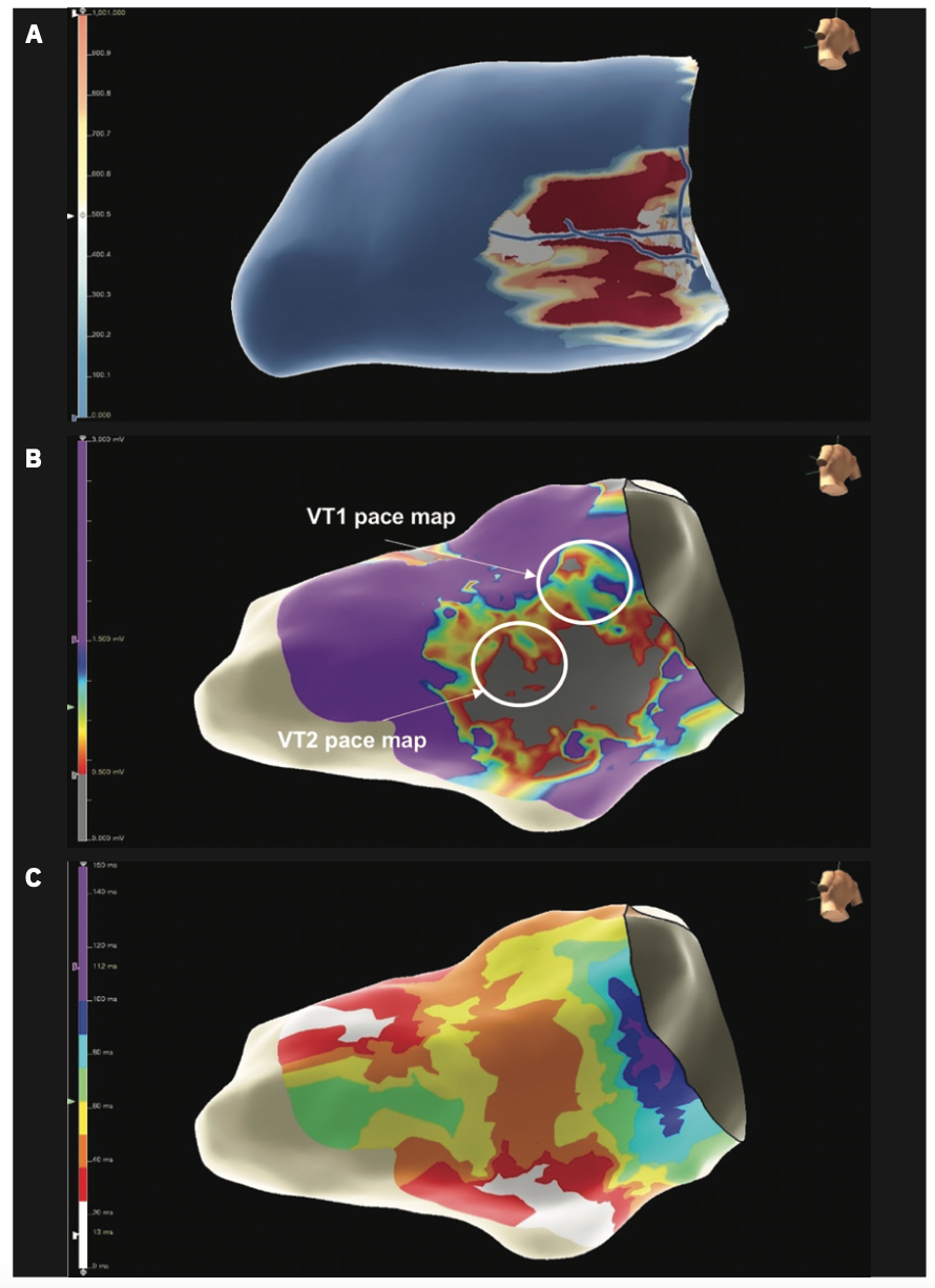
A 70-year-old man with a history of coronary artery disease, coronary artery bypass graft surgery, and secondary prevention implantable cardioverter-defibrillator (ICD) implantation, presented with recurrent episodes of monomorphic VT requiring ICD shocks despite escalating doses of amiodarone. He was referred for VT ablation. Cardiac MRI examination revealed extensive transmural LGE accompanied by wall thinning and akinesis, specifically affecting the basal inferolateral wall of the left ventricle, a 3D model of which was generated using ADAS 3D (Figure 1A). His left ventricular ejection fraction (LVEF) was measured at 41%.
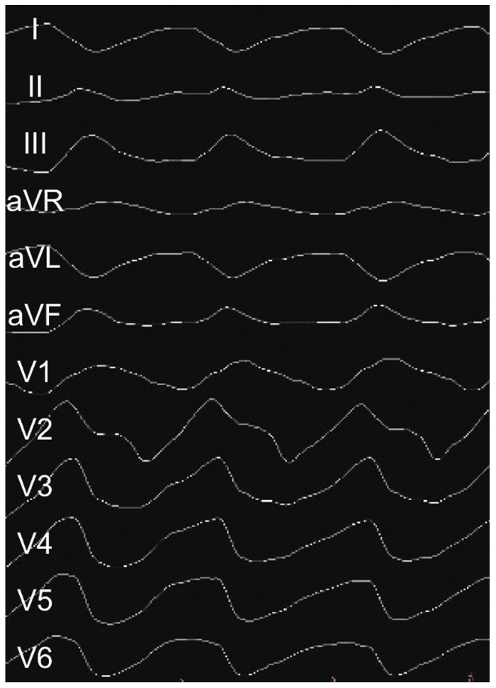
After initiation of general anesthesia with high frequency jet ventilation, vascular access was obtained and a TactiFlex Ablation Catheter (Abbott) was advanced to the right heart to create initial geometry and collect fiducial points to facilitate merge of EAM geometry with imaging-based substrate map and ECGi. The ablation catheter was then used for programmed electrical stimulation, resulting in induction of VT1 with a cycle length of 200 milliseconds, inferior axis, concordantly positive in precordial leads, and negative in lead I (Figure 2). Burst pacing to terminate VT was unsuccessful and the patient was cardioverted to restore sinus rhythm.
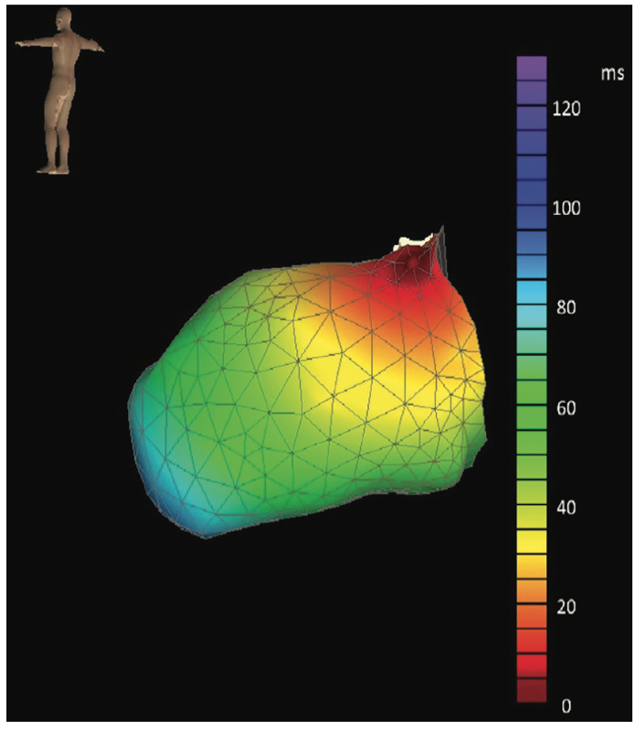
During transseptal puncture, the substrate map generated by ADAS 3D was merged with the EnSite X (Abbott) electroanatomic anatomy, and a 12-lead ECG was exported to VIVO for creation of ECGi to localize VT1 exit site (Figure 3). Detailed, high-density, focused omnipolar mapping of basal, lateral LV was performed, confirming low voltage (Figure 1B) and isochronal late activation mapping (ILAM) deceleration zones identified by the presence of “isochronal crowding,” defined as 3 colors within ~1 cm (Figure 1C), in region corresponding with LGE and possible channels on ADAS 3D map (Figure 1A). Pace match mapping at exit site identified by VIVO confirmed likely exit site of VT1 at basal anterolateral aspect of scar. Ablation lesions were strategically delivered to render region of likely VT1 exit and deceleration zones within lateral LV scar unexcitable to high output pacing.
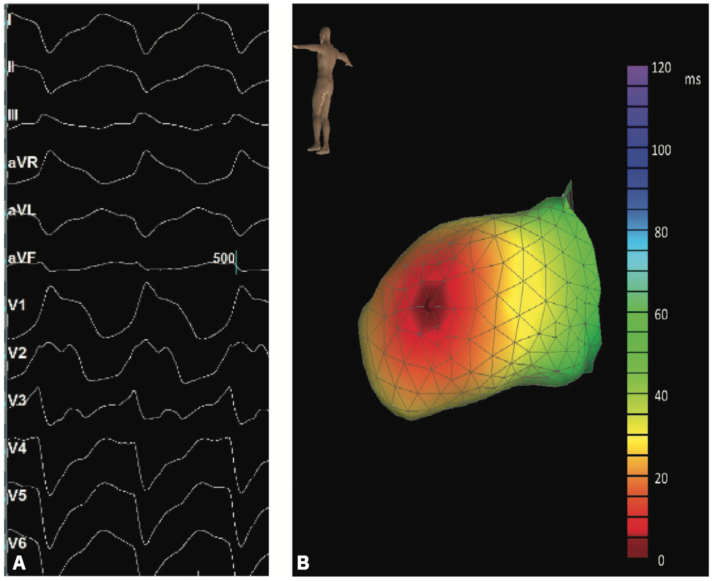
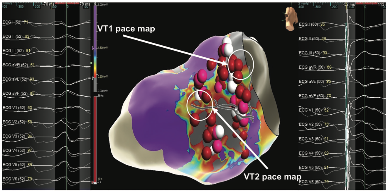
Repeat programmed electrical stimulation with up to triple extrastimuli with isoproterenol infusion confirmed noninducibility of VT1; however, VT2 with cycle length 242 milliseconds, right superior axis, and right bundle morphology with V3-V4 transition was induced (Figure 4A). VIVO ECGi exit site was at apical inferior aspect of lateral scar (Figure 4B). The lesion set was expanded to incorporate the more apical VT exit site, which was also confirmed with pace mapping (Figure 5). Of note, both exit sites for induced VTs were within channels identified by ADAS 3D analysis of cardiac MRI (Figure 5). Following radiofrequency application to render the VT2 exit site unexcitable to high output pacing, programmed stimulation with up to 3 extrastimuli with isoproterenol infusion was repeated, and no VT was inducible. The patient continues to remain free from VT at 6-month follow-up following catheter ablation.
Discussion
This case demonstrates the feasibility and potential usage of combined noninvasive substrate and activation mapping to facilitate catheter ablation of scar-related VT. There was excellent agreement between noninvasive mapping data and confirmatory traditional mapping in the presented case. Additional research is needed to ascertain the optimal criteria for defining substrate noninvasively, develop best practices for synchronization of noninvasive and invasive mapping data, and ultimately determine the degree to which confirmatory invasive mapping is required. Noninvasive ECGi activation mapping was previously limited by the need for a specialized vest and dedicated preprocedural CT scan. The ECGi approach used in the present case accurately localized VT using data and imaging readily available during a conventional ablation procedure. While the accuracy of ECGi for localization of VT exit sites for scar-related arrhythmia requires prospective validation, the appeal of immediate exit site identification, particularly in cases with multiple induced VTs without the need for multiple inductions of sustained VT, is clear. The combination of noninvasive substrate and activation mapping holds great potential as a means of improving the efficiency, safety, and effectiveness of VT ablation procedures.
Disclosure: The authors have completed and returned the ICMJE Form for Disclosure of Potential Conflicts of Interest, and report no conflicts of interest regarding the content herein. Dr Barbhaiya reports consulting fees from Abbott and Biosense Webster, Inc, and payment or honoraria for lectures, presentations, speakers bureaus, manuscript writing, or educational events from ZOLL Medical.
References
1. Vlachos K, Letsas KP, Srinivasan NT, et al. The value of functional substrate mapping in ventricular tachycardia ablation. Heart Rhythm O2. 2023;4(2):134-146. doi:10.1016/j.hroo.2022.10.013
2. Khan H, Bonvissuto MR, Rosinski E, et al. PO-622-06 Relative utility of omnipolar substrate mapping for ventricular tachycardia ablation. Heart Rhythm. 2022;19(5):S137. doi:10.1016/j.hrthm.2022.03.848
3. Santangeli P, Muser D, Zado ES, et al. Acute hemodynamic decompensation during catheter ablation of scar-related ventricular tachycardia: incidence, predictors, and impact on mortality. Circ Arrhythmia Electrophysiol. 2015;8(1):68-75. doi:10.1161/CIRCEP.114.002155
4. Wu E, Judd RM, Vargas JD, et al. Visualisation of presence, location, and transmural extent of healed Q-wave and non-Q-wave myocardial infarction. Lancet. 2001;357(9249):21-28. doi:10.1016/S0140-6736(00)03567-4
5. Berte B, Bogun FM, Santangeli P, et al. Image-integration duringVT ablation results in major procedural shortening: results from the international music consortium. Heart Rhythm. 2022;19(5):S248. doi:10.1016/j.hrthm.2022.03.237
6. Berte B, Cochet H, Dang L, et al. Image-guided ablation of scar-related ventricular tachycardia: towards a shorter and more predictable procedure. J Interv Card Electrophysiol. 2020;59(3):535-544. doi:10.1007/s10840-019-00686-w
7. Andreu D, Ortiz-Pérez JT, Fernández-Armenta J, et al. 3D delayed-enhanced magnetic resonance sequences improve conducting channel delineation prior to ventricular tachycardia ablation. EP Europace. 2015;17(6):938-945. doi:10.1093/europace/euu310
8. Soto-Iglesias D, Penela D, Jáuregui B, et al. Cardiac magnetic resonance-guided ventricular tachycardia substrate ablation. JACC Clin Electrophysiol. 2020;6(4):436-447. doi:10.1016/j.jacep.2019.11.004
9. Vázquez-Calvo S, Casanovas JM, Garre P, et al. Evolution of deceleration zones during ventricular tachycardia ablation and relation with cardiac magnetic resonance. JACC Clin Electrophysiol. 2023;9(6):779-789. doi:10.1016/j.jacep.2022.12.015
10. Graham AJ, Orini M, Zacur E, et al. Evaluation of ECG imaging to map hemodynamically stable and unstable ventricular arrhythmias. Circ Arrhythm Electrophysiol. 2020;13(2):E007377. doi:10.1161/CIRCEP.119.007377
11. Misra S, van Dam P, Chrispin J, et al. Initial validation of a novel ECGI system for localization of premature ventricular contractions and ventricular tachycardia in structurally normal and abnormal hearts. J Electrocardiol. 2018;51(5):801-808. doi:10.1016/j.jelectrocard.2018.05.018

