Current and Future Strategies for Left Atrial Appendage Closure in Patients With Atrial Fibrillation: Experience at Two Centers
EP Lab Digest. 2023;23(3):1,12-15.
The left atrial appendage (LAA) is an anatomical structure that sits within the anterolateral LA. It has many theorized physiologic functions; however, it is most infamously known for its large role in cardioembolic phenomenon related to atrial fibrillation (AF).
Percutaneous left atrial appendage closure (LAAC) devices have been designed for use in patients with nonvalvular AF who cannot tolerate long-term oral anticoagulation (OAC). In this article, we will highlight the strategies for LAAC implantation at 2 metropolitan Detroit institutions. We will discuss each LAAC program and how care is delivered at both institutions.
About Our Programs
McLaren Macomb is a 288-bed teaching hospital in Mount Clemens, Michigan. The LAAC program at McLaren Macomb started in 2020, with more than 158 LAAC implants taking place since then. Experience at McLaren Macomb has solely been with the Watchman 2.5 and Watchman FLX device (Boston Scientific). We use a structural heart team approach for each implant with a dedicated Watchman coordinator, structural heart imaging cardiologist, dedicated structural heart sonographer, and anesthesiology. We initially used a dual-operator system with cardiac electrophysiology and interventional cardiology; however, we have transitioned to a single implanter using only interventional cardiology.
Ascension Providence is a 576-bed teaching hospital in Southfield, Michigan. The LAAC program here started in 2017, with more than 849 LAAC implants taking place since then. We have transitioned to the exclusive use of the Amplatzer Amulet LAA Occluder (Abbott), of which 290 have been implanted. We also use a structural heart team approach organized through our coordinators. We perform implants using a coprimary operator system using an electrophysiologist and interventional cardiologist, as well as a cardiac imager, sonographer, and anesthesiologist.
Experience at McLaren Macomb
The Watchman, a percutaneous LAAC device designed for use in patients with nonvalvular AF who cannot tolerate long-term OAC, is one of the 2 major strategies for LAAC used in clinical practice. The first human implant occurred in August 2002. The PROTECT-AF1 and PREVAIL2 trials demonstrated that LAA occlusion is a reasonable alternative to warfarin therapy for stroke prevention in patients with nonvalvular AF. Our laboratory at McLaren Macomb currently uses only the Watchman FLX device. We aim to provide the reader with an overview of our program and methods for implantation.
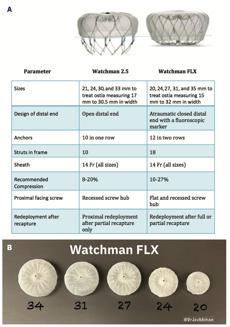
The Watchman is a parachute-shaped device made out of a nitinol cage with a polyethylene terephthalate fabric membrane cap. The original Watchman 2.5 device had an open design with a single row of 10 active fixation anchors, while the newly designed Watchman FLX device has a closed design with a dual row of 18 fixation anchors (Figure 1A). The Watchman FLX comes in 5 sizes, which are selected based on depth and ostial sizes of the LAA (Figure 1B).
After a patient with AF undergoes a workup demonstrating the inability to tolerate long-term OAC, they are referred to our Watchman coordinator, who organizes preprocedural clinic visits and additional testing if necessary. Once the workup is complete, the patient presents to our facility as an outpatient. The patient is then placed under general anesthesia and “day of” preprocedural transesophageal echocardiography (TEE) is performed to confirm the absence of LAA thrombus as well as to assist in device sizing. Occasionally, sizing is also provided by computed tomography (CT) angiography, which may have been completed during the initial workup.
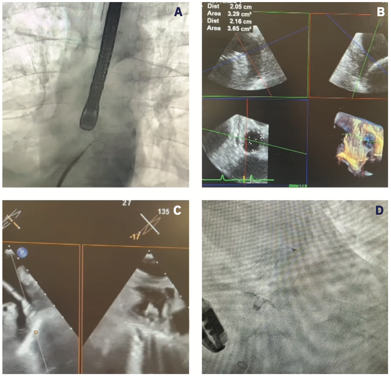
If sizing is acceptable and thrombus is ruled out, ultrasound-guided micropuncture access is achieved and the Watchman 12 French (F) TruSeal Access System (Boston Scientific) with the VersaCross Connect Transseptal Dilator (Baylis Medical) is placed into the superior vena cava. Systemic heparin is administered and therapeutic activating clotting time is achieved. The sheath is pulled down to the fossa ovalis, and a mid and posterior crossing position is confirmed with TEE. Using the VersaCross Transseptal Platform, radiofrequency is used to cross the septum and the sheath is advanced into the LA (Figure 2A/Video 1). LA pressure is obtained. Next, a pigtail catheter is placed into the LAA. CT angiography of the LAA is performed in a right anterior oblique 30º caudal position. Sizing of the device is determined by LAA assessment on TEE (Figure 2B/Video 1). Then, the pigtail catheter is removed, and the Watchman system is prepared and delivered to the LAA. The device is passively exposed to form a “FLX-Ball,” which is advanced and deployed under TEE guidance (Figure 2C/Video 1). The system is held in place and the device is fully deployed. Using TEE, device assessment is performed to evaluate for PASS criteria (positioning, anchoring, size and seal), which must be met before the device is released. If PASS criteria is not met, the device can be recaptured (as many times as needed) and repositioned or exchanged for an alternatively sized device.
Video 1
Video 1. Watchman FLX deployment using multimodality imaging. (A) Fluoroscopy demonstrating a mid and posterior transseptal puncture with the VersaCross Connect System (Baylis Medical) (video). (B) Two-dimensional and 3-dimensional TEE imaging demonstrating measurements of the LAA. (C) Watchman “FLX-Ball” being advanced and positioned for deployment under TEE guidance (video). (D) Watchman FLX #24 device deployed on fluoroscopy.
Once an acceptable result is achieved, the device is released and the system is removed from the body (Figure 2D/Video 1). Hemostasis is achieved with a figure-of-8 suture and stop-cock method. The patient is monitored for 4-6 hours and usually discharged home on the same day as the indexed procedure. Dual antiplatelet medications with aspirin 81 mg and clopidogrel 75 mg once a day are used for 6 months postimplant. Imaging is repeated at 45 days postimplant with either TEE or CT to assess for any possible device-related complications.
Experience at Ascension Providence
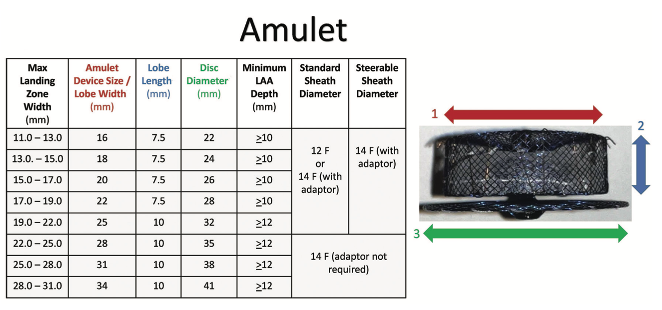
In this section, we will provide an overview of our periprocedural process and experience with the Amulet occluder at Ascension Providence. The Amulet occluder received US Food and Drug Administration approval based on the Amulet IDE trial.3,4 The Amulet occluder is a double-disc device consisting of a nitinol mesh with polyester fabric cover. Unique to the device is the dual-seal function using the lobe component placed at the LAA ostium and the disc positioned at the orifice. A single row of fixation anchors are positioned circumferentially around the distal lobe. There is a range of sizes available in lobe length (diameter range 16-34 mm), with concomitant gradation in lobe length and disc sizes (Figure 3).
Candidates for Amulet occluder implant are selected in the same manner as described above. TEE use, hemostasis, and transseptal access vary; however, they generally mirror the same practices outlined. The Amplatzer Steerable Delivery Sheath became commercially available in the United States in April 2022. Measurement of the LAA orifice, landing zone, and desired depth help determine choice of the appropriate lobe size (Figure 4A).
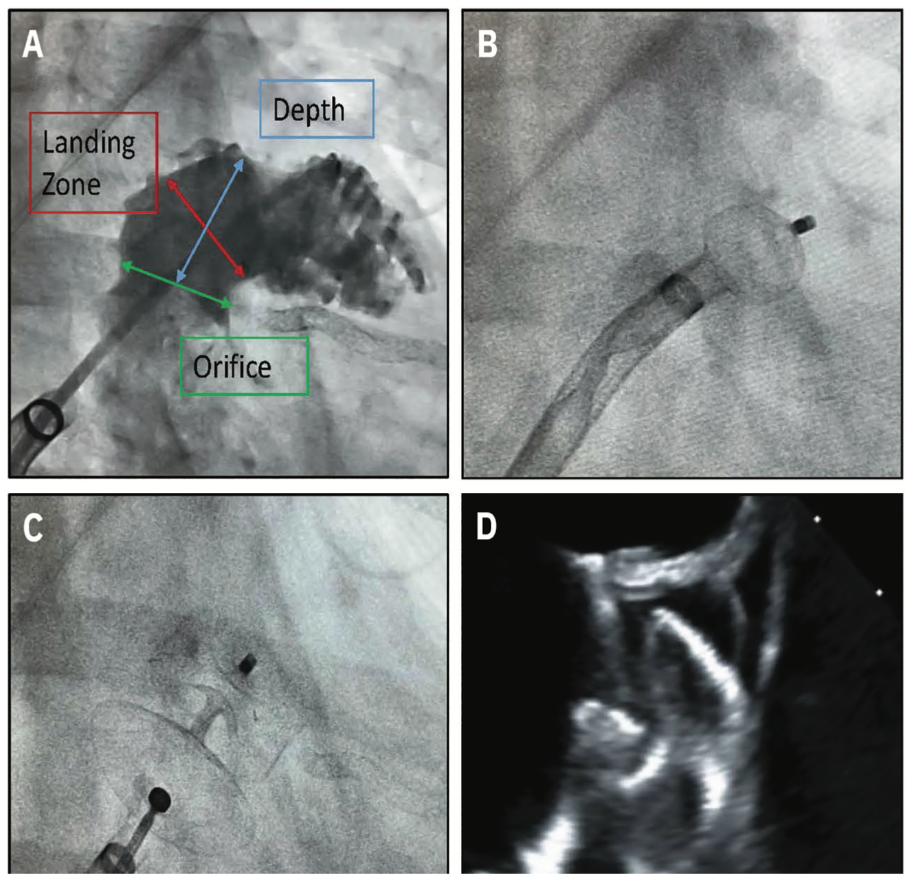
The device is unsheathed to a “ball” position (Figure 4B), which allows for rotational movement. Once proper coaxial alignment is obtained with the sheath and ball, forward pressure can be applied to transition the device from the “ball” to an intermediate configuration of “triangle” if more depth is required. The triangle configuration allows for the force at the tip of the device to be radially distributed, thereby making advancement safer. Once the “ball” or “triangle” is in the correct position, the lobe is deployed with concomitant unsheathing and forward pressure.
The body of the lobe should be two-thirds past the circumflex artery, with some compression within the LAA, and oriented perpendicular to the long access of the LAA. Next, the sheath is retracted to expose the disc and create separation between the lobe (Figure 4C). The disc should not excessively overlay the coumadin ridge of the pulmonary vein and mitral valve, respectively. If the orientation is suitable and the tug test stable, the device is released.
Based on our experience, the Amulet occluder has required connections between the device loader and delivery sheath, which include a hemostatic valve and a sheath adapter for connecting a 14F sheath to devices sized 16-25 mm. Extra care should be taken to eliminate air entry with Amulet sheath preparation.
Regarding sizing, the Amulet occluder has a shorter lobe length, which may facilitate placement in those with limited LAA depth or challenging side lobe anatomy. Another advantage is the ability to close proximal LAA lobes with the disc. To minimize thrombus, the disk should overlay the coumadin ridge to the mitral valve. The Amulet occluder carries no suggested percentage of compression. Although some compression is required, we resist the urge to significantly oversize the Amulet occluder. During deployment, the Amulet occluder objectively requires less forward pressure while unsheathing. The Amulet occluder may only be partially recaptured up to 3 times.
Dual antiplatelet therapy is administered postoperatively, and our general practice is to obtain a TEE (Figure 4D) or CT at the 6-month mark for medication de-escalation.
Future Directions in LAA Occlusion
There have been significant developments in LAAC since its inception in the early 2000s. Each of our institutional experiences demonstrate how 2 LAAC programs in a similar area can function in alternative but effective capacity. The most important aspect of a strong LAAC program revolves around a dedicated team approach. Anatomical considerations also need to be included when considering device implantation. To accommodate variable anatomies, both the Watchman and Amulet occluder device should ideally be available for use; however, institutional policies and price may limit accessibility.
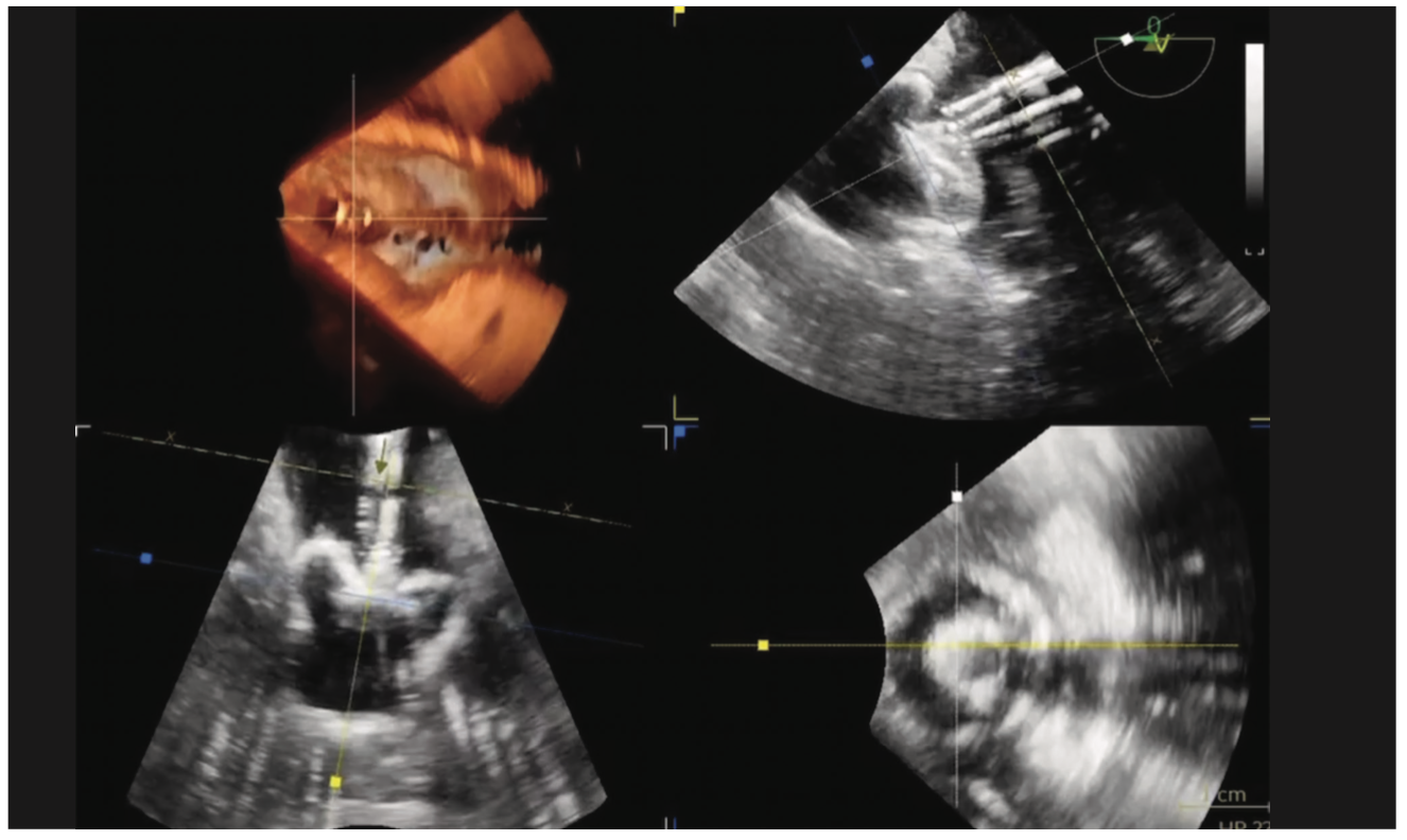
Advanced cardiac imaging is a foundation for any successful LAAC program. Institutions looking to start a LAAC should possess a strong and dedicated cardiac imaging program with TEE. The availability of dedicated anesthesia staff, an advanced imager, and TEE resources may limit the growth of some LAAC programs. Intracardiac echocardiography (ICE) imaging5 with conscious sedation may draw further interest to expand access to growing programs (Figure 5).
Video 2
Video 2. ICE imaging of the LAA for Watchman deployment.5
The selection of appropriate LAAC candidates will continue to evolve. Two large randomized clinical trials aim to study novel OAC therapy vs the Watchman FLX and Amulet occluder devices via the CHAMPION-AF6 and CATALYST7 trials, respectively. The results of these trials are eagerly awaited, as they may expand the role of LAAC to beyond those intolerant to OAC.
Discussion Questions:
1. Is your LAAC center using Watchman and/or Amulet in clinical practice?
2. What is your imaging strategy for preoperative, intraoperative, and postoperative imaging for LAAC?
Disclosures: The authors have completed and returned the ICMJE Form for Disclosure of Potential Conflicts of Interest. Dr Mohan has no conflicts of interest to report regarding the content herein. Dr Bradley reports provision of limited study materials from Abbott. Outside the submitted work, Dr Bradley reports consulting fees and support for attending meetings and/or travel from Abbott.
References
1. Reddy VY, Sievert H, Halperin J, et al. Percutaneous left atrial appendage closure vs warfarin for atrial fibrillation: a randomized clinical trial. JAMA. 2014;312(19):1988-1998. doi:10.1001/jama.2014.15192
2. Holmes DR Jr, Kar S, Price MJ, et al. Prospective randomized evaluation of the Watchman left atrial appendage closure device in patients with atrial fibrillation versus long-term warfarin therapy: the PREVAIL trial. J Am Coll Cardiol. 2014;64(1):1-12. doi:10.1016/j.jacc.2014.04.029
3. Lakkireddy DJ, Thaler D, Ellis CR, et al. Amplatzer Amulet Left Atrial Appendage Occluder versus Watchman device for stroke prophylaxis (Amulet IDE): a randomized, controlled trial. Circulation. 2021;144(19):1543-1552. doi:10.1161/CIRCULATIONAHA.121.057063
4. Abbott strengthens left atrial appendage closure leadership with U.S. availability of Amplatzer™ Steerable Delivery Sheath for the company’s Amulet™ device. Abbott. April 28, 2022. Accessed November 30, 2022. https://abbott.mediaroom.com/2022-04-28-Abbott-Strengthens-Left-Atrial-Appendage-Closure-Leadership-With-U-S-Availability-of-Amplatzer-TM-Steerable-Delivery-Sheath-for-the-Companys-Amulet-TM-Device
5. @Dr_Santangeli. First @PennMedicine cases of LAA occlusion using Nuvision 4D ICE. Groundbreaking advancement in ICE imaging technology @BiosenseWebster. February 22, 2022. Accessed November 30, 2022. https://twitter.com/dr_santangeli/status/1497232210365071365?s=46&t=MO1KW-LSHxxftH7wjS81gg
6. CHAMPION-AF Clinical Trial. ClinicalTrials.gov. November 14, 2022. Accessed November 30, 2022. https://clinicaltrials.gov/ct2/show/NCT04394546
7. Amplatzer Amulet LAAO vs. NOAC (CATALYST). ClinicalTrials.gov. November 14, 2022. Accessed November 30, 2022. https://clinicaltrials.gov/ct2/show/NCT04226547
Related Content