Unexpected Complication Involving a Retrosternal Biloma: A Guide to Managing Sternal Wound Infections
© 2024 HMP Global. All Rights Reserved.
Any views and opinions expressed are those of the author(s) and/or participants and do not necessarily reflect the views, policy, or position of ePlasty or HMP Global, their employees, and affiliates.
Questions
1. What are the incidence and mortality rates of sternal wound infections?
2. What are the most common causes of sternal wound infections?
3. What are the various options for sternal reconstructions?
4. What are potential complications of the various muscle flaps for sternal reconstruction, and what does the postoperative management entail?
Case Description
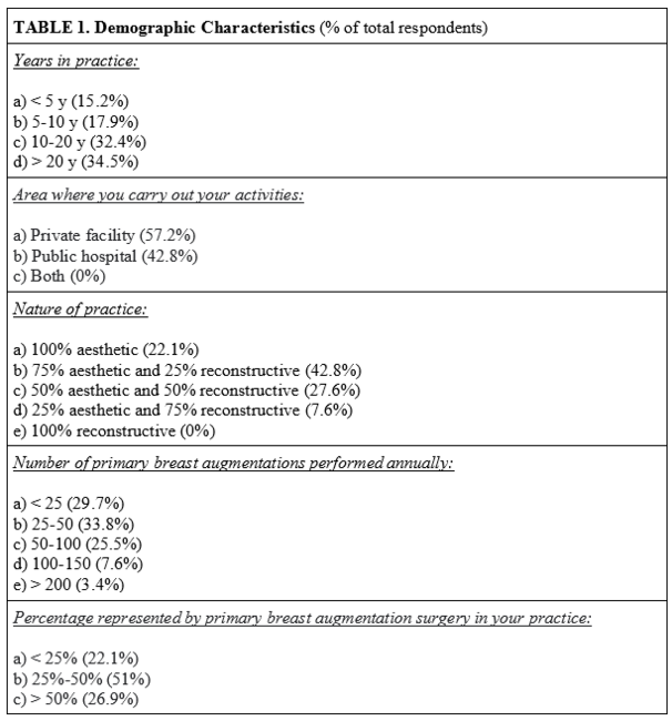
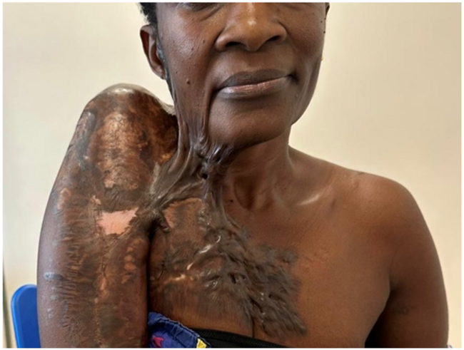
A 60-year-old male patient with a complex medical history developed a right-sided pleural effusion and a dehisced, infected sternal wound following a coronary artery bypass graft (CABG) for single-vessel coronary artery disease. The pleural effusion was addressed with a tube thoracostomy, leading to the drainage of bilious fluid, which notably resembled the fluid within the sternal wound (Figure 1). A subsequent computed tomography (CT) scan revealed a liver tear and a large retrosternal biloma, also leaking bilious fluid, leading to several consultations, including the Plastic and Reconstructive Surgery (PRS) team (Figure 2).
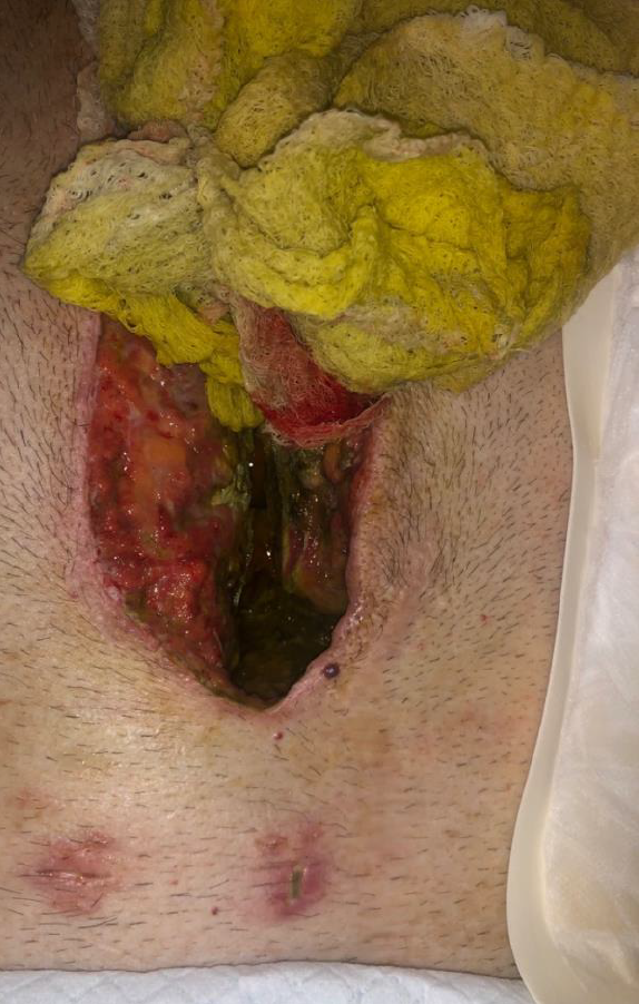
Figure 1. Bilious fluid emerging from the sternal wound.
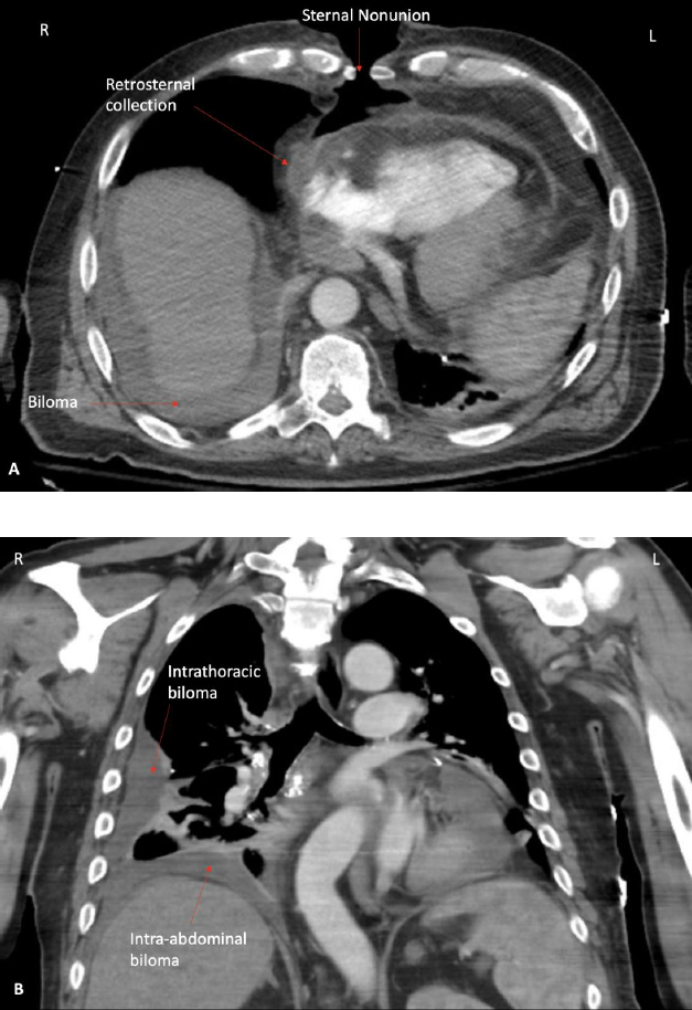
Figure 2. CT Image. (A) Axial image showing biloma surrounding liver, sternal nonunion, and substernal fluid collection. (B) Coronal image showing both intrathoracic and intraabdominal fluid collection secondary to hepatic laceration and diaphragmatic injury.
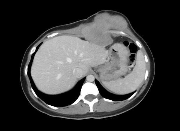
A multidisciplinary strategy was employed to treat the patient. General Surgery initiated the process by conducting a partial hepatectomy to address the biloma source. Thoracic Surgery repaired a diaphragmatic injury and debrided the sternum concurrently, followed by application of a vacuum-assisted closure (VAC) device over the wound (Figure 3). One week later, PRS reconstructed the sternum using sternal plating and bilateral pectoralis major myocutaneous advancement flaps. An incisional VAC was placed to reduce fluid build-up, ensure sterility, and aid wound healing.
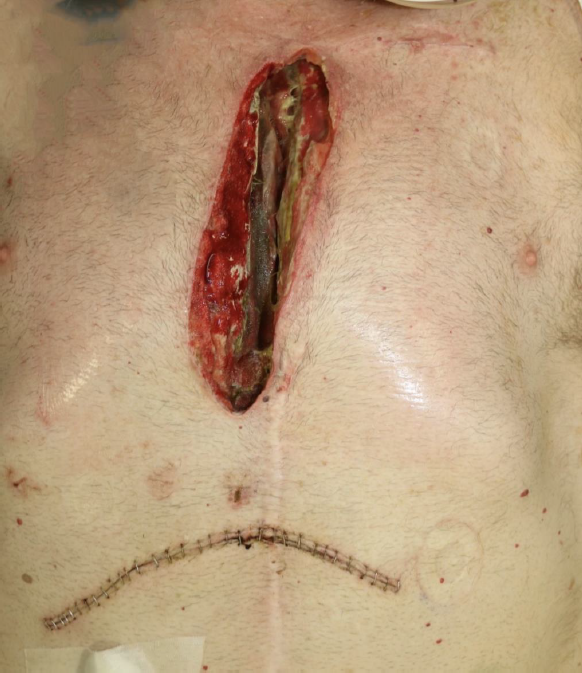
Figure 3. Sternal wound following negative pressure wound therapy and partial hepatectomy.
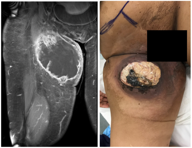
Postoperatively, the patient was placed under strict sternal precautions to avoid strain on the repaired sternum. The recovery was successful with no complications (Figure 4).
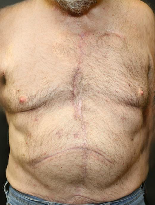
Figure 4. Five months postoperative following reconstruction.
Q1. What are the incidence and mortality rates of sternal wound infections?
The incidence of surgical site infection (SSI) following cardiac surgery varies between 1% and 3%, with some values falling as low as 0.5% and as high as 10%.1 These SSIs can be further divided into 2 groups: deep sternal wound infections (DSWIs) and superficial sternal wound infections (SSWIs).
DSWIs involving the deeper structures of the thorax (eg, the mediastinum) have an incidence rate following sternotomy of 1 to 5% and an associated mortality rate of 10 to 47%.2,3 SSWIs involve the skin, subcutaneous tissue, and pectoralis fascia with no bone involvement. While SSWIs have a slight increase in incidence, 0.5% to 8%, when compared with DWSIs, they are associated with significantly better outcomes, with a mortality rate of 0.5 to 9%.2 As would be expected, DSWIs require a more aggressive treatment plan, including surgical debridement and long-term systemic antibiotic therapy.
Although DSWIs are relatively infrequent in patients who undergo cardiac surgery, the higher mortality rate makes them clinically relevant.4
Q2. What are the most common causes of sternal wound infections?
The most common cause of sternal wound infections is postoperative complications following a median sternotomy for cardiac surgery. However, the exact etiology is multifactorial. Postoperative sternal wound infection is clearly associated with sternal dehiscence, but it is not clear whether the sternal dehiscence causes the sternal wound infection or the sternal wound infection causes the sternal dehiscence.5
There are numerous risk factors that contribute to sternotomy wound complications. Preoperative risk factors include older age, increased body mass index (BMI), smoking, and the presence of comorbidities such as compromised immunity, diabetes, irradiation, reoperation, and chronic lung and kidney disease. Perioperative risk factors that increase the risk for sternal dehiscence include prolonged operative time, the need for chest compressions, and the need for transfusions. Postoperative risk factors include pronged ventilator support, the need for tracheostomy and/or inotropes, postoperative bleeding, and delayed chest closure.5
The most common organism cultured from sternal wounds is Staphylococcus aureus, followed by other species of coagulase-negative Staphylococcus.2 Infrequently, dehiscence of the sternal wound may be seen in the absence of a causative pathogen; these are termed noninfectious sternal dehiscence, and Olbrecht et al described an associated incidence rate of 0.39%.6 Often, noninfectious sternal dehiscence is associated with sternal instability.7 Sternal instability, defined as abnormal mobility of the sternum, often occurs due to primarily mechanical forces, most notably by pectoral flexion but also due to expansion of the thoracic cage or flexion of the rectus abdominus muscles causing displacement or failure of sternal fixation hardware.
Q3. What are the various options for sternal reconstructions?
Several options for sternal reconstructions exist which include the use of several muscle flaps; however, no uniform treatment protocol exists, producing variability in surgical preference.3,8-11 Piwnica-Worms et al listed pectoralis muscle flaps as the most common muscle flaps, followed by rectus abdominis and omental flaps.12 Since the pectoralis muscles are positioned proximal to the wound, they exhibit easier accessibility, which provides increased stability without hindering patients’ postoperative range of motion.10,11 Some sternal reconstructions, on the contrary, require further caudal sternal coverage via a rectus abdominal flap, though lung function may decline postoperatively.10 Latissimus dorsi flaps are utilized in more sparse cases since they cannot cover a sternal defect in the caudal third of the sternum but still exist as an option.10,11 Pedicled omental flaps and free flaps exist as other reconstructive alternatives, mostly in the case of larger wounds.13
In the event of extensive loss, bone grafts or sternal plating may be necessary. A titanium sternal plating system was first introduced in 2005 to achieve sternal stability for complex reconstructions.8,10 In a case report, Danker et al described a 3D-printed sternal plate utilized in a 64-year-old patient that yielded successful results, demonstrating the potential for innovation within the field.8
In addition, debridement and negative pressure therapy are common adjuncts to the sternal reconstructive practice. Negative pressure has been proven to promote wound healing and is highly effective in decreasing mortality, replacing the previous method of vacuum-assisted closure.10,11 With the numerous techniques that exist, muscle flaps continue to be the gold standard for sternal reconstructions; however, it is ultimately the patient’s specific presentation that guides the treatment.8
Q4. What are potential complications of the various muscle flaps for sternal reconstruction, and what does the postoperative management entail?
Surgical management for sternal wound complications following cardiac surgery include debridement, deep wound culture, reclosure after delayed closure, sternal dressings, negative pressure wound therapy, and a variety of sternal flap closures as described above.12
The pectoralis major myocutaneous flap is the most utilized flap for sternal reconstruction because there are no limitations in shoulder mobility nor observed contour irregularities. The pectoralis major turnover, or split turnover flap that is used for lower one-third sternal defects, requires division of humeral insertion. This often leads to raised contour deformity of the anterior chest wall, causing tension on the skin closure in addition to decreased pectoralis major function. Since the omental flap requires a laparotomy, it can lead to intra-abdominal infections, as well as an increased risk for abdominal hernia formation. An abdominal hernia is also a potential complication of the rectus abdominus flap, but this risk can be minimized by leaving the rectus fascia in place and closing in 2 layers.
It is especially important to pay attention to other general complications that coexist with sternal wound infections. The greatest immediate danger is a laceration of the right ventricle by an unsecured portion of the sternum. Other complications include osteomyelitis, sternal necrosis, recurrent dehiscence, hardware failure, and flap failure.2,13 Additionally, hematomas, which almost always require reoperation, may be avoided by meticulous hemostasis and delaying the readministration of anticoagulation until 72 hours after reconstruction. Seromas have been reported in less than 2% of cases with single muscle pedicled flaps and approximately 30% when bilateral, and utilizing drain in the immediate postoperative time period may decrease the incidence.13
Following sternal reconstruction, postoperative protocol should include avoidance of forceful coughing or activation of the pectoralis major muscle while transferring or standing, especially if reconstruction includes a pectoralis major myocutaneous flap. In addition, a chest binder or surgical bra may be warranted to reduce tension from the midline.14
Acknowledgments
Authors: Alexander L. Mostovych, MS1; Milind D. Kachare, MD2; Anthony Azzolini, MD2; Alec Moore, BA1; Lauren A. Tranthem, BS1; Claire Fell, BS1; Carter Prewitt, BA1; Ryan L. Shapiro, MD2
Affiliations: 1University of Louisville School of Medicine, Louisville, Kentucky; 2Division of Plastic and Reconstructive Surgery, Department of Surgery, University of Louisville, Louisville, Kentucky
Correspondence: Milind D. Kachare, MD; Milind.Kachare@gmail.com
Disclosures: The authors disclose no relevant conflict of interest or financial disclosures for this study.
References
1. Lemaignen A, Birgand G, Ghodhbane W, et al. Sternal wound infection after cardiac surgery: incidence and risk factors according to clinical presentation. Clin Microbiol Infect. 2015;21(7):674.e11-674.e6.74E18. doi:10.1016/j.cmi.2015.03.025
2. Singh K, Anderson E, Harper JG. Overview and management of sternal wound infection. Semin Plast Surg. 2011;25(1):25-33. doi:10.1055/s-0031-1275168
3. Kaul P. Sternal reconstruction after post-sternotomy mediastinitis. J Cardiothorac Surg. 2017;12(1):94. Published 2017 Nov 2. doi:10.1186/s13019-017-0656-7
4. Mauermann WJ, Sampathkumar P, Thompson RL. Sternal wound infections. Best Pract Res Clin Anaesthesiol. 2008;22(3):423-436. doi:10.1016/j.bpa.2008.04.003
5. Alebrahim K, Al-Ebrahim E. Prevention, classification and management review of deep sternal wound infection. Heart Surg Forum. 2020;23(5):E652-E657. doi:10.1532/hsf.3153
6. Olbrecht VA, Barreiro CJ, Bonde PN, et al. Clinical outcomes of noninfectious sternal dehiscence after median sternotomy. Ann Thorac Surg. 2006;82(3):902-907. doi:10.1016/j.athoracsur.2006.04.058
7. Ståhle E, Tammelin A, Bergström R, Hambreus A, Nyström SO, Hansson HE. Sternal wound complications--incidence, microbiology and risk factors. Eur J Cardiothorac Surg. 1997;11(6):1146-1153. doi:10.1016/s1010-7940(97)01210-4
8. Danker SJ, Mericli AF, Rice DC, Santos DA, Butler CE. Custom 3D-printed titanium implant for reconstruction of a composite chest and abdominal wall defect. Plast Reconstr Surg Glob Open. 2021;9(11):e3885. Published 2021 Nov 30. doi:10.1097/GOX.0000000000003885
9. Grapow M, Haug M, Tschung C, et al. Therapy options in deep sternal wound infection: Sternal plating versus muscle flap. PLoS One. 2017;12(6):e0180024. Published 2017 Jun 30. doi:10.1371/journal.pone.0180024
10. Hamaguchi R, Shekar PS, Johnson JA, Orgill DP. current management of sternal wounds. Plast Reconstr Surg. 2021;148(6):1012e-1025e.
11. Juhl AA, Koudahl V, Damsgaard TE. Deep sternal wound infection after open heart surgery--reconstructive options. Scand Cardiovasc J. 2012;46(5):254-261. doi:10.3109/14017431.2012.674549
12. Piwnica-Worms W, Azoury SC, Kozak G, et al. Flap reconstruction for deep sternal wound infections: factors influencing morbidity and mortality. Ann Thorac Surg. 2020;109(5):1584-1590. doi:10.1016/j.athoracsur.2019.12.014
13. Orgill, DP. Surgical management of sternal wound complications. Colwell AS, ed. In: UpToDate. Wolter Kluwers, Waltham, MA. Accessed on May 21, 2023. https://www.uptodate.com/contents/surgical-management-of-sternal-wound-complications
14. Jones G, Jurkiewicz MJ, Bostwick J, et al. Management of the infected median sternotomy wound with muscle flaps. The Emory 20-year experience. Ann Surg. 1997;225(6):766-778. doi:10.1097/00000658-199706000-00014