Solitary Phalangeal Osteochondroma in a 12-Year-Old
Case Description
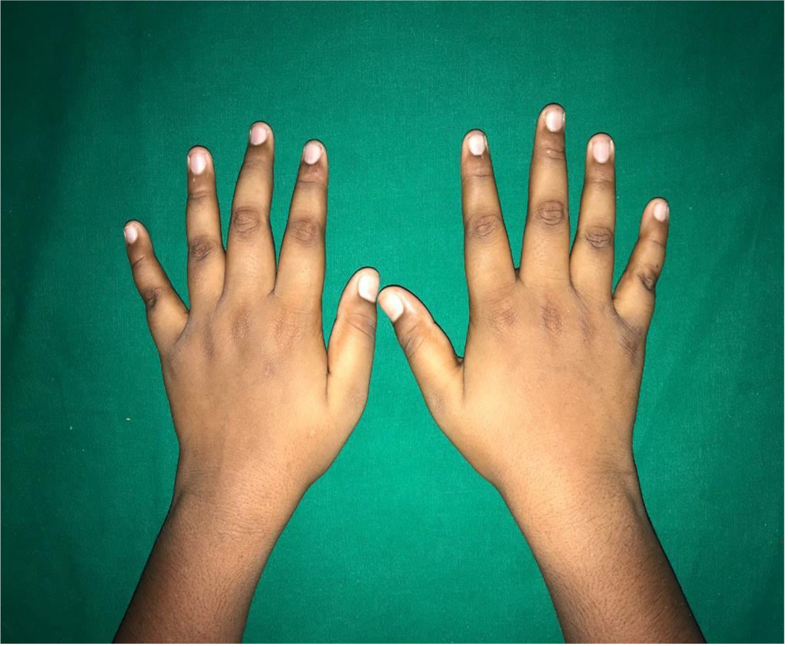
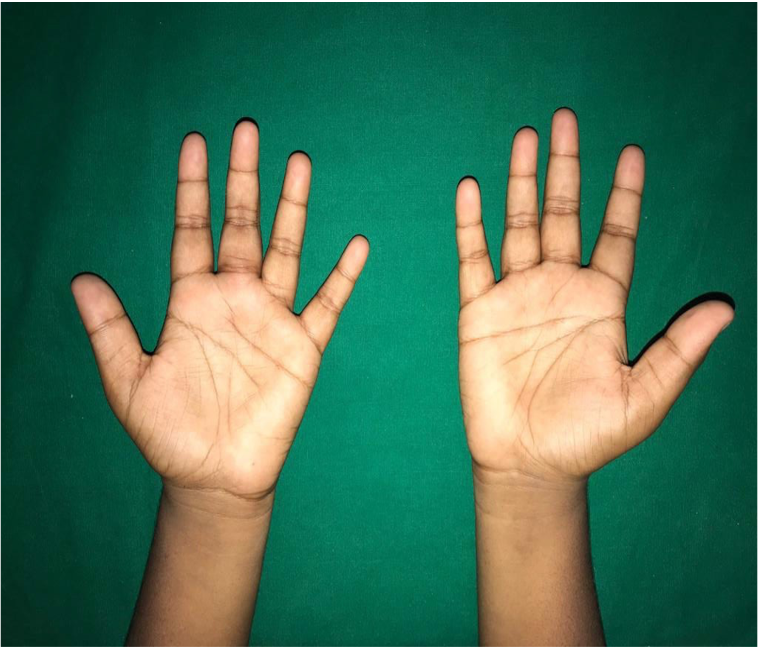
A 12-year-old-boy accompanied by his mother presented with the complaint of swelling of his left middle finger of 3 months’ duration associated with restricted movements. He is a right-handed school-aged boy. There was no history of trauma or similar swellings elsewhere or similar complaints in the past. The swelling had not changed in size and was painful while performing activities.
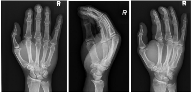
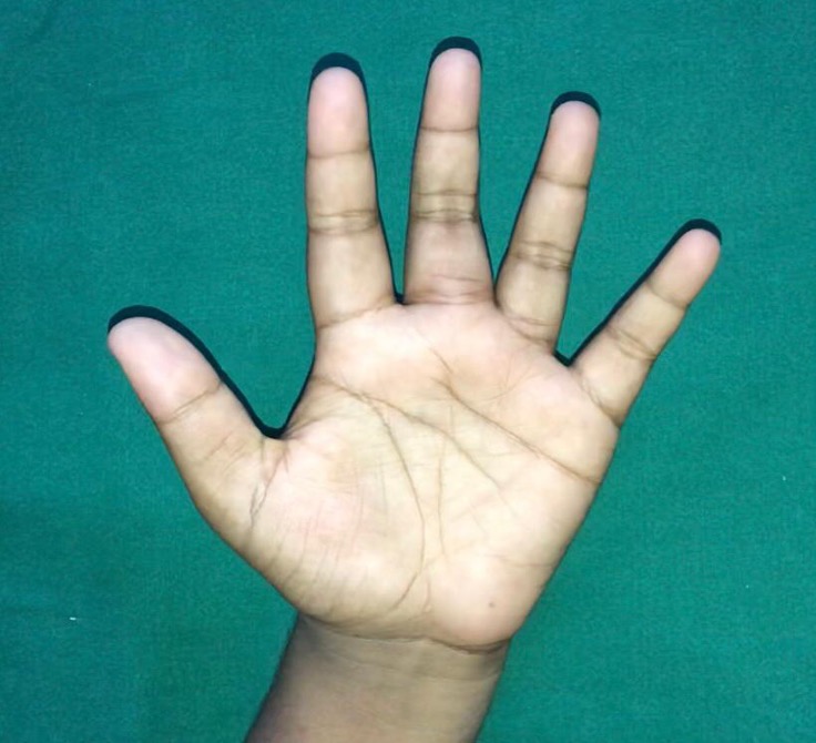
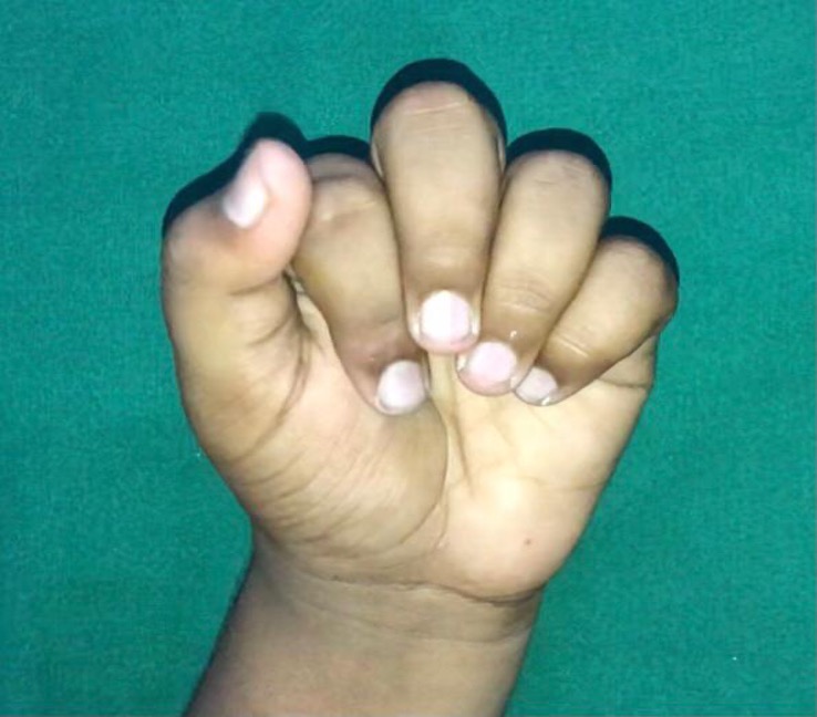
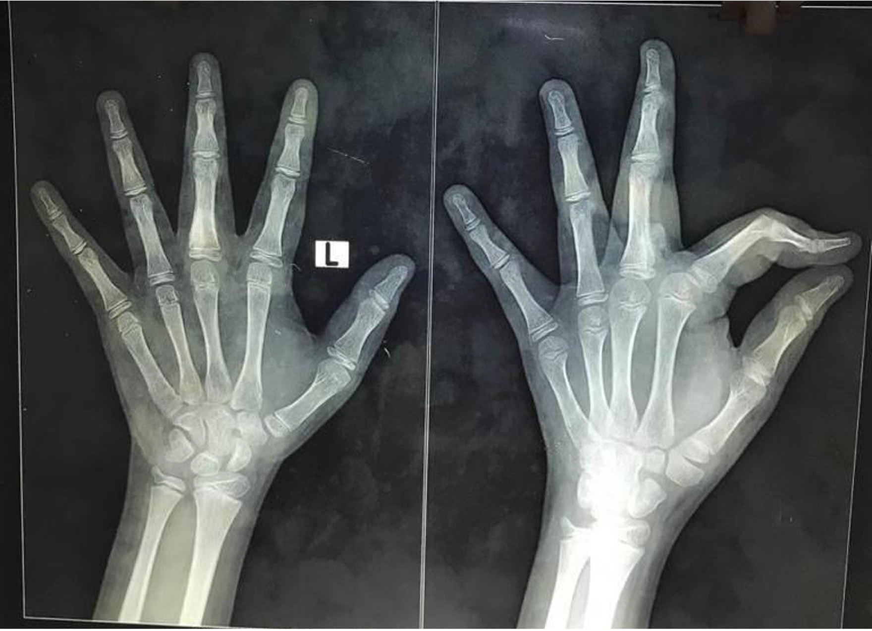
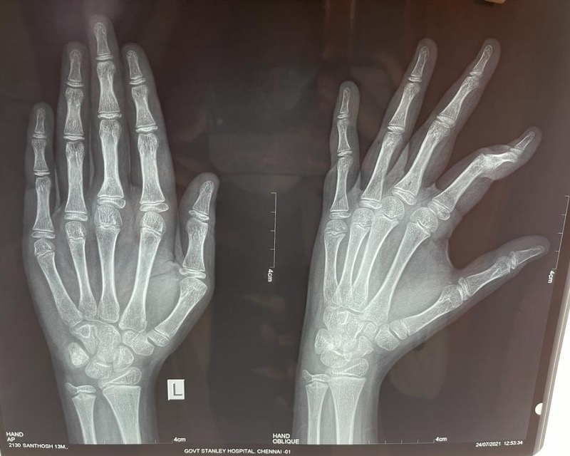
On examination, there was a well-defined single firm-to-hard non-tender swelling of about 2 x 1 cm in size on the volar aspect of his left middle finger extending from base to mid-shaft region of proximal phalanx (Figures 1,2,3). There was no associated angulation or shortening deformity. It was associated with minimal pain on deep palpation. There was restriction of flexion of the middle finger metacarpophalangeal joint (Figure 4; Video 1). Sensation was intact. He had no other swellings in his hands or rest of the body. X-ray showed a single irregular radiopaque lesion on the volar aspect of proximal phalanx of about 2 x 1 cm in size with no obvious angulation (Figure 5).
Video 1. Preop from HMP on Vimeo.
Questions
- What other investigations were done to confirm the diagnosis?
- What was the further plan of management?
- What were the steps of the procedure, the difficulties encountered, the process for circumventing those difficulties, and the immediate post-op protocol?
- What was the follow-up protocol and outcome?
Q1: What other investigations were done to confirm the diagnosis?
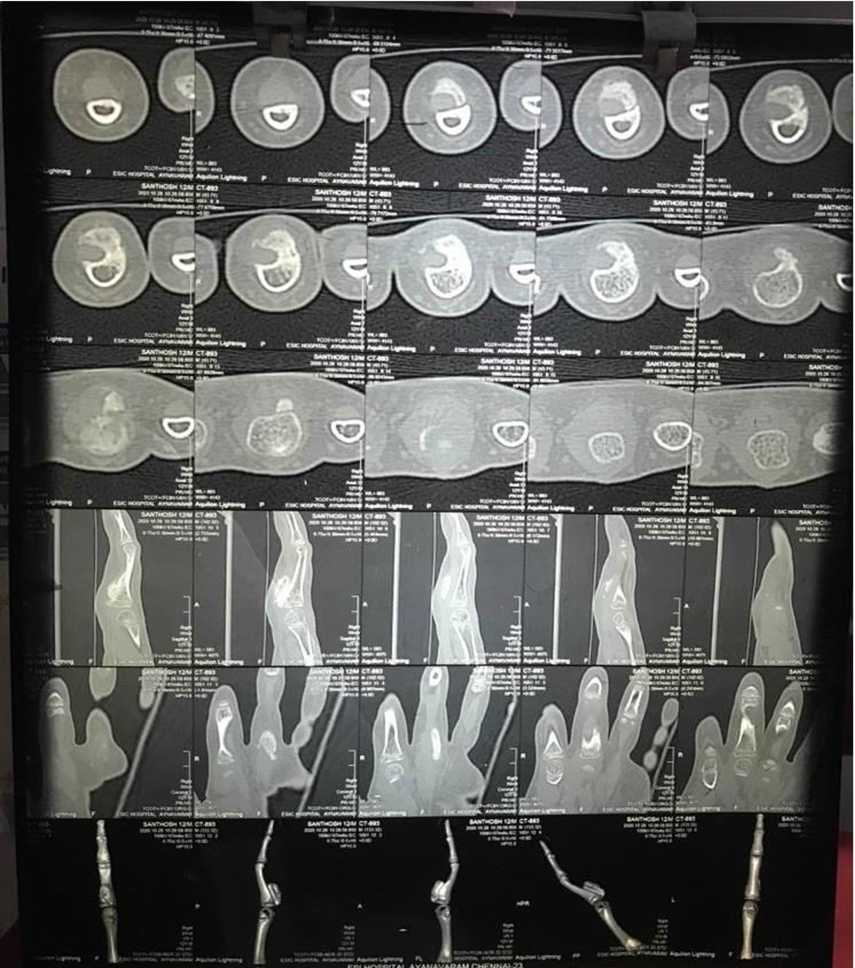
Ultrasound and computerized tomography (CT) of the hand were performed (Figure 6).
- Ultrasound of the hand reported an ill-defined bony outgrowth noted in proximal phalanx of middle finger with cartilage cap. The lesion extends superiorly displacing the above tendon—possibly osteochondroma of proximal phalanx of middle finger.
- CT of the hand report was a solitary pedunculated bony lesion with cortex and medulla of the lesion in continuity with proximal phalanx of middle finger. The lesion measures 1.8 x 0.7 cm, and base of lesion is 7.1 mm. Cartilage cap thickness is 2.2 mm. The rest of the phalanx of all fingers and metacarpals appear normal. Impression was osteochondroma of proximal phalanx of left middle finger.
Q2: What was the further plan of management?
Firstly, adequate counseling of parents was done explaining the nature of the lesion, need for surgical excision, proximity of the lesion to tendon, neurovascular bundle and growth plate, and need for histopathological confirmation and extensive postoperative physiotherapy to regain maximum movement.
The possible complications of surgery were explained to the parents including recurrence, malignant degeneration, restriction of joint movements, and probable adherence of tumor to flexor tendons or tendon sheath.
Q3: What were the steps of the procedure, the difficulties encountered, the process for circumventing those difficulties, and the immediate post-op protocol?
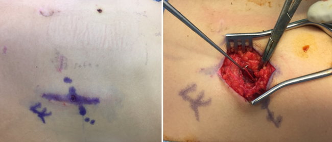
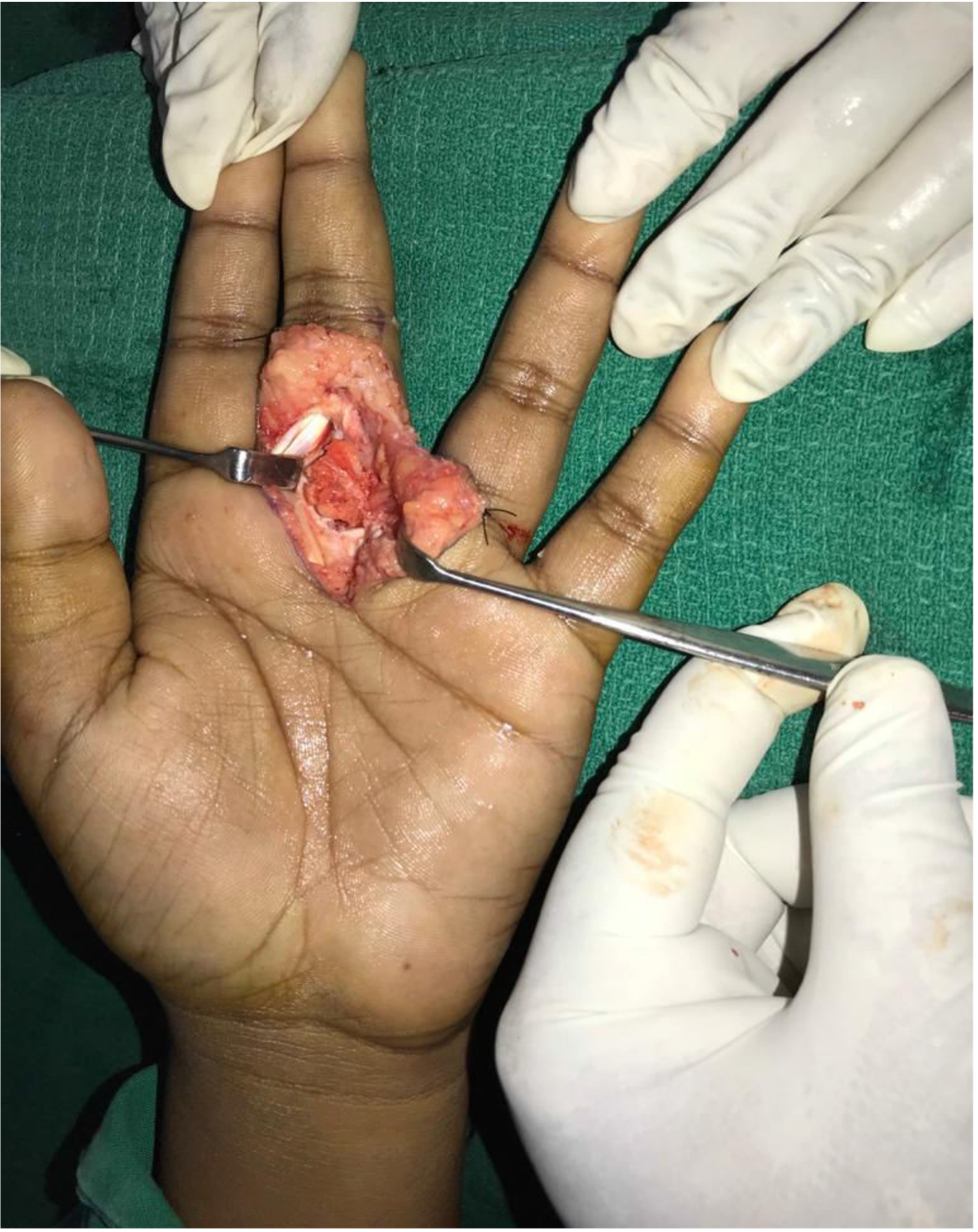
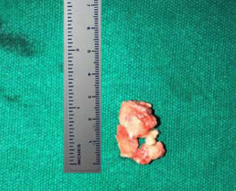
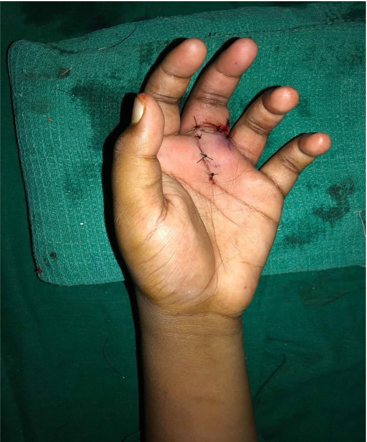
Under supraclavicular block and tourniquet control, a liberal incision was given first on the ulnar aspect–mid neutral line of proximal phalanx extending transversely over the proximal digital crease line to the distal transverse crease line on the palm. The incision was deepened, and the bilateral neurovascular pedicles were identified and secured. The swelling displaced the flexor tendon sheath. The sheath was opened and tendons retracted radially (Figure 7). The stretched-out pseudo capsule around the swelling was opened vertically, and the tumor was visualized. Using periosteal elevator, a plane was created around the tumor; the tumor was found to be very hard and was removed using a mallet and osteotome in toto (Figure 8). The epiphyseal plate was left behind intact. The stretched-out capsule was redraped after excising the excess tissue. The A2 pulley was repaired. Passive unrestricted gliding of flexor tendons was ensured. Tourniquet was released, hemostasis secured, and closure done (Figure 9).
A dorsal hand pop was given, and early active mobilization within the pop was started after 48 hours as soon as patient was pain free and local edema had reduced.
Q4: What was the follow-up protocol and outcome?
Patient was started on early gentle active mobilization exercises within the dorsal pop under the supervision of trained hand therapist. Post-op x-ray showed complete tumor clearance (Figure 10). He was discharged on the 3rd postoperative day advising strict compliance to physiotherapy protocol.
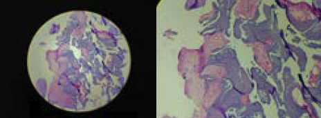
He was reviewed on the 5th and 10th postoperative days. Sutures were removed on the 10th day. The pop was removed after 2 weeks. Histopathology report indicated multiple fragments of mature lamellar bony trabeculae with adjoining lobules of mature hyaline cartilage. Excised bone features were consistent with osteochondroma (Figures 11 and 12).
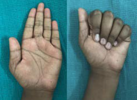
Passive mobilization and passive stretching exercises were advised after 3 weeks. Initial follow-up showed an extensor lag of the proximal interphalangeal joint. With further physiotherapy and splinting, patient now has near-normal pain-free range of movements on 6 months' follow-up (Figures 13 and 14; Video 2).
Video 2. Postop from HMP on Vimeo.
Summary
Ganglion cysts, foreign bodies, and vascular malformations are some of the most common causes of a palpable mass in the pediatric hand.1,2 Osteochondromas are the most common cartilaginous tumors seen in the general skeleton accounting for 30-35%, but in the hand they are less common than enchondromas.3,4 These lesions are osseous growths with a hyaline cartilage cap thought to originate from the physis or regions of tendon insertion. Enchondromas are benign cartilaginous lesions that account for as many as 90% of bone tumors seen in the hand.4 When osteochondromas present in the hand, they may be in isolation or part of multiple hereditary exostosis, in which case they are usually multiple, bilateral, and associated with deformities of phalanges. There have been reports of spontaneous resolution in early stages of the disease.5,6 Hereditary multiple osteochondromas are inherited in an autosomal dominant manner. Penetrance is approximately 96% in female patients and 100% in male patients. In 10% of affected individuals hereditary multiple osteochondromas are the result of a de novo pathogenic variant. Offspring of an affected individual are at a 50% risk of inheriting the pathogenic variant.7 There have been reports of osteochondroma arising from carpal bones in children.8,9 The patient in this study, a 12-year-old boy, had a unilateral solitary tumor confined to the middle finger proximal phalanx of non-dominant hand without any angulation, rotation deformity, or swellings elsewhere.5,6 There is a 0.5% to 1% chance of malignant degeneration to chondrosarcoma.10 Histopathologically, osteochondromas show a regular arrangement of bone trabeculae oriented at 90° to the cartilage cap.11 Differential diagnosis include periostitis ossificans, bizarre paraosteal osteochondromatous proliferation (Nora lesions), and Turret exostosis, which have all been proposed to be variants of a lesional spectrum.12 The differentiation between Nora lesions and osteochondroma is made from histological examination.13
Acknnowledgments
Affiliations: The Institute for Research and Rehabilitation of Hand and Department of Plastic Surgery, Stanley Medical College and Govt. Stanley Hospital, India
Correspondence: Shalini Salim; shalini_salim@yahoo.com
Disclosures: The authors disclose no financial or other conflicts of interest.
References
1. Colon F, Upton J. Pediatric hand tumors. A review of 349 cases. Hand Clin. 1995;11(2):223-243.
2. Civan O, Cavit A, Pota K, Özcanlı H. Tumorous conditions of the pediatric hand and wrist: Ten-year experience of a single center. Jt Dis Relat Surg. 2020;31(2):341-345. doi:10.5606/ehc.2020.74972
3. Lam Y. Bone tumors: Benign bone tumors. FP Essent. 2020;493:11-21.PMID: 32573182
4. Edward A Nathanasian. Bone and Soft Tissue Tumors. In: Scott W. Wolfe, ed. Green’s Operative Hand Surgery. 6th ed. Elsevier Churchill Livingstone; 201:2176-2179.
5. Woodside JC, Ganey T, Gaston RG. Multiple osteochondroma of the hand: initial and long-term follow-up study. Hand (N Y). 2015;10(4):616-620. doi:10.1007/s11552-015-9775-6
6. Sreenivas T, Lokare NB, Jagdish M, Nataraj AR. Multiple osteochondroma of the hand in a 6 year old child- a case report. J Hand Microsurg. 2012;4(2):81-83. doi:10.1007/s12593-011-0055-6
7. Wuyts W, Schmale GA, Chansky HA, et al. Hereditary Multiple Osteochondromas. 2000 Aug 3 [Updated 2020 Aug 6]. In: Adam MP, Ardinger HH, Pagon RA, et al., eds. GeneReviews® [Internet]. University of Washington, Seattle; 1993-2021.
8. van Alphen JC, te Slaa RL, Eulderink F, Obermann WR. Solitary osteochondroma of the scaphoid: a case report. J Hand Surg Am. 1996;21(3):423-425. doi:10.1016/S0363-5023(96)80356-X
9. Laliotis NA, Crysanthou CK, Konstandinidis PA. Solitary osteochondroma of the capitate, in a child. J Clin Orthop Trauma. 2018;9(Suppl 1):S136-S139. doi:10.1016/j.jcot.2018.01.005
10. Chao J, Brummund D, Datiashvilli R. Osteochondroma of the Distal Volar Thumb. Eplasty. 2019;19:ic17. Published 2019 Sep 30.
11. De Lange EE, Pope TL Jr, Fechner RE, et al. Case Report 428: Bizarre Parosteal Osteochondromatous Proliferation (BPOP). Skeletal Radiol. 1987;16:481-483
12. Michelsen H, Abramovici L, Steiner G, Posner MA. Bizarre parosteal osteochondromatous proliferation (Nora's lesion) in the hand. J Hand Surg Am. 2004;29(3):520-525. doi:10.1016/j.jhsa.2004.02.002