Biomechanical Evaluation of Additional Surgical Maneuvers to Improve Symmetry in Performing Nuss Procedure for Asymmetric Pectus Excavatum
© 2023 HMP Global. All Rights Reserved.
Any views and opinions expressed are those of the author(s) and/or participants and do not necessarily reflect the views, policy, or position of ePlasty or HMP Global, their employees, and affiliates.
Abstract
Background. Severing part of the thorax prior to bar placement is effective to improve outcomes in performing the Nuss procedure for asymmetric pectus excavatum. This study aims to elucidate the patterns of severing to provide an ideal outcome.
Methods. Three-dimensional biomechanical computer models were produced simulating the thoraxes of 10 actual patients with asymmetric pectus excavatum. Virtual surgical operation was performed on the 10 models in 4 patterns: group 1—no part of the thorax was severed (default group). Group 2—The sternum was severed (sternum-severing group). Group 3—The ribs on the affected side were severed (rib-severing group). Group 4—Both the sternum and ribs on the affected side were severed (sternum/rib-severing group). After performing this preparation, simulation of bar placement was performed. Comparing the pre- and postoperative shapes of the models, we examined whether symmetry improved for each group.
Results. Symmetry of the chest wall improved for rib-severing group and sternum/rib-severing group. Asymmetry remained for default group and sternum-severing group.
Conclusions. Performance of the Nuss procedure for asymmetric pectus excavatum does not greatly improve symmetry of the chest wall. Severing the ribs as an additional maneuver is effective to improve symmetry.
Introduction
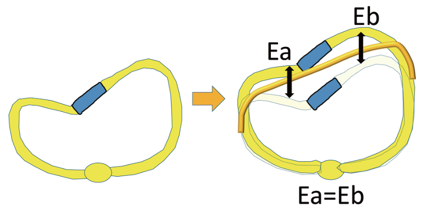
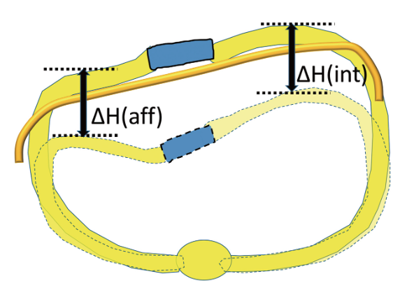
Pectus excavatum is the most common congenital deformity of the chest, occurring in one in 300 to 1000 births.1 In the last century, the Ravitch procedure was mainly used to correct pectus excavatum.2-3 The appearance of the Nuss procedure at the end of the last century drastically changed the treatment of pectus excavatum due to its reduced invasiveness and technical ease.4-5 However, the Nuss procedure often does not bring about satisfactory outcomes for asymmetric cases because correction bars push up the whole chest wall, both the side with minor deformity (defined as the “intact side” in this paper) and that with major deformity (“affected side”). Herein, let’s assume that bar-placement elevates the affected side by Ea and elevates the intact side by Eb (Figure 1). When the 2 sides are elevated by the same distance (Ea = Eb), asymmetry of the chest wall remains, even though the chest wall becomes less concave. To avoid this complication and achieve optimal results, we sever part of the thorax prior to placing bars. This biomechanical study—using computer simulation—aims to elucidate how the thorax should be severed to achieve symmetry.
Methods
Model Production
When we treat pectus excavatum patients, we measure the thicknesses of their left and right hemithoraxes at their most protruding point and compare the thicknesses. When the thickness of one side is smaller than 85% of the other side, we define it as an asymmetric thorax. We randomly selected 10 patients from 72 asymmetric pectus excavatum patients we treated in the past 5 years. The 10 patients consist of 3 males (29.3 ± 5.6 years of age) and 7 females (30.4 ± 4.2 years of age). We collected digital imaging and communications in medicine (DICOM) data of the thoraxes of these patients. The data of the 10 thoraxes were transformed into 10 3-dimensional CAD (computer-aided design) models. The 10 CAD models were further transformed into 10 finite-element models consisting of 145,000 to 186,000 tetrahedron elements. We calculated Young’s modulus of the bone and cartilage based on Kopperdahl’s equation, referring to computed tomography (CT) density of these tissues,6 and allotted the material properties to the corresponding part of the models. For each of the 10 finite element models, simulation surgery severing some parts of the thorax was performed.7-10
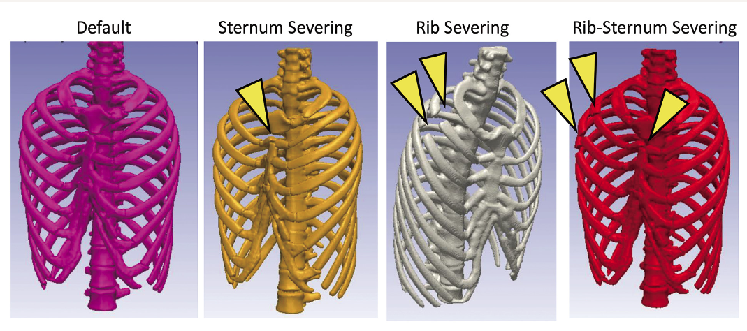
These simulation maneuvers were conducted using Simpleware (Simpleware Inc). Severing of bones was performed in 4 ways (Figure 2), producing 4 groups, each consisting of 10 models, as follows.
Default group: No parts of the thorax are severed.
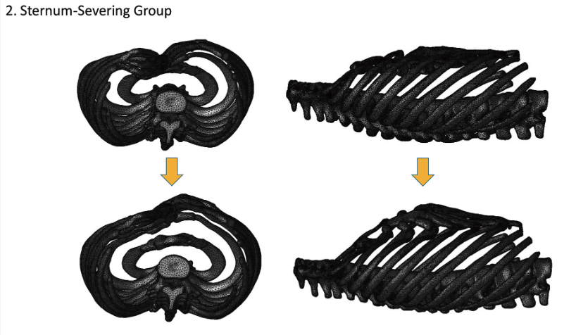
Sternum-severing group: The sternum is severed.
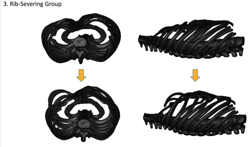
Rib-severing group: The second and third ribs on the affected side (the side with major deformity) were severed.
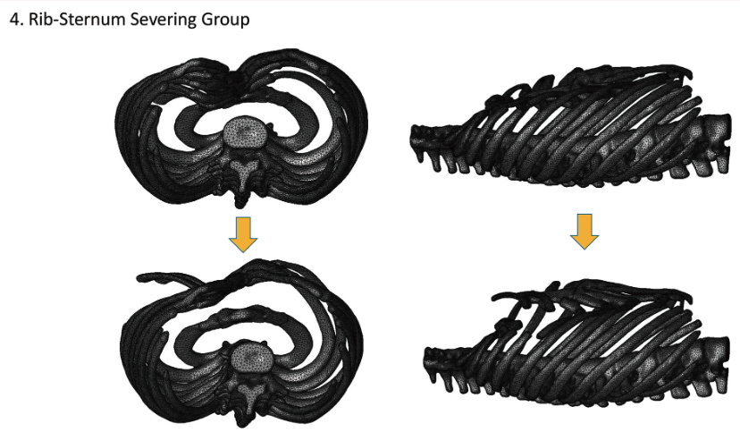
Sternum/rib-severing group: The second and third ribs on the affected side and the sternum are severed.
Simulation of Operation
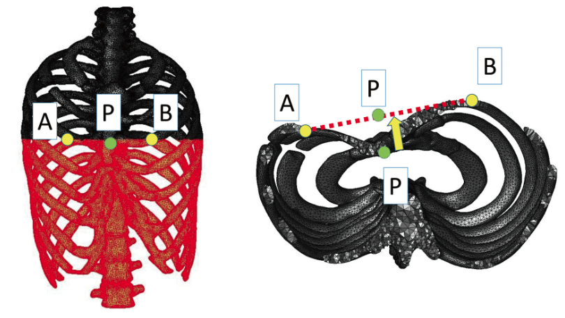
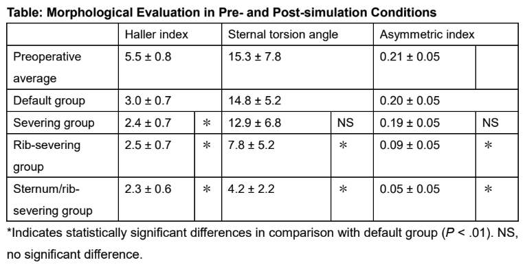
For each 3-dimensional thorax CAD model, we identified a point at which the sternum presents the greatest concavity (point P in Figure 3). Then we identified the pair of ribs closest to point P. We further identified the most protruding points on the pair of ribs (point A and point B in Figure 3). We applied forces to these points so that P moves onto the segment connecting point A and point B. As a result of this force application, point P was elevated by 26 to 50 mm, depending on the model.
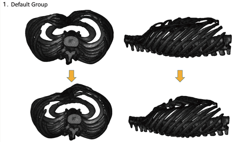
The transformations of the thorax under these dynamic conditions were calculated using LS-DYNA (Ansys). Figure 4 shows the shapes of a model in the default group before and after the (virtual) placement of the bar. Similarly, Figure 5, Figure 6, and Figure 7 show before and after shapes of a model belonging to the sternum-severing group, rib-severing group, and rib/sternum-severing group, respectively.
Evaluation
Degree of Thoracic Wall Elevation. The degree of elevation differs for all points on the thorax. We identified the point that deviates the most for the affected-side hemithorax and the intact-side hemithorax of each model. We defined the deviations of these points as ΔH(aff) and ΔH(int), respectively (Figure 8). With the 10 models in each of the 4 groups (default, sternum-severing, rib-severing, and sternum/rib-severing groups), statistical comparison was performed between ΔH(aff) and ΔH(int) by means of the Mann-Whitney test.
Haller Index, Sternum Torsion Angle, and Asymmetry Index. We measured the Haller index,11 sternum torsion angle, and asymmetry index12 of the 10 thorax models in the preoperative condition. After performing simulation of surgery, we measured the values again. We compared the data of default group and other 3 groups using t test to evaluate the effectiveness of bone-severing techniques.
SPSS (IBM) was used for statistical analyses; P values smaller than .01 were considered to be statistically significant.
Results
Degree of Thoracic Wall Elevation
Default Group. ΔH(aff) (27.2 ± 10.2 mm) and ΔH(int) (28.0 ± 8.5 mm) do not present a statistically significant difference, meaning asymmetry persists after bar placement.
Sternum-Severing Group. ΔH(aff) (31.4 ± 10.0 mm) and ΔH(int) (32.6 ± 9.8 mm) do not present a statistically significant difference, meaning asymmetry persists after bar placement.
Rib-Severing Group. ΔH(aff) (40.6 ± 9.2 mm) was greater than ΔH(int) (32.2 ± 9.4 mm), meaning symmetry improves after bar placement.
Sternum/Rib-Severing Group. ΔH(aff) (41.0 ± 8.0 mm) was greater than ΔH(int) (32.6 ± 8.3 mm), meaning symmetry improves after bar placement.
Haller Index, Sternum Torsion Angle, Asymmetry Index
Haller Index. The Haller index values were smaller for all 3 groups than those of the default group, meaning the thickness of the thorax increases by merely severing the sternum, by severing the ribs, or by severing both (Table).
Sternal Torsion Angle. The sternal torsion angle did not present statistically significant differences between those of the default group and the sternum-severing group, meaning merely severing the sternum does not improve symmetry. On the other hand, the rib-severing group and the sternum/rib-severing group presented smaller sternal torsion angles than the default group, meaning severing ribs-even solely―contributes to improved symmetry (Table).
Asymmetry Index. The asymmetry index did not present statistically significant differences between the default group and the sternum-severing group, meaning merely severing the sternum does not contribute to improved symmetry. On the other hand, the rib-severing group and the sternum/rib-severing group present smaller asymmetry index values than those for the default group, meaning severing ribs contributes to improved symmetry (Table).
Discussion
The standard Nuss procedure is not necessarily suited to treating asymmetric cases. In asymmetric cases, the chest wall presents with minor deformity on one side (the right side in most patients) and major deformity on the other side (usually the left side). To achieve optimal results, the side with major deformity must be elevated more than the other side. However, this cannot be done with the standard Nuss procedure because the simple placement of correction bars is incapable of such selective elevation. So, we sever part of the thorax in advance-before we place correction bars―to elevate the chest wall selectively. Depending on which parts of the thorax we sever, the effect of the selective elevation can differ. We conducted this study to elucidate the severing patterns that effectively achieve the selective elevation.
The present study employs the finite element method. It may be argued that the purpose of this study can be achieved by evaluating operative outcomes in actual clinical cases. However, applying various operative maneuvers on an identical thorax is impossible in evaluations of actual cases. Furthermore, from an ethical point of view, randomly changing operative patterns for patients is not acceptable. In contrast, with surgical simulation, we can try various surgical procedures on the same thorax without ethical restrictions. Thus, we conducted the present study by using virtual surgery on finite element models. The finite element method is a technique of dynamic analysis. The finite element method uses computer calculation to predict how an object transforms when external forces work on the object.7-10 The reliability of the finite element method has been established in medical research, and the method is used for biomechanical analyses of various organs, such as the thorax,13-14 facial bones,15-18 and skin.19
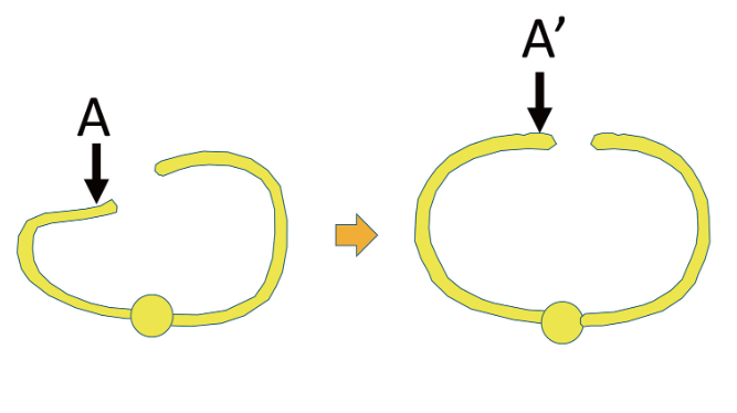
In treating asymmetric pectus excavatum, 2 problems need to be solved to improve the symmetry of the chest wall. The first problem is the deformity of the sternum. In asymmetric cases, the sternum inclines to the affected side. It needs to be rotated toward the intact side (Figure 9). The second problem is the asymmetry of the ribs. The anterior portion of the ribs on the affected side is positioned posterior to the ribs on the intact side. This part must be repositioned to an anterior position (Figure 10).
The fourth to the sixth ribs present the greatest deformity in a majority of pectus excavatum patients. However, we assumed severing the second and third ribs for the rib-severing group and the sternum/rib-severing group. We assumed so for the following reasons: The shapes of the fourth to sixth ribs can be easily corrected by simply placing bars at the third to fifth intercostal spaces. On the other hand, correction of asymmetry in the upper chest regions (the area around the clavicle) is difficult to achieve because placement of bars at the second (or even third) intercostal space presents the danger of thorax outlet syndrome.20 Instead, we mobilize the second and third ribs by severing them and let them move with the sternum as the sternum is elevated by bars. The effectiveness of severing the second and third ribs has been published elsewhere.21
Initially, we hypothesized that severing the sternum and severing the ribs would both solve both problems. Herein, the validity of these independent hypotheses is reviewed by referring to the results.
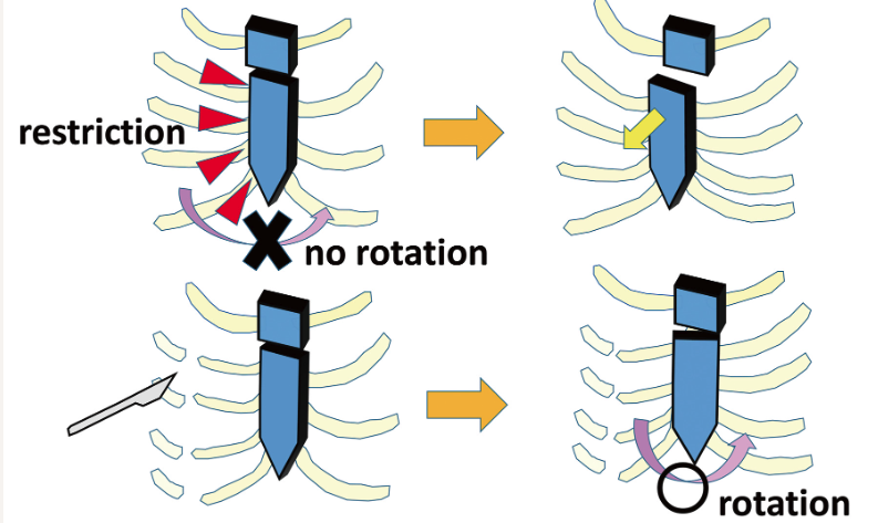
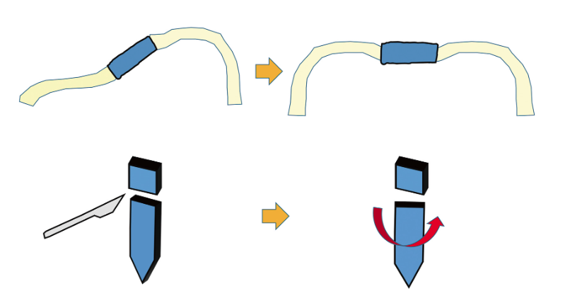
First, we review the effect of severing the sternum. Before conducting this study, we had hypothesized that severing the sternum at the junction between the manubrium and the body should release the body, and that the inclination of the body could be corrected as shown in Figure 9. However, symmetry of the chest wall of the sternum-severing group stays at the same level as that of the default group. This means that solely severing the sternum is not effective in improving symmetry. The sternum cannot be rotated enough because it is restricted by the ribs attached to the edge of its affected side (Figure 11). The sternum is simply pushed forward, and the inclination of the sternum is not corrected.
On the other hand, symmetry improved in the rib-severing group and the sternum/rib-severing group. This means that severing ribs (whether solely or combined with severing the sternum) is effective to make the chest wall symmetric. Then, why does severing the ribs contribute to increasing symmetry? We explain this with 2 mechanisms. First, it releases the sternum and allows its rotation. As shown in Figure 11, the sternum cannot rotate to the intact side, restricted by the ribs attached to it. Severing the ribs releases this restriction, and the sternum becomes able to rotate to the intact side.
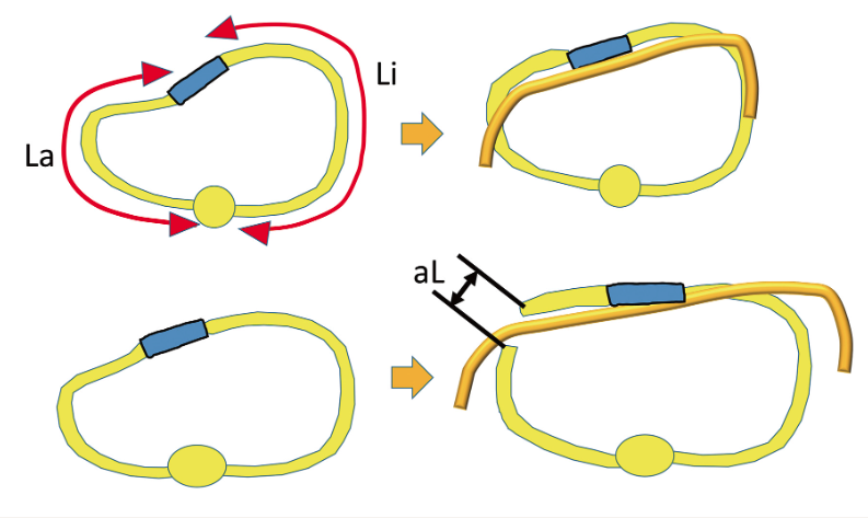
Second, severing ribs allows an increase in the circumference of the affected side. It would be ideal if we could correct the symmetry of the circumference as shown in Figure 10. However, this does not occur because the circumferences of the ribs on the affected side are shorter than those on the intact side. In the 10 patients included in the study, the circumferences of the affected sides (35.3 ± 3.4 cm: La in Figure 12) were significantly smaller than those of the intact sides (38.2 ± 3.2 cm: Li in Figure 12). Whether or not this difference exists with asymmetric cases in general is to be elucidated in future studies. In these patients, even if part of the rib is elevated, the thorax still remains asymmetrical. Severing the ribs adds length to the ribs of the affected side, allowing a symmetric contour (aL in Figure 12). In essence, the result demonstrates that severing the ribs is necessary to achieving symmetry.
Prior to performing the present study, we had assumed that severing the sternum would also contribute to improved symmetry. However, the results demonstrate that severing the sternum is not essential to improving symmetry. Viewed from aesthetic standpoints, this finding is encouraging. To approach the sternum, surgeons need to incise the skin above the sternum. Making scars in this region is aesthetically disadvantageous for patients. In particular, when female patients wear dresses that expose the anterior part of the chest (often exposing the clavicular part, too), conspicuous scars in this region impair their appearance. Surgeons should avoid incising the skin of this region. Since severing the sternum is not necessarily essential to achieving symmetry, surgeons do not need to create scars on these aesthetically important regions.
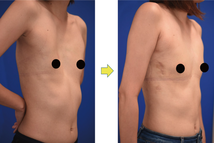
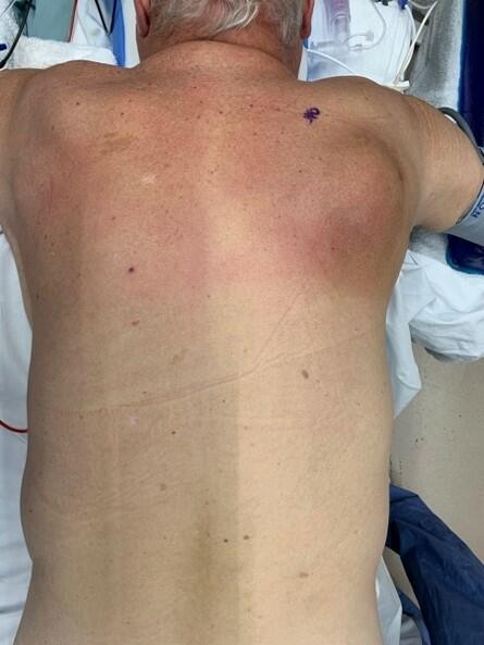
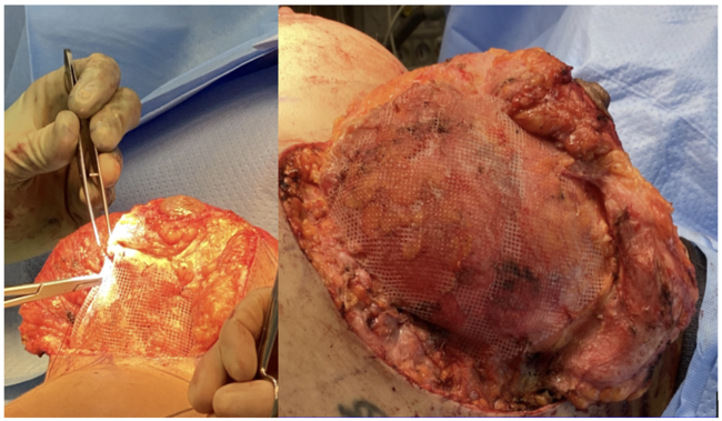
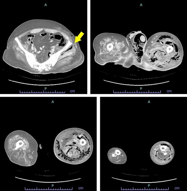
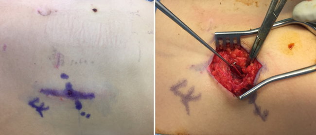
On the other hand, surgeons do not need to incise the visible part of the chest to sever the ribs. Ribs can be approached through skin incisions made posterior to the axillary fold. The resultant scars are not conspicuous, which improves the patient’s quality of life. Severing ribs prior to placing correction bars is an original surgical method invented by the authors. Figure 13 demonstrates a case operated on using the authors’ method. An optimal result could be achieved. Currently, there is no universally accepted definition for asymmetry in pectus excavatum. However, if we temporarily define asymmetry as conditions in which the asymmetry index is 0.15 or greater, 72 patients(32.7%)among 220 patients the authors have treated in the past 5 years were categorized as asymmetric, suggesting a considerable ratio of pectus excavatum patients have asymmetric thoraxes.
Limitations
We conducted the present study using biomechanical (finite element) models. Although finite element analyses are an established method in medical research, and we believe the results of this study are reliable, it is preferable that the findings of the present study are confirmed by clinical research.
Conclusions
Application of the standard Nuss procedure on asymmetric cases does not necessarily bring about satisfactory outcomes. The present study demonstrates that severing ribs on the affected side (the side with major deformity) enhances the elevation of the affected side. Severing ribs of the affected side is an effective surgical method to improve outcomes for asymmetric cases of pectus excavatum.
Acknowledgments
Affiliations: 1Department of Plastic and Reconstructive Surgery, Faculty of Medicine/Graduate School of Medicine, Kagawa University, Takamatsu, Japan; 2Department of Plastic and Reconstructive Surgery, Faculty of Medicine/Graduate School of Medicine, Keio University, Tokyo, Japan
Correspondence: Tomohisa Nagasao, MD, PhD; nagasao.tomohisa@kagawa-u.ac.jp
Funding: Part of this study was supported by JSPS KAKENHI Grant-in-Aid for Scientific Research (C) 22K12783.
Ethics: This study’s design was reviewed and approved by the institutional review board of Kagawa University (Approval #2019-138). Patients provided consent for use of images.
Disclosures: The authors disclose no financial or other conflicts of interest.
References
1. Kelly RE Jr, Lawson ML, Paidas CN, Hruban RH. Pectus excavatum in a 112-year autopsy series: anatomic findings and the effect on survival. J Pediatr Surg. 2005;40(8):1275-1278. doi:10.1016/j.jpedsurg.2005.05.010
2. Ravitch MM. The operative treatment of pectus excavatum. Ann Surg. 1949;129(4):429-444. doi:10.1097/00000658-194904000-00002
3. RAVITCH MM. The operative treatment of pectus excavatum. J Pediatr. 1956;48(4):465-472. doi:10.1016/s0022-3476(56)80075-9
4. Hanna WC, Ko MA, Blitz M, Shargall Y, Compeau CG. Thoracoscopic Nuss procedure for young adults with pectus excavatum: excellent midterm results and patient satisfaction. Ann Thorac Surg. 2013;96(3):1033-1038. doi:10.1016/j.athoracsur.2013.04.093
5. Guo L, Mei J, Ding F, et al. Modified Nuss procedure in the treatment of recurrent pectus excavatum after open repair. Interact Cardiovasc Thorac Surg. 2013;17(2):258-262. doi:10.1093/icvts/ivt150
6. Kopperdahl DL, Pearlman JL, Keaveny TM. Biomechanical consequences of an isolated overload on the human vertebral body. J Orthop Res. 2000;18(5):685-690. doi:10.1002/jor.1100180502
7. Aizezi N, Nagasao T, Morotomi T, Tamai M, Imajo K. Separation patterns of orbital wall and risk of optic canal injury in Le Fort 3 osteotomy. J Craniomaxillofac Surg. 2018;46(5):795-801. doi:10.1016/j.jcms.2018.03.011
8. Nagasao T, Morotomi T, Kuriyama M, Tamai M, Sakamoto Y, Takano N. Biomechanical analysis of likelihood of optic canal damage in peri-orbital fracture. Comput Assist Surg (Abingdon). 2018;23(1):1-7. doi:10.1080/24699322.2018.1460401
9. Nagasao T, Miyanagi T, Wu L, Hatano A, Morotomi T. Hardness of artificial bone and vulnerability of reconstructed skull-A biomechanical study. Eplasty. 2022;22:e41. Published 2022 Sep 15.
10. Nagasao T, Itamiya T, Sakamoto Y, et al. Not only "nurture", but also "nature", influence the outcome of zygoma repair. J Plast Surg Hand Surg. 2013;47(6):484-488. doi:10.3109/2000656X.2012.738422
11. Haller JA Jr, Kramer SS, Lietman SA. Use of CT scans in selection of patients for pectus excavatum surgery: a preliminary report. J Pediatr Surg. 1987;22(10):904-906. doi:10.1016/s0022-3468(87)80585-7
12. Sesia SB, Heitzelmann M, Schaedelin S, et al. Standardized Haller and asymmetry index combined for a more accurate assessment of pectus excavatum. Ann Thorac Surg. 2019;107(1):271-276. doi:10.1016/j.athoracsur.2018.07.086
13. Nagasao T, Hamamoto Y, Tamai M, et al. Scoring of deformed costal cartilages reduces postoperative pain after Nuss procedure for pectus excavatum. Thorac Cardiovasc Surg. 2016;64(1):62-69. doi:10.1055/s-0035-1552924
14. Nagasao T, Miyamoto J, Ichihara K, Jiang H, Jin H, Tamaki T. Age-related change of postoperative pain location after Nuss procedure for pectus excavatum. Eur J Cardiothorac Surg. 2010;38(2):203-208. doi:10.1016/j.ejcts.2009.12.047
15. Huempfner-Hierl H, Schaller A, Hierl T. Biomechanical investigation of the supraorbital arch - a transient FEA study on the impact of physical blows. Head Face Med. 2014;10:13. Published 2014 Apr 21. doi:10.1186/1746-160X-10-13
16. Huempfner-Hierl H, Bohne A, Wollny G, Sterker I, Hierl T. Blunt forehead trauma and optic canal involvement: finite element analysis of anterior skull base and orbit on causes of vision impairment. Br J Ophthalmol. 2015;99(10):1430-1434. doi:10.1136/bjophthalmol-2015-306646
17. Nagasao T, Miyamoto J, Nagasao M, et al. The effect of striking angle on the buckling mechanism in blowout fracture. Plast Reconstr Surg. 2006;117(7):2373-2381. doi:10.1097/01.prs.0000218792.70483.1f
18. Nagasao T, Miyanagi T, Tamai M, Hatano A, Sakamoto Y, Takano N. How partial skull defect affects vulnerability of the skull in traumatic situations: A biomechanical study. Eplasty. 2022;22:e13. Published 2022 May 12.
19. Miyamoto J, Nagasao T, Miyamoto S. Biomechanical analysis of surgical correction of syndactyly. Plast Reconstr Surg. 2010;125(3):963-968. doi:10.1097/PRS.0b013e3181cb6743
20. Nagasao T, Morotomi T, Kuriyama M, et al. Thoracic outlet syndrome after the Nuss procedure for pectus excavatum: Is it a rare complication?. J Plast Reconstr Aesthet Surg. 2017;70(10):1433-1439. doi:10.1016/j.bjps.2017.05.043
21. Uemura S, Yoshida A, Kuyama H. Rib osteotomy with the Nuss procedure for the repair of adult pectus excavatum. Gen Thorac Cardiovasc Surg. 2021;69(2):409-411. doi:10.1007/s11748-020-01463-7