Cryoablation of Breast Cancer
ABSTRACT: Cryoablation, while not a new therapy, is increasingly being used for breast cancer treatment due to new advances in imaging technology and recently released interim results from the ICE3 clinical trial on cryoablation of small, low-risk breast cancers without excision. This article explains why cryoablation is a promising choice for breast cancer treatment, provides an overview of the cryoablation technique, and describes proper patient selection. In addition, 2 cases are presented that illustrate common mammographic findings before and after treatment with cryoablation.
IO Learning. 2021;9:E8-E12. Epub 2021 August 5.
Key words: ablation, cryoablation, interventional oncology
Introduction
Cryoablation for the treatment of breast cancer has been discussed for many years, but only recently has come to the forefront as a potentially viable procedure for a certain subset of patients due to significant improvements in imaging. We now have high-resolution ultrasound that can find smaller and smaller lesions, along with magnetic resonance imaging and digital breast tomosynthesis. In many radiology and surgical training programs, the focus is now on image-guided procedures.
Prior studies used all-comer populations, but current investigations are more refined; they are designed with proper patient selection, realistic limitations, and specific treatment guidelines. This article will outline why cryoablation is a promising choice for breast cancer treatment, describe proper patient selection and techniques for breast cancer cryoablation, and present 2 cases that illustrate mammographic findings post treatment with cryoablation as part of the ICE3 clinical trial.
Cryoablation Mechanism
Cryoablation uses either argon gas or liquid nitrogen to cool a disposable cryoprobe and create an ice ball around a lesion via a closed system; nothing is injected into the breast. The area around the ice ball is cooled down to -40 °C. The ice formation alters tissue osmosis and causes cell membrane damage, resulting in lysis and cell death. This occurs in the cell both immediately and over time, and the body resorbs the dead tissue.
Patient Selection
Proper patient selection for this procedure is the key to success. Cryoablation is ideal for patients with low-grade (grade 1 or 2) cancers that are relatively small (around 1.5 cm or less). Cryotherapy is also a good option for estrogen receptor/progesterone receptor positive, human epidermal growth factor receptor 2 negative tumors. Conversely, ductal carcinoma in situ and lobular tumors are not amenable to cryotherapy, because we cannot see the margins effectively on ultrasound. An ideal tumor for cryoablation treatment has a margin of at least 5 mm between the tumor and the skin, and a visible residual target lesion after ultrasound-guided core-needle biopsy procedures. A typical cryotherapy patient is illustrated in Figure 1A.
Technique
Cryoablation techniques are similar to those used for ultrasound-guided core biopsy of the breast. First, the field is sterilized, and 10 mL of 1% lidocaine is applied; no general anesthesia or conscious sedation is necessary. A small, 5 mm skin incision is made and the cryoprobe is placed through the center of the lesion (Figure 2). The key to successful cryoablation is ultrasound-guided cryoprobe placement as close to the center of the lesion as possible; cryoprobe placement is optimized with the ProSense™ Cryoablation System (IceCure Medical), which provides a computer algorithm to determine accurate measurements (Figure 1B). Once the cryoprobe is correctly placed, a freeze-thaw-freeze cycle is performed; the time is variable based on lesion size, but 8 minutes for each part of the cycle is the norm (Figures 1C and 1D). Finally, the cryoprobe is extracted and a sterile dressing is applied.
Helpful tips. Optimally, the freezing process results in a 10 mm margin of ice around the tumor. It is important to maintain a 5 mm margin from the ice ball to the skin in order to prevent skin injury or frostbite. Usually, an 18 or 20 gauge spinal needle is placed between the ice ball and the skin to inject saline and create a buffer zone. If ablation is performed near the chest wall and the patient feels some discomfort, it is important to lift the probe from the chest wall, which usually provides instant relief to the patient (the operator can actually lift the ice ball off of the chest wall while it is forming). A typical room set-up and procedural materials are illustrated in Figure 3.
Postprocedure care. After cryoablation, steri-strip adhesive bandages and a loose gauze dressing are applied to the wound, and patients usually resume their normal activities immediately, often returning to work the same day. The patient will feel the lump in the breast on the day of the procedure, as well as for weeks to months afterward. Imaging follow-up is done with a mammogram at 6 months and 12 months, and then yearly thereafter. Cryoablation is very well tolerated, with excellent patient satisfaction. Postprocedure complications are rare, and may include minor hematoma.
Postablation Imaging
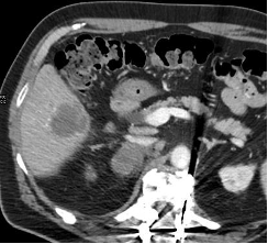
Common mammography findings. Tumors are not removed from the breast during cryotherapy; they are still in the breast, so an inflammatory response is normal and will be visible on mammography. A cryohalo and some fat necrosis are also common. Often, an increasing rim of density at the ablation site and dystrophic calcification are also apparent, so it is important to recognize these findings as normal, in order to avoid unnecessary biopsies in these patients. Case examples showing typical mammography findings post ablation and throughout follow-up are presented in Figure 4 and Figure 5.
Conclusion
This article suggests that cryoablation for breast cancer is a feasible and safe procedure that takes about an hour or less in an outpatient setting. It can be performed with local anesthesia, with no general anesthesia or conscious sedation necessary. The most common mammographic findings after cryoablation are fat necrosis and a cryohalo with fibrosis. Physicians who wish to perform cryoablation must learn to recognize the “new normal” findings on mammograms in order to prevent unnecessary biopsies. Further studies are needed to confirm cryoablation as an alternative to surgical intervention.
Affiliations and Disclosures
From Princeton Radiology CSMC, Department of Radiology, Freehold, New Jersey.
Disclosure:The author has completed and returned the ICMJE Form for Disclosure of Potential Conflicts of Interest. Dr Tomkovich reports honoraria and travel expenses; member of the advisory board (non-paid position) from IceCure Medical. He is the co-principal investigator of the ICE3 trial, for which IceCure provided study materials and equipment.
Address for Correspondence: Kenneth Tomkovich, MD, Princeton Radiology CSMC - Dept. of Radiology, 901 West Main Street, Freehold, NJ 07728. Email: KTomkovich@princetonradiology.com