Angio-Guidewire-Ultrasound (AGU) Guidance for Femoral Access in Procedures Requiring Large Sheaths
Abstract: Many techniques are based on fluoroscopic, angiographic, and echocardiographic guidance to achieve the ideal femoral artery puncture, which is important to decrease vascular-related complications. We report an original technique for femoral access integrating angiographic, guidewire, and ultrasound (AGU) guidance, working together to obtain the best femoral artery stick according to individual patient’s anatomy. This technique is designed to facilitate large-sheath femoral access in procedures requiring ancillary arterial access.
Key words: ultrasound, AGU, angio-guidewire-ultrasound guidance
The use of femoral artery access is decreasing for coronary, diagnostic, and interventional procedures due to radial access adoption, but is still required for an increasing number of endovascular procedures. Most transfemoral percutaneous endovascular procedures, such as transcatheter aortic valve implantation (TAVI), left ventricular percutaneous support, aortic repair, etc, require quite large sheaths. Not surprisingly, such procedures are burdened by a remarkable risk of vascular complications; the incidence of vascular complications after TAVI in the recent PARTNER II trial1 was 7.9%.
The common femoral artery (CFA) is the prolongation of the external iliac artery and begins midway behind the inguinal ligament (which is directed from the anterior-superior spine of the ilium to the symphysis pubis). The CFA, approximately 3-4 cm distal to the inguinal ligament, gives the profundal femoris artery and continues into the superficial femoral artery (SFA). This bifurcation is usually at, or below, the bottom of the femoral head. Of note, in approximately 25% of cases, the femoral bifurcation occurs above this point (occasionally being located as high as above the ligament). Usually, the CFA over the femoral head is regarded as the ideal stick zone, since its large lumen may accommodate a wider range of sheath sizes and the underlining femoral head may facilitate external compression efficacy.2,3 High puncture above the level of the inguinal ligament is associated with higher incidence of retroperitoneal hematoma, which is a fatal complication because this site is not compressable.4 Low puncture below the femoral bifurcation is associated with higher incidence of pseudoaneurysm and arteriovenous fistula.5-7
For the interventionalist, an important vascular-anatomic marker of the relationship between external iliac, CFA, and inguinal ligament is represented by the inferior epigastric artery. Indeed, the inferior epigastric artery arises from the external iliac artery immediately above the inguinal ligament, initially descends without crossing it, and soon ascends to the abdominal wall, crossing the ligament at the lowest point of its course.
Techniques for Femoral Artery Puncture With Large Sheaths
Fluoroscopic guidance. In this method, CFA access is obtained under fluoroscopic guidance while aiming the needle tip at a “target zone” that is located immediately below the center of the femoral head. Due to the variability of femoral artery bifurcation, this technique is completely unreliable when the individual patient’s anatomy is unknown. Moreover, direct radiation exposure on the operator’s hand is needed.8 A possible trick to reduce radiation exposure is to mark the ideal puncture level by putting a hemostat over the area where the femoral head has been identified by fluoroscopy.
Ultrasound guidance. Ultrasound guidance is an emerging technique for femoral catheterization, since it does not require x-ray exposition, and may facilitate procedure success and reduce complications.9 The main drawbacks of this technique are poor visibility of the femoral head and the absence of a clear landmark for the inguinal ligament.
Micropuncture kit. Recently, some operators started using a micropuncture kit that is designed to allow access to the femoral artery with a very small needle (21 gauge; “external diameter, 0.819 mm”) as compared with a regular needle (18 gauge; “external diameter, 1.27 mm”). When the puncture is achieved while guided by either fluoroscopy or ultrasound, a 4 Fr sheath is inserted and angiogram is done to confirm correct positioning. If the position is good, the sheath can be exchanged over a .035˝ wire for the bigger sheath; however, if the position is not good, the sheath is removed, manual compression is applied, and a better puncture is attempted.10 The main drawbacks include the possibility of multiple trials to obtain the perfect femoral artery puncture, and this method still requires either fluoroscopic or ultrasound guidance to localize the femoral artery.
Guidewire guidance. A recent evolution of angiographic guidance is represented by the advancement of a guidewire (usually .035˝) in the CFA from an ancillary access point (such as via radial access). The J-tip of the guidewire is placed in the “ideal” CFA site using the best angiographic view (which is usually oblique). The artery is then punctured under fluoroscopic guidance in postero-anterior view, advancing the needle toward the guidewire’s J tip. This technique does not reduce the risk of direct radiation exposure to the operator’s hand and does not provide any information regarding the CFA wall structure.
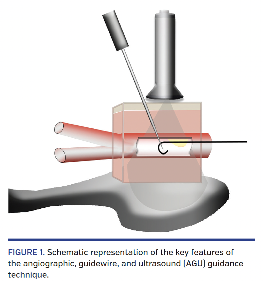
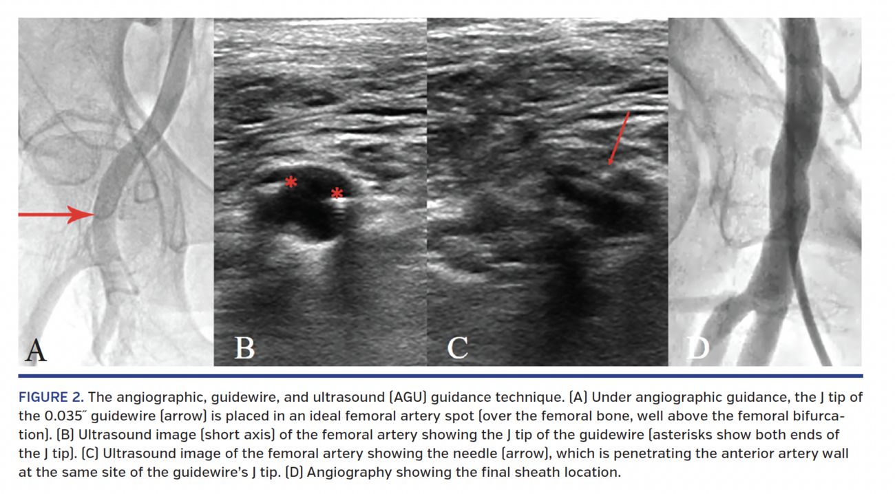
Angiographic, guidewire, and ultrasound (AGU) guidance. We integrated several different techniques to create the AGU guidance technique (see Figure 1 for schematic representation), which is specifically designed with the aim of facilitating the achievement of a precise femoral entry in patients with complex anatomy undergoing procedures requiring large femoral sheaths with simultaneous use of other arterial accesses. The AGU guidance technique consists of the following steps (Figure 2):
- Secondary arterial access (preferably radial access) is taken first and is used to advance a catheter into the iliac artery of the side selected for the large femoral access procedure.
- Under angiographic guidance, a .035˝ J-tip guidewire is placed in the “angiographically ideal” site (wider femoral artery diameter, no calcium, etc) for femoral artery access (Figure 2A).
- The ultrasound probe is used to scan the femoral artery (especially in the longitudinal view) to identify the site of the femoral bifurcation.
- The J-tip of the guidewire is identified inside the femoral artery (Figure 2B) by ultrasound (confirming the vessel quality at the spot identified on angiography), which is also used to guide the advancement of the needle toward the anterior wall (Figure 2C).
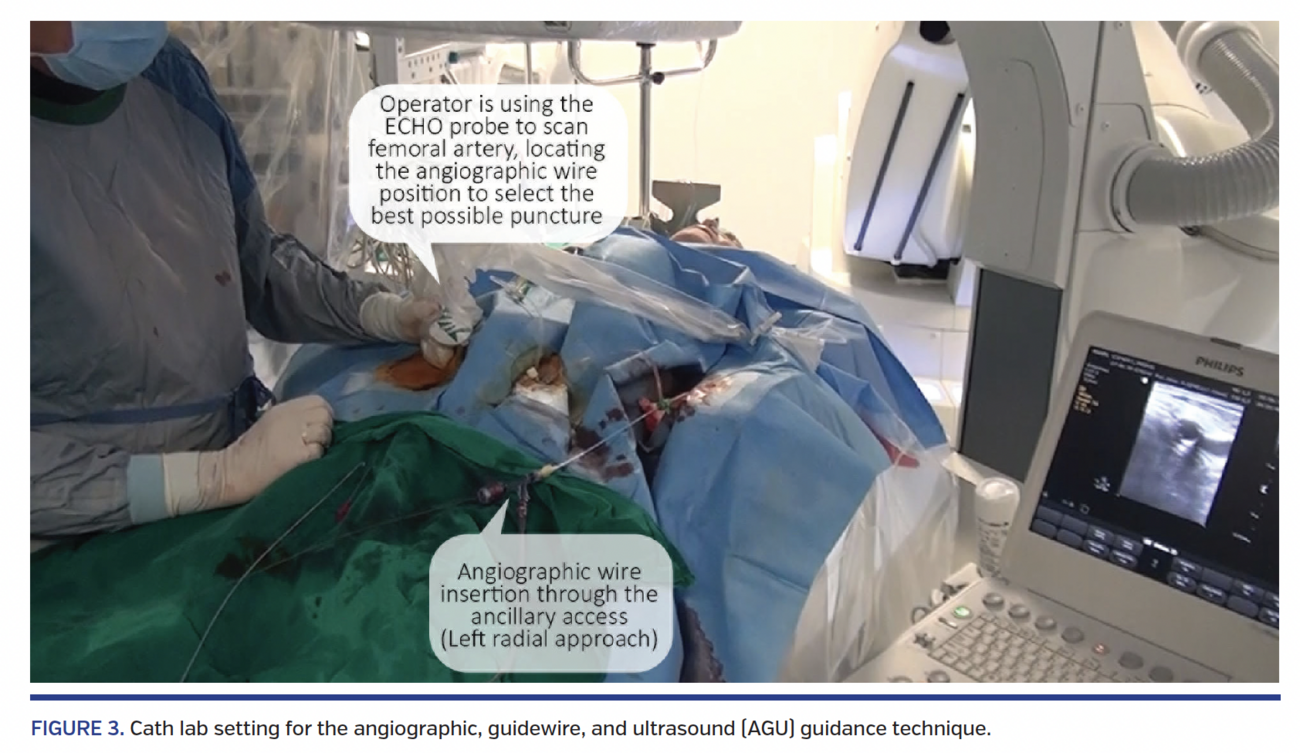
Figure 3 shows a typical catheterization laboratory set-up while practicing the AGU technique.
Conclusion
This novel technique has the potential to facilitate the systematic achievement of the best femoral entry in patients with different vascular anatomy undergoing large femoral access percutaneous procedures. Specifically designed, prospective studies are needed to establish the clinical impact of this new technique.
References
- Leon MB, Smith CR, Mack MJ, et al. Transcatheter or surgical aortic-valve replacement in intermediate-risk patients. N Engl J Med. 2016;374:1609-1620.
- Sandoval Y, Burke MN, Lobo AS et al. Contemporary arterial access in the cardiac catheterization laboratory. JACC Cardiovasc Interv. 2017;10:2233-2241.
- Turi ZG. An evidence-based approach to femoral arterial access and closure. Rev Cardiovasc Med. 2008;9:7-18.
- Raphael M, Hartnell G. Femoral artery catheterization and retroperitoneal haematoma formation. Clin Radiol. 2001;56:933-934; author reply, pp. 934-935.
- Altin RS, Flicker S, Naidech HJ. Pseudoaneurysm and arteriovenous fistula after femoral artery catheterization: association with low femoral punctures. AJR Am J Roentgenol. 1989;152:629-631.
- Gabriel M, Pawlaczyk K, Waliszewski K, Krasinski Z, Majewski W. Location of femoral artery puncture site and the risk of postcatheterization pseudoaneurysm formation. Int J Cardiol. 2007;120:167-171.
- Kim D, Orron DE, Skillman JJ et al. Role of superficial femoral artery puncture in the development of pseudoaneurysm and arteriovenous fistula complicating percutaneous transfemoral cardiac catheterization. Catheter Cardiovasc Diagn. 1992;25:91-97.
- Turi ZG. Fluoroscopy guided vascular access: asking the right question, but getting the wrong answer? Catheter Cardiovasc Interv. 2009;74:540-542.
- Sobolev M, Slovut DP, Lee Chang A, Shiloh AL, Eisen LA. Ultrasound-guided catheterization of the femoral artery: a systematic review and meta-analysis of randomized controlled trials. J Invasive Cardiol. 2015;27:318-323.
- Cilingiroglu M, Feldman T, Salinger MH, Levisay J, Turi ZG. Fluoroscopically-guided micropuncture femoral artery access for large-caliber sheath insertion. J Invasive Cardiol. 2011;23:157-161.
From the Institute of Cardiology, Fondazione Policlinico Universitario A. Gemelli IRCCS, Università Cattolica del Sacro Cuore, Rome, Italy; *on behalf of absence from the Cardiology Department, Tanta University, Tanta, Egypt.
Disclosure: The authors have completed and returned the ICMJE Form for Disclosure of Potential Conflicts of Interest. Dr Burzotta reports advisory board and/or speaker fees from Medtronic, Abbott, and Abiomed. Dr Trani reports advisory board and/or speaker fees from St. Jude Medical, Abiomed, and Biotronic. Dr Aurigemma reports advisory board activities for Biotronic. The remaining authors report no conflicts of interest regarding the content herein.
The authors report that patient consent was provided for publication of the images used herein.
Manuscript submitted July 17, 2018, provisional acceptance given July 23, 2018, final version accepted August 7, 2018.
Address for correspondence: Francesco Burzotta, MD, Universita Cattolica del Sacro Cuore, L. Augosto Gemelli, Roma 00168, Italy. Email: francescoburzotta@gmail.com