

Antecubital Fossa Venous Access For Right Heart Catheterization
Abstract: With the advance of radial access and ulnar access there has been an increased interest in performing right heart catheterization (RHC) and right-heart based procedures via antecubital venous access. Our purpose is to describe the venous anatomy of the upper extremities, technique, equipment, and cost for employing this approach. Reported also is the international experience based on publications assessing procedural success, complications, fluoroscopy time and radiation dose, access-site compression time, and time to ambulation. We conclude that antecubital-venous-access based RHC carries satisfactory success rates, requires a short learning curve, and is exceptionally safe even when performed with full anticoagulation. It is our hope that industry will provide us with even better tools to extend the practice of antecubital-venous-access based procedures and interventions.
J INVASIVE CARDIOL 2017;29(5):169-174.
Key words: radial access, venous anatomy
Anatomy
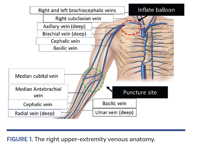
The right upper-extremity venous anatomy is shown in Figure 1. The deep venous system of the forearm is composed of the radial and ulnar veins, which accompany the radial and ulnar arteries (on the lateral and medial aspect of the arm, respectively). These two arteries form the brachial vein at the elbow level. The superficial venous circulation of the forearm is composed of three major superficial veins that are very well interconnected: (1) the basilic vein on the medial aspect of the anterior forearm and arm; (2) the cephalic vein traveling on the lateral aspect of the anterior forearm; and (3) the median antebrachial vein traveling in the middle of the anterior forearm. At the level of the antecubital fossae, these three veins tend to run superficially and create interconnections; the median antebrachial vein connects to the basilic vein and the cephalic vein via the median cephalic and median basilic veins, respectively. Occasionally, the cephalic vein will connect directly with the basilic vein by the median cubital vein. At the level of the elbow (or distal humerus) the basilic vein drains into the deeper brachial vein, which later becomes the axillary vein and subclavian vein. The cephalic vein continues as a more superficial and lateral aspect of the arm and within the deltopectoral groove and triangle (between the pectoralis major and deltoid muscles and the clavicle) to join the subclavian vein. When the internal jugular joins the subclavian vein, they form the right brachiocephalic vein. The left brachiocephalic merges into the right brachiocephalic vein to create the superior vena cava (SVC), which drains into the superior pole of the right atrium.
Anatomical Variants
There are several congenital anatomical variations of the central venous system that can lead to problems or complications during cardiac interventions, especially when performed from the left-side antecubital venous access. Most literature on this subject reflects issues related to electrophysiological procedures, but can probably be extrapolated to RHC. The most widely known anomaly is a persistent left SVC draining into the coronary sinus. Persistent left SVC is found in <0.5% of the general population, but occurs in up to 4% of patients with congenital heart disease. The left brachiocephalic vein is absent and the right SVC is smaller than the left in the majority of these patients.1 Persistent left SVC almost always drains into the coronary sinus. In a minority of the patients, the persistent left SVC drains into the left atrium. In most subjects, the persistent left SVC is an incidental asymptomatic finding on imaging (computed tomography, chest x-ray, or echocardiogram). Persistent left SVC may present technical challenges during RHC when performed from the right-sided approach, as the catheter may be difficult to pass through a dilated coronary sinus and exit the sinus at an acute angle toward the right ventricle. This can be overcome by the use of contrast injections to create a “roadmap,” coronary 0.014˝ wires, and flow-directed catheters.2 Similarly, presence of Thebesian valve at the coronary sinus exit can make left-sided approach to the right heart difficult. Although Thebesian valves are present in >70% of patients, only a small minority of these valves result in significant narrowing of the coronary sinus ostium.3,4 Variation of left brachiocephalic vein may also affect catheter placement for right heart monitoring. The angle at which the left brachiocephalic vein (or left innominate vein) joins the right brachiocephalic vein (or right innominate vein) to form the SVC is variable. Acute angulations at this junction may lead to suboptimal positioning of indwelling central venous catheters inserted from the left side with risk of catheter thrombosis or perforation.5 Rarely, a left subclavian vein has an anomalous course with connection to the left atrium, which may potentially lead to inadvertent advancement of catheters or leads into the left ventricle.
Even more challenging is the presence of totally anomalous venous return typically draining into the coronary sinus.
Procedure
Choice of antecubital venous access side. The preferred antecubital side will typically be the right. Considerations taken into account are: (1) restricted, injured, or painful arm; (2) other devices within the venous circulation; (3) difficulties or concerns encountered with the venous anatomy during previous procedures or imaging studies, respectively; (4) patient height (attempting left antecubital venous access in tall patients may result in the inability to reach the pulmonary artery with our conventional 110 cm pulmonary artery catheters); (5) convenience of set-up, patient and operator comfort in case simultaneous arterial access is required (ipsilateral side is advantageous for both patient and operator).
Venous access (Table 1). Venous access to an antecubital vein will usually be obtained in the preprocedural holding area by the nursing staff. Occasionally, when venous access is not attainable in the holding area, a venous tourniquet will be placed on the arm; under local anesthesia and employing ultrasound guidance, a venous puncture should be attempted with a 21 g Seldinger needle. The most accessible and largest antecubital vein should be targeted by an anterior wall stick. Ideal veins should be ≥6 mm in diameter (3-6 mm is borderline size) and depth should not exceed 10 mm (10-16 mm depth is borderline). Once puncture is performed and venous blood return is observed, the 0.018˝, 45 cm-long, soft-tipped wire (present in most radial kits) should be advanced through the needle into the proximal venous system with no encountered resistance. Before withdrawing the needle, a small superficial nick adjacent to the needle puncture site (with a #11 blade) should be done and the venous tourniquet removed. Practically all superficial veins of the antecubital fossa can be used whether they drain into the cephalic or basilic veins.6
In case venous access was obtained in the preprocedural holding area, after prepping, draping the arm, and disinfecting the intravenous cannula, under local anesthesia the intravenous cannula cap should be removed and discarded and the 0.018˝, 45 cm wire should be advanced through the intravenous cannula. The access site should be superficially nicked with a #11 blade and the intravenous cannula should be removed over the wire and discarded. A 5 Fr radial sheath should be advanced over the wire. The choice of venous sheath size varies among operators. We typically use a 5 Fr sheath since we do not see the advantage in using a larger-sheath diameter, which could potentially allow a larger-diameter pulmonary artery catheter (PAC) to be used; the most frequent cause of procedure failure is the inability to advance the PAC across the tortuous, calcified, and stenosed venous segment (typically in the cephalic, axillary, or subclavian vein). While the 0.014˝ wire can almost always easily negotiate the stenosed or tortuous segment, the bulkier large-diameter PAC can be more of a disadvantage than an advantage.
Once the sheath is within the vein, the dilator and wire are removed and discarded. Gloves should be exchanged and the sheath is flushed with saline via its side-port. If you can’t get blood return from the sheath side-port, you can flush the sheath via its valve using a saline-filled dilator or 0.014˝ wire introducer attached to a syringe, or by insertion of the distal 2-3 cm tip of the previously flushed PAC. The access site is secured by Tegaderm (3M), which is nicked by the blade to allow access to the venous sheath valve.
Pulmonary artery catheter. The choice of PAC depends on whether thermodilution cardiac outputs are required or if the patient has history of latex allergy. If thermodilution cardiac output is warranted or the patient has latex allergy, the preferred catheter is the 5 Fr, 110 cm TS105FS (Edwards Lifesciences). In any other scenario, we prefer to use a 5 Fr, 110 cm AI-017124 (Arrow), which costs less and will less frequently result in pressure dampening, inability to draw blood samples, or difficulty advancing wires through the catheter lumen. Both of these catheters have relatively small balloons at their tips (0.75 mL), which occasionally will make acquisition of optimal pulmonary wedge pressure quite a challenge.
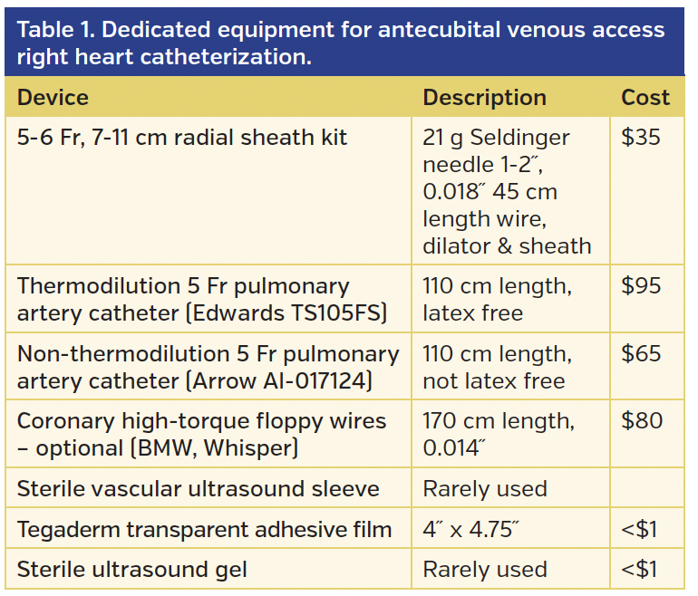
Catheterization. The initial 30-40 cm insertion of the PAC should be done without fluoroscopy while the balloon tip is deflated. There should be no resistance to the advancement of the PAC. If resistance is encountered, fluoroscopy with optional venography (to delineate a venous roadmap) is suggested. Within 30-40 cm, the PAC should reach the axillary and subclavian veins (Figure 2). At that point, advancing the catheter should be done under fluoroscopic guidance with the option of balloon inflation. Whenever there is any uncertainty about the patient anatomy, injection of contrast (via the major port of the PAC while the balloon is deflated) should be considered.
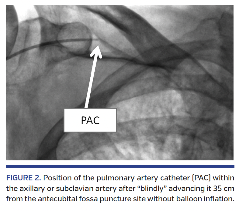
The advancement of the PAC is sometimes challenging, especially in older subjects harboring previous or current venous devices or leads in their venous system. Besides angiography that better defines the anatomy (Figure 3), one can use 0.014˝ or 0.018˝ soft-tip coronary or peripheral wires (170 cm length) to negotiate the tortuosity and challenging angulations of venous and cardiac structures. The typical tip of the wire should be prepared by the operator (our preferred starting angle is 45° at 1-2 cm from the distal tip of the wire. Typical friction point is the 90° entry point of the cephalic vein into the axillary vein and the right ventricular outflow track.
Difficulty advancing PAC. When encountering resistance in advancing the PAC, one should consider the following options: (1) angiography to better delineate the venous anatomy and delineate alternative venous routes; (2) using a 0.014-0.018˝ soft-tip wire to negotiate the anatomy; (3) intravenous nitroglycerin to optimize venodilation; (4) patient asked to “take a deep breath” or other positional maneuvers to enhance flow and modify the anatomy; (5) increase sheath size to 6 Fr and use two wires (one as a buddy wire); (6) use a long sheath and dilator; (7) if pulmonary wedge pressure is not a must, one can use diagnostic 4 Fr catheters (multipurpose or pigtail) or even low-profile catheters like Renegade (Boston Scientific) to obtain pressures and blood samples; and finally (8) consider seeking alternative venous access.
Complications
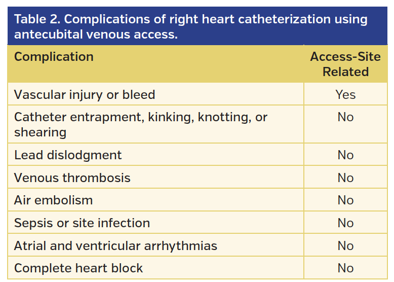
Table 2 shows that access-site related complications occurring during antecubital-venous-access based RHC are mostly local bleeding complications related to the attempt to obtain venous access and inadvertently injure the brachial or other antecubital arteries in the process. Other non-access site potential complications, such as major bleed, major vascular injury, air embolism, bacteremia, antecubital venous access-site infection, catheter knotting, entrapment or shearing, lead dislodgment, venous thrombosis, heart block or ventricular or atrial arrhythmias, and hemoptysis, are exceedingly rare and can potentially be encountered in RHC using any access site.
Incomplete Procedure and Venous Access Crossover
Incomplete procedure is by far the most common limitation of RHC via antecubital venous access and varies greatly based on populations studied. While incomplete procedures are an extremely rare occurrence in younger subjects (age <65 years), incomplete procedure rate may reach 5%-10% in our octogenarian and nonagenarian subjects, especially with the presence of current or former venous leads, devices, and interventions and the presence of advanced calcific, tortuous, and degenerated venous system.
While certain authors report success rates approaching 100%, a more realistic success rate map is drawn in a report by Roule et al,6 who evaluated their procedural outcomes during 1007 consecutive RHC procedures. They found that 43 subjects (4.3% of the study population) were not suitable for antecubital venous access. Of the 964 patients (95.7% of study cohort) in which RHC was attempted by antecubital venous access, only 80.7% had RHC completed via the initial antecubital venous access site (84.3% of those attempted). Hence, the initial antecubital venous access-site failure rate was 16.7%. Of the 151 patients (15% of study cohort) crossing over to an additional RHC access site, 82 patients (8.1%) crossed over to the contralateral antecubital venous access site and 69 patients (6.9%) crossed over to femoral vein access.
Advantages, Limitations, and Contraindications
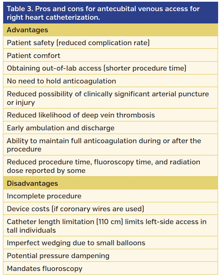
The advantages and limitations of RHC via antecubital venous access are delineated in Table 3. In the era of “patient-centered care” where patient safety and comfort take center stage, antecubital-venous-access based RHC should be the preferred approach for most subjects requiring RHC. In conjunction with radial-based or ulnar-based arterial access and the associated anticoagulation requirements, as well as early ambulation and discharge, antecubital-venous-access based RHC seems more compatible, especially with ambulatory-based diagnostic and therapeutic procedures.
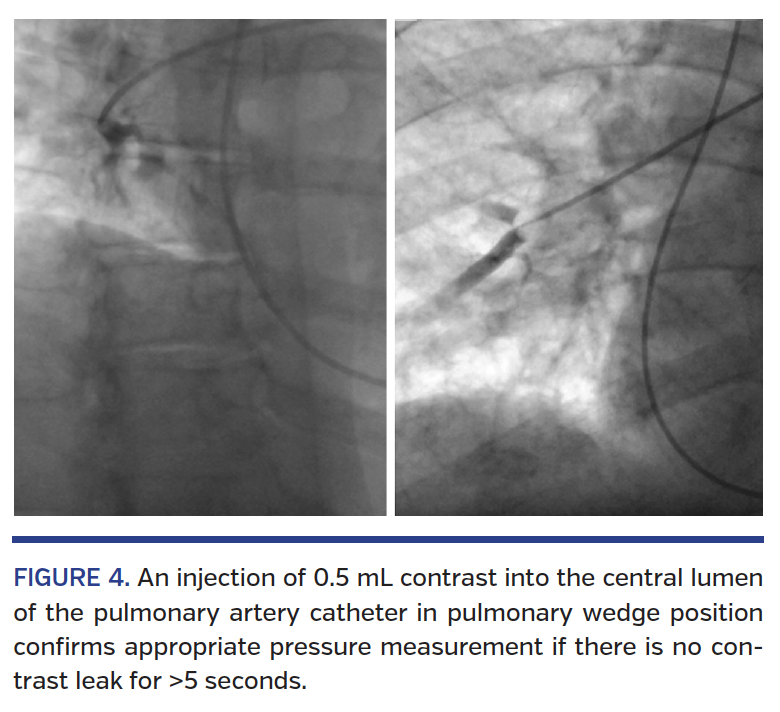
One disadvantage of the currently available 5 Fr PAC is the relatively short length (110 cm) and small balloon, which may yield imperfect pulmonary wedge pressure due to suboptimal occlusion of the pulmonary artery. Besides obtaining pulmonary capillary wedge saturation and looking carefully at the pressure tracings, we have made it a habit to inject 0.5 mL contrast and document that there is no escape of the contrast for at least 5 seconds (Figure 4).
Although venous thromboembolism after antecubital venous access practically never results in any clinical sequelae, there are no follow-up data on the patency of the veins used for antecubital venous access.
Contraindications
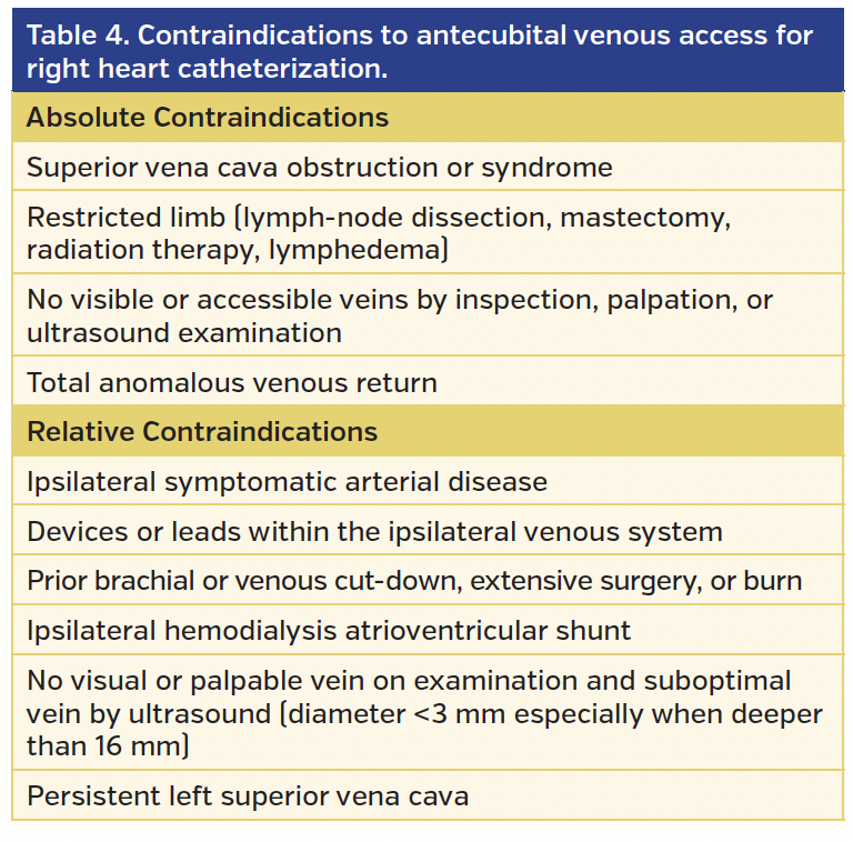
Absolute and relative contraindications are listed in Table 4.
International Experience
International experience is shown in Table 5. The reported success rate approaches 100% by some authors; however, others report initial site failure as high as 16% when RHC via antecubital venous access is used as the exclusive RHC strategy. Complication rates (especially site-specific complication rates) are uniformly extremely low and are typically limited to small access-site hematomas.
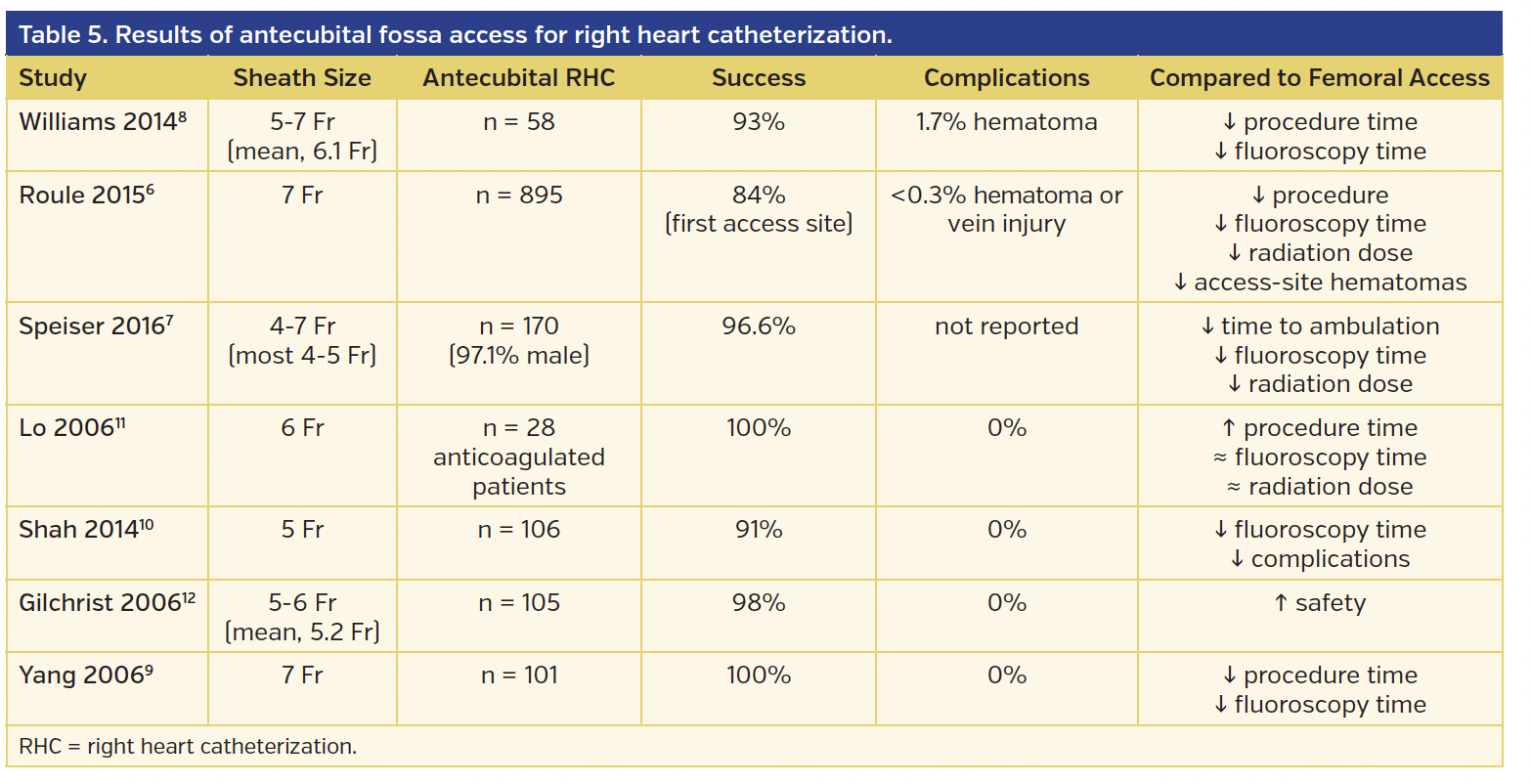
Antecubital-venous-access based RHC can be done safely on anticoagulated6,7 patients without interrupting the anticoagulation. This is particularly important in conjunction with the prevailing practice of radial or ulnar angiography and intervention (where anticoagulation is mandatory) and early ambulation and discharge are used as quality and/or performance measures. Most of the reported series suggest that RHC via antecubital venous access is associated with reduced fluoroscopy dosage and procedure times,6,8,9 reduced radiation exposure site compression time,6,7 and reduced time to ambulation,7 while a few suggest reduced complication rate.6,10 The procedure can be executed very well by physicians in training6 and has a relatively brief learning curve.
Conclusion
Antecubital-venous-access based RHC should probably be the preferred method to perform RHC in view of the fact that this procedure is greatly preferred by most patients and is associated with an excellent access-site safety profile while requiring a very short learning curve. In conjunction with transradial or ulnar angiography and interventions (which mandate full anticoagulation) and at times when performance measures include early ambulation and discharge, antecubital-venous-access based RHC appears to be a better choice for most patients.
Refinement in dedicated equipment for this procedure may not only further improve success rates and procedural safety, but also allow us to perform many procedures that are currently performed only via larger venous access, including biopsy of the endomyocardium, biopsy of endocardial masses, transcatheter interventions in pulmonary embolism and deep vein thrombosis, insertion and removal of inferior vena cava filters, transvenous pacing, and even structural heart interventions.
References
1. Biffi M, Boriani G, Frabetti L, Bronzetti G, Branzi A. Left superior vena cava persistence in patients undergoing pacemaker or cardioverter-defibrillator implantation: a 10-year experience. Chest. 2001;120:139-144.
2. Rubenfire M, Evangelista J, Wajszczuk WJ, Kantrowitz A. Implication of a persistent left superior vena cava in transvenous pacemaker therapy and cardiac hemodynamic monitoring. Chest. 1974;65:145-147.
3. Mak GS, Hill AJ, Moisiuc F, Krishnan SC. Variations in Thebesian valve anatomy and coronary sinus ostium: implications for invasive electrophysiology procedures. Europace. 2009;11:1188-1192.
4. Hołda MK, Klimek-Piotrowska W, Koziej M, Mazur M. Anatomical variations of the coronary sinus valve (Thebesian valve): implications for electrocardiological procedures. Europace. 2015;17:921-927.
5. Stonelake PA, Bodenham AR. The carina as a radiological landmark for central venous catheter tip position. Br J Anaesth. 2006;96:335-340.
6. Roule V, Ailem S, Legallois D, et al. Antecubital vs femoral venous access for right heart catheterization: benefits of a flashback. Can J Cardiol. 2015;31:1497.e1-e6.
7. Speiser B, Pearson K, Xie H, Shroff AR, Vidovich MI. Compared to femoral venous access, upper extremity right heart catheterization reduces time to ambulation: a single-center experience. Catheter Cardiovasc Interv. 2017;89:658-664. Epub 2016 May 19.
8. Williams PD, Palmer S, Judkins C, et al. Right and left heart catheterization via an antecubital fossa vein and the radial artery – a prospective study. J Invasive Cardiol. 2014;26:669-673.
9. Yang CH, Guo GB, Yip HK, et al. Bilateral cardiac catheterizations: the safety and feasibility of a superficial forearm venous and transradial arterial approach. Int Heart J. 2006;47:21-27.
10. Shah S, Boyd G, Pyne CT, et al. Right heart catheterization using antecubital venous access: feasibility, safety and adoption rate in a tertiary center. Catheter Cardiovasc Interv. 2014;84:70-74.
11. Lo TS, Buch AN, Hall IR, Hildick-Smith DJ, Nolan J. Percutaneous left and right heart catheterization in fully anticoagulated patients utilizing the radial artery and forearm vein: a two-center experience. J Intervent Cardiol. 2006;19:258-263.
12. Gilchrist IC, Moyer CD, Gascho JA. Transradial right and left heart catheterizations: a comparison to traditional femoral approach. Catheter Cardiovasc Interv. 2006;67:585-588.
From the Division of Cardiology, Robert Packer Hospital, Guthrie Health Systems, Sayre, Pennsylvania; and the Commonwealth Medical College, Scranton, Pennsylvania.
Disclosure: The authors have completed and returned the ICMJE Form for Disclosure of Potential Conflicts of Interest. The authors report no conflicts of interest regarding the content herein.
Manuscript submitted December 10, 2016, provisional acceptance given December 16, 2016, final version accepted January 6, 2017.
Address for correspondence: Edo Kaluski, MD, FACC, FESC, FSCAI, Guthrie Health Systems, 1 Guthrie Square, Sayre, PA 18840. Emails: ekaluski@gmail.com and Kaluski_edo@guthrie.org


