


Amplatzer or Figulla Flex II Occluder: A Comparative Study of Outcomes After Transcatheter Patent Foramen Ovale Closure
© 2024 HMP Global. All Rights Reserved.
Any views and opinions expressed are those of the author(s) and/or participants and do not necessarily reflect the views, policy, or position of the Journal of Invasive Cardiology or HMP Global, their employees, and affiliates.
Abstract
Objectives. Percutaneous closure of a patent foramen ovale (PFO) for the prevention of recurrent paradoxical thromboembolic events has been shown to be safe and effective in randomized controlled trials. However, it remains uncertain if differences in the structure and design of the occluder devices impact the outcomes. The aim of this study was to compare results of percutaneous PFO closure using 2 widely used double-disc occluders.
Methods. Consecutive patients who underwent percutaneous PFO closure with the Abbott Amplatzer occluder (APO) or the Occlutech Figulla-Flex-II occluder (OPO) at the Heart Center Lucerne between February 2017 and December 2022 were included in a registry. The primary endpoint was effective closure of the PFO, defined as a residual shunt grade 0 or 1, assessed by contrast echocardiogram at 6-month follow-up. Secondary endpoints included procedural efficacy/safety and major adverse cardiovascular events during the hospital stay and at 6-month follow-up.
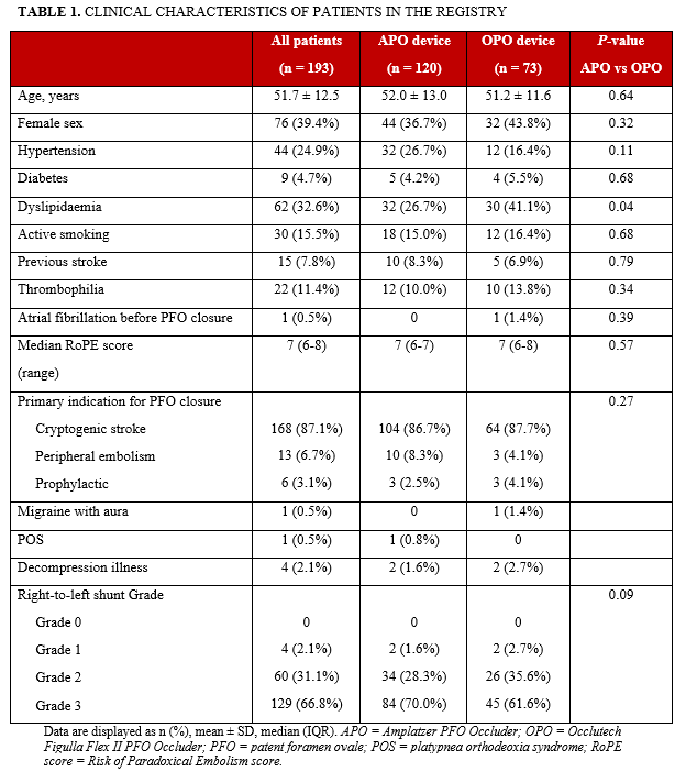
Results. One hundred ninety-three consecutive patients (mean age 51.7 ± 12.5 years; 39% women; Risk of Paradoxical Embolism (RoPE) score = 7, interquartile range [IQR] = 6-8) underwent percutaneous PFO closure with the APO (120 patients, 62.2%) or the OPO (73 patients, 37.8%). Main indications for closure were crypotogenic stroke in 168 patients (87.1%) and peripheral embolism in 13 patients (6.7%). At baseline, right-to-left shunt (RLS) greater than or equal to grade 2 was present in 189 patients (97.9%). Immediate procedural success was 99.5%. In 1 patient, an air embolism occurred during positioning of the APO occluder with transient chest pain and electrocardiogram changes, but without further sequelae to the patient. At 6-month follow-up, effective closure was achieved in 185 patients (95.8%; APO: 96.6% vs OPO: 94.5%, P = .30). Rates of atrial fibrillation and recurrent thromboembolic events were 4.2% and 0.5%, respectively.
Conclusions. PFO closure is safe and effective when performed with either the self-expanding Abbott Amplatzer or Occlutech Figulla Flex II PFO occluder.
Introduction
Multiple randomized trials have shown that, in patients with a history of cryptogenic stroke, percutaneuous patent foramen ovale (PFO) closure is superior to medical therapy for the prevention of recurrent events.1-4 As a consequence, self-expanding double-disc occluders have been increasingly implanted during the past years. However, it remains uncertain whether differences in the structure and design of the device impacts the outcome. The present report compares results of 2 widely used, self-expanding, double-disc occluders: the Abbott Amplatzer PFO occluder (APO) and the Occlutech Figulla Flex II PFO occluder (OPO). The comparison is based on procedural data as well as mid-term echocardiographic and clinical data from an ongoing Swiss real-world registry.
Methods
Patients and study design. The single-center registry was initiated by the investigators and conducted from February 2017 to December 2022 at the Heart Center Lucerne (Switzerland). The registry included consecutive patients undergoing transcatheter PFO closure, using either the APO or the OPO. The study was approved by the Swiss Ethics Committee (PAL-Registry ID: 2022-02034). Treatment was conducted according to standard-of-care at our institution. The choice between the 2 occluders was at the operator’s discretion; no anatomical or clinical considerations were involved. Before PFO-closure, patients underwent a thorough work-up, including an accurate clinical history, transthoracic echocardiography (TTE) and transoesophageal echocardiography (TOE), echocardiogram (ECG) monitoring over 1 to 7 days, and screening for thrombophilia. Patients with stroke also underwent brain magnetic resonance imaging (MRI) or a computed tomography (CT) scan. All patients underwent clinical and echocardiographic follow-up 6 months after the procedure.
The primary efficacy endpoint was effective closure of the PFO, defined as a residual shunt grade 0 or 1, assessed by contrast ECG at 6-month follow-up. Secondary endpoints included mortality, stroke, bleeding, and vascular complications during hospital stay and through 6-month follow-up.
Echocardiography. All patients underwent TTE and TOE prior to PFO closure. TTE +/- TOE was repeated 6 months after the procedure to assess procedural efficacy. Right-to-left shunt (RLS) was assessed semi-quantitatively, using agitated saline at rest and after the Valsalva maneuver. RLS was graded according to the number of microbubbles detected in the left atrium after shunting from the right atrium. A still frame, identified during the first 3 cardiac cycles of contrast entering the right atrium and the RLS, was graded as follows: Grade 0 (no bubbles); Grade 1 (small RLS, ≤ 5 bubbles seen in the left heart); Grade 2 (moderate RLS, obvious shunt with 6-20 bubbles seen in the left heart); or Grade 3 (large RLS, > 20 bubbles with partial or complete opacification of the left heart).2 The highest RLS grade was used for the analysis.
PFO closure devices. The APO is a self-expandable, double-disc device made from a nitinol wire mesh (Figure 1). The nitinol wire is etched using a patented Intaglio treatment, which reduces the amount of nickel leached from the device. The 2 discs are linked together by a short connecting waist. In order to increase endothelialization capability, the discs contain thin polyester fabric, which are sewn to each disc by polyester thread. The delivery system consists of a delivery sheath with a Tuohy-Borst adapter, dilator, loading device, and delivery cable. The occluder is connected to the delivery cable via a screw mechanism. Once in position, the occluder is released by a counter-clockwise turn on the delivery cable. The device comes in 4 sizes, with the right atrial disc ranging from 18 to 35 mm.
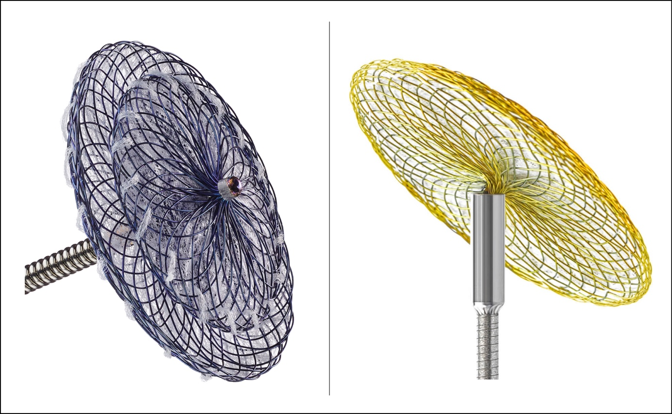
The OPO is a self-expandable, double-disc device made from a nitinol wire mesh (Figure 1). A biocompatible titanium oxide surface reduces nickel leaching. The 2 discs are linked together by a short connecting waist. Each disc contains a polyethylene terephthalate (PET) patch to facilitate endothelialization. The delivery system consists of a delivery sheath with Tuohy-Borst adapter, dilator, loading device, and a shapeable pusher. The occluder is attatched to the pusher via a ball-forceps connection, allowing angulation up to 50°. Once the occluder is in position, it is released by opening the forceps. The device comes in 4 sizes, with the right atrial disc ranging from 18 to 35 mm. Compared to the APO, the OPO has a larger left atrial disc for each size.
PFO closure procedure. The implantation procedure has been described in detail for both the APO2 and the OPO.5,6 PFO closure was performed under local anesthesia, using fluoroscopy guidance. TOE was used at the operator's discretion. In 4 patients (younger age [< 25 years], with concern about radiation),7 implantation was performed solely under TOE guidance. After puncture of a femoral vein, the PFO was crossed using a 5-French (Fr) multipurpose catheter (Cordis). The catheter was then exchanged with an 8- or 9-Fr transseptal sheath and parked in the left atrium. Size of the occluder was chosen according to the TOE results. The APO or OPO device was then advanced into the left atrium. The position of the occluder was held in the left atrium, and the transseptal sheath was pulled back to develop the left atrial disc of the occluder. The device was then pulled back to the interatrial septum, and the right atrial disc was developed by further pulling back the transseptal sheath. The stability of the occluder was ascertained with a gentle push/pull test. The device position was confirmed by fluoroscopy/TOE, and if satisfactory, the delivery system was disconnected from the occluder. Following implantation, correct positioning was documented by fluoroscopy and echocardiography. The procedure was successful if the occluder was in the correct position and no or minimal residual shunt was detected by ECG. After removal of the delivery sheath, manual compression was applied to the access side until hemostasis was achieved, followed by application of a compression bandage for 6 hours. All patients were treated on an outpatient basis.
Medical treatment. Conscious sedation was used in all patients. Patients received heparin intravenously (80 units/kg body weight) during the implantation procedure, to achieve an activated clotting time (ACT) of greater than 250 seconds. Amoxicillin, or vancomycin in cases of known allergy, was used for periprocedural antibiotic prophylaxis. Antibiotic prophylaxis of endocarditis was recommended for 6 months after the procedure. Dual antiplatelet therapy with aspirin (100 mg once daily) and clopidogrel (75 mg once daily) was recommended for all patients during the first 3 months after PFO closure, followed by 3 months of aspirin monotherapy.
Statistics. Data are presented as mean ± SD for continuous variables, and as numbers and frequencies for categorical variables. Unpaired and paired continuous variables were compared using unpaired and paired Student’s t-tests or the Wilcoxon rank sum test, as appropriate. Comparison of categorical variables was performed using chi-square or Fisher’s exact tests, as appropriate. Statistical analyses were conducted with STATA´s statistical software package (Version 16.1, StataCorp). A P-value of less than .05 was considered statistically significant.
Results
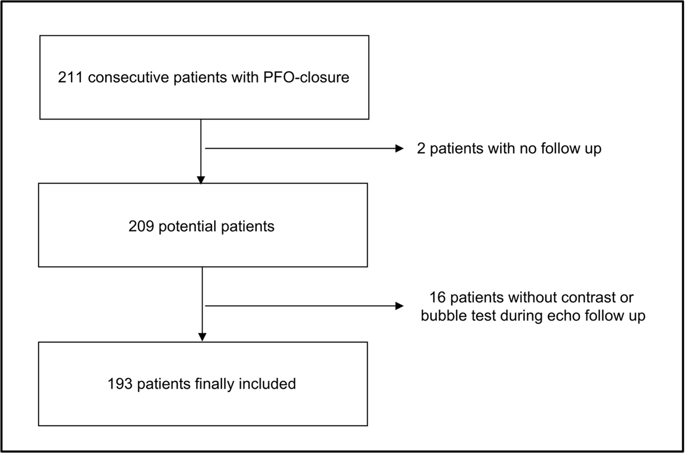
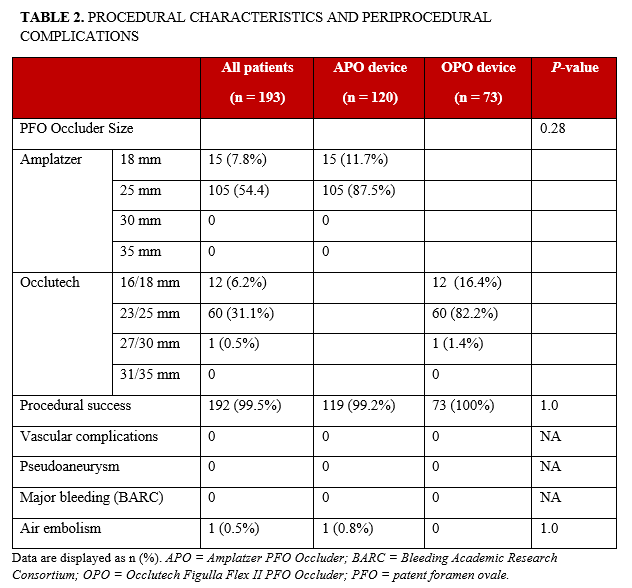
A total of 193 consecutive patients undergoing PFO closure were analyzed (Figure 2). The APO device was used in 120 patients, and the OPO device was used in 73 (Table 1). Mean age was 51.7 ± 12.5 years, and 39% of patients were women. Prior paradoxical embolization was the indication for PFO closure in 181 patients (93.8%). Median Risk of Paradoxical Embolism score (RoPE score) was 7 (range, 6-7). A grade 2 or 3 RLS was documented in 189 patients (97.9%). Acute procedural success was achieved in 99.5% of patients (Table 2). In 1 patient (APO group), the right atrial disc was partially deployed on the left atrial side. Following a snare maneuver, the device was properly positioned as documented by fluoroscopy and TOE. In 1 patient, transient chest pain occurred, accompanied by ST-elevation in the inferior ECG leads. Immediate coronary angiography documented air embolism of the distal right artery, which resolved spontaneously without any sequelae to the patient. One patient was readmitted within the first month due to new onset atrial fibrillation.
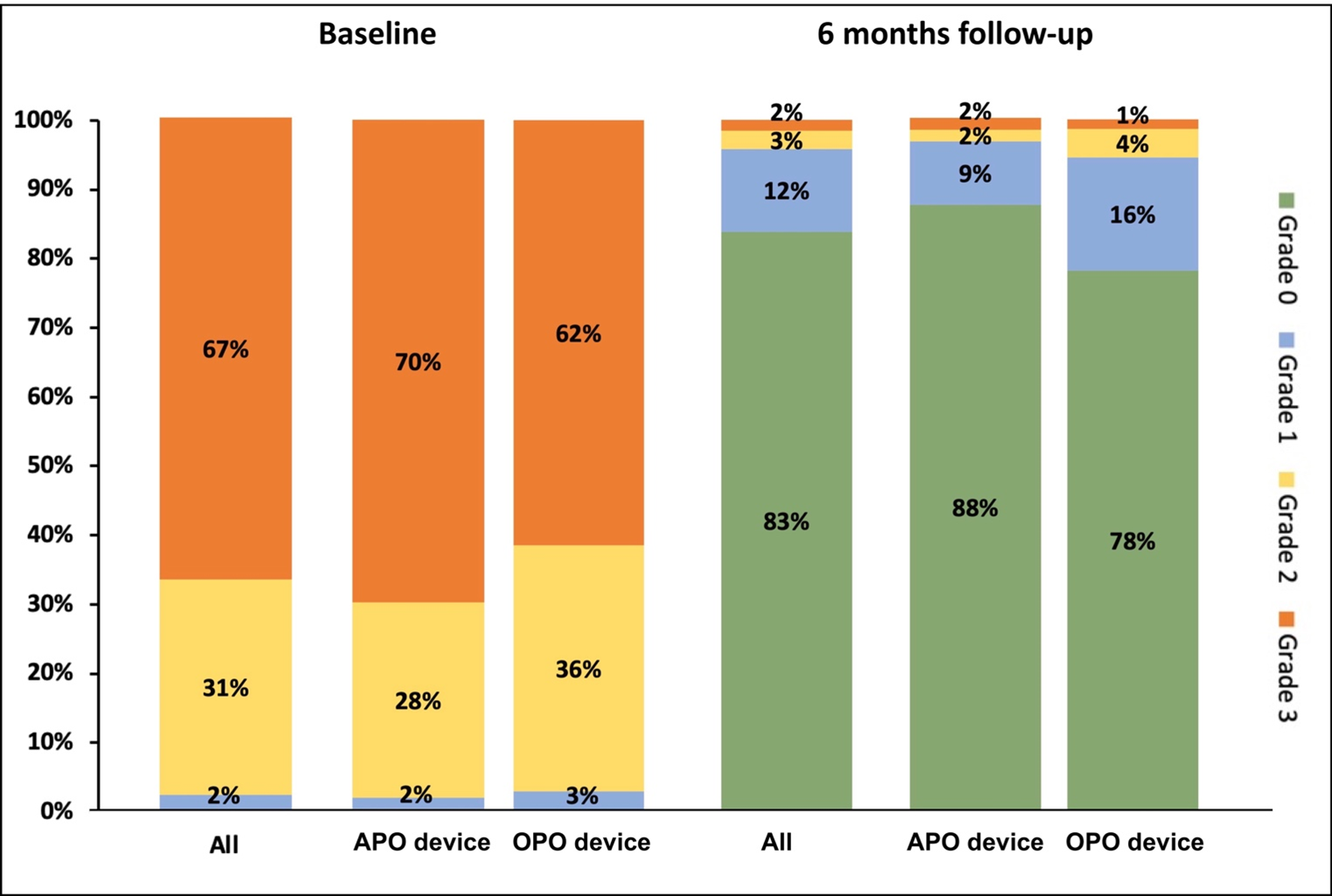
ECG outcomes. At 6-month follow-up, echocardiography documented an RLS of grade 0 or 1 (effective closure) in 185 patients (95.8%). There was no statistical difference between the APO and OPO group (Figure 3). No pericardial effusion or thrombus on the PFO device was noted during follow-up.
Clinical outcomes. No death was recorded during the 6-month follow-up. One stroke occurred in a patient in the APO group (0.5%). This patient showed a mild RLS without evidence of thrombus during ECG follow-up and no evidence of atrial fibrillation. The patient recovered, and life-long aspirin monotherapy was recommended. Atrial fibrillation was documented in 8 patients (4.2%) during follow-up, 5 from the APO group and 3 from the OPO group (P = .96). These patients were subsequently treated with oral anticoagulation, and antiplatelet therapy was stopped.
Discussion
This is the first report that directly compares Occlutech’s third generation PFO occluder device with the APO. Six-month outcomes of patients undergoing PFO closure with the APO or OPO confirmed high procedural success and a low periprocedural complication rate for both occluders. ECG follow-up showed adequate closure of the PFO with no or only mild RLS (effective closure), and there was no statistical difference between the 2 occluder devices.
Periprocedural outcome. The high procedural success (99.5%) for both the APO and OPO occluders is line with previous, mostly single-arm, observational studies,5,8-10 and is similar to results of the large randomized RESPECT (Randomized Evaluation of Recurrent Stroke Comparing PFO Closure to Established Current Standard of Care Treatment) trial, which used the Amplatzer device in the PFO closure group.2 In the current study, air was embolized in 1 patient during the procedure, but there were no other major periprocedural complications, similar to results of other studies.5,9-12 In the past, only one registry using the Amplatzer device reported outcomes of patients treated in an outpatient setting.13 Most patients have been treated during a short hospital stay, regardless of the closure device used.2,5,8-10,12 In those studies, a substantial number of patients (15%) had to be readmitted to the emergency department shortly after the PFO closure procedure. In our registry, only one patient had to be readmitted within the first month after the procedure, due to new onset of atrial fibrillation. Further data on safety of outpatient treatment is strongly warranted, as same-day discharge has the potential to reduce constraints on our health system.
Residual RLS during ECG follow-up. The evidence of no or mild RLS (grade 0 or 1) is frequently used during ECG follow-up to demonstrate effective PFO closure, as only moderate and large residual RLS seem to be linked to high rates of cerebrovascular events. Effective closure rates for the APO and OPO devices ranged from 79% to 100% in single-arm observational studies.5,8-10 Data from randomized, controlled clinical trials is only available for the APO device: The RESPECT and PC (Clinical Trial Comparing Percutaneous Closure of Patent Foramen Ovale [PFO] Using the Amplatzer PFO Occluder with Medical Treatment in Patients with Cryptogenic Embolism) trials reported effective closure in 93.5% and 95.9% of patients, respectively.14,15 In our registry, the rate of effective closure was 95.8%.
The APO and OPO devices have been directly compared in only 2 observational studies, which solely16 or partly12 used previous-generation OPO devices. One small study (20 APO vs 20 OPO) suggested that the OPO device had a higher rate of large residual RLS at 6-month ECG follow-up compared to the APO device.16 However, a larger study (179 APO vs 227 OPO) found similar effective closure rates (95.5%) for the APO and OPO devices at 6-month follow-up,12 which is consistent with our results.
Clinical outcome during mid-term follow-up. Atrial fibrillation after PFO closure is a major clinical concern. In our registry, atrial fibrillation was documented in 8 patients (4.2%) during follow-up. The rate of new onset of atrial fibrillation ranged from 0% to 7.6% in observational studies for both occluders.5,8-10 The RESPECT and PC trials reported new onset of atrial fibrillation in 3% and 2.9%, respectively, of patients treated with the Amplatzer device.14,15 Aggregated evidence from randomized clinical trials indicates that the risk of new onset atrial fibrillation is significantly increased with device closure compared to medical therapy (risk ratio 4.68, absolute risk 4.3% at mean follow-up of 4 years).4
Limitations. The current registry has some limitations. It is an observational study. Echocardiography and other assessments were carried out according to the standards of our center. The choice between the 2 occluders was at the operator’s discretion. However, no anatomical or clinical considerations were involved. ECG outcomes and adverse events were based on site-reported data. Nevertheless, the present study is the largest comparison of the newest generation of OPO and APO devices to date, and therefore, provides useful efficacy and safety information for clinicians.
Conclusions
PFO closure with both the self-expanding APO and OPO PFO occluders seems effective and safe when examined through mid-term follow-up.
Affiliations and Disclosures
From the Heart Center Lucerne, Luzerner Kantonsspital, Lucerne, Switzerland.
Data availability statement: The data that support the findings of this study are available from the corresponding author upon reasonable request.
Disclosures: Dr Toggweiler serves as a consultant and proctor for Medtronic, Boston Scientific, Edwards Lifesciences, and Biosensors; as a proctor for Edwards Lifesciences and Abbott Vascular; as a consultant for Medira, Shockwave, Teleflex, AtHeart Medical, Veosource, and Polares Medical; has received institutional research grants from Boston Scientific, Fumedica, and Novartis; and holds equity in Hi-D Imaging. Dr Bossard has received speaker and consulting fees from Abiomed, Abbott Vascular, Astra Zeneca, Amarin, Amgen, Novartis, OmPharma, Terumo, and SIS Medical. Dr Wolfrum serves as a proctor for Biosensors. The remaining authors report no financial relationships or conflicts of interest regarding the content herein.
Address for correspondence: Mathias Wolfrum, MD, Cardiology Division, Heart Center Lucerne, Luzerner Kantonsspital, Spitalstrasse 16, 6000 Luzern, Switzerland. E-mail: mathias.wolfrum@luks.ch; X: @WolfrumMathias
References
1. Mas JL, Derumeaux G, Guillon B, et al; CLOSE Investigators. Patent foramen ovale closure or anticoagulation vs. antiplatelets after stroke. N Engl J Med. 2017;377(11):1011-1021. doi: 10.1056/NEJMoa1705915
2. Saver JL, Carroll JD, Thaler DE, et al; RESPECT Investigators. Long-term outcomes of patent foramen ovale closure or medical therapy after stroke. N Engl J Med. 2017;377(11):1022-1032. doi: 10.1056/NEJMoa1610057
3. Søndergaard L, Kasner SE, Rhodes JF, et al; Gore REDUCE Clinical Study Investigators. Patent foramen ovale closure or antiplatelet therapy for cryptogenic stroke. N Engl J Med. 2017;377(11):1033-1042. doi: 10.1056/NEJMoa1707404
4. Ahmad Y, Howard JP, Arnold A, et al. Patent foramen ovale closure vs. medical therapy for cryptogenic stroke: a meta-analysis of randomized controlled trials. Eur Heart J. 2018;39(18):1638-1649. doi: 10.1093/eurheartj/ehy121
5. Neuser J, Akin M, Bavendiek U, Kempf T, Bauersachs J, Widder JD. Mid-term results of interventional closure of patent foramen ovale with the Occlutech Figulla Flex II Occluder. BMC Cardiovasc Disord. 2016;16(1):217. doi: 10.1186/s12872-016-0391-3
6. Pedra C, Pedra S, Costa R, Ribeiro M. The Figulla-Occlutech device. In: Sievert H, Qureshi SA, Wilson N, Hijazi ZM, eds. Interventions in Structural, Valvular and Congenital Heart Disease. CRC Press; 2014.
7. Han Y, Zhang X, Zhang F. Patent foramen ovale closure by using transesophageal echocardiography for cryptogenic stroke: single center experience in 132 consecutive patients. J Cardiothorac Surg. 2020;15(1):11. doi: 10.1186/s13019-020-1042-4
8. Hildick-Smith D, Williams T, MacCarthy P, et al. Occlutech percutaneous patent foramen ovale closure: Safety and efficacy registry (OPPOSE). Int J Cardiol. 2017;245:99-104. doi: 10.1016/j.ijcard.2017.07.058
9. Krizanic F, Sievert H, Pfeiffer D, Konorza T, Ferrari M, Figulla HR. Clinical evaluation of a novel occluder device (Occlutech) for percutaneous transcatheter closure of patent foramen ovale (PFO). Clin Res Cardiol. 2008;97(12):872-877. doi: 10.1007/s00392-008-0699-9
10. Thanopoulos BVD, Dardas PD, Karanasios E, Mezilis N. Transcatheter closure versus medical therapy of patent foramen ovale and cryptogenic stroke. Catheter Cardiovasc Interv. 2006;68(5):741-746. doi: 10.1002/ccd.20868
11. Higgins JPT, Thompson SG, Spiegelhalter DJ. A re-evaluation of random-effects meta-analysis. J R Stat Soc Ser A Stat Soc. 2009;172(1):137-159. doi: 10.1111/j.1467-985X.2008.00552.x
12. Trabattoni D, Gaspardone A, Sgueglia GA, et al. AMPLATZER versus Figulla occluder for transcatheter patent foramen ovale closure. EuroIntervention. 2017;12(17):2092-2099. doi: 10.4244/EIJ-D-15-00499
13. Abrahamyan L, Barker M, Dharma C, et al. Real world long-term outcomes among adults undergoing transcatheter patent foramen closure with amplatzer PFO occluder. Int J Cardiol. 2023;371:109-115. doi: 10.1016/j.ijcard.2022.09.033
14. Carroll JD, Saver JL, Thaler DE, et al; RESPECT Investigators. Closure of patent foramen ovale versus medical therapy after cryptogenic stroke. N Engl J Med. 2013;368(12):1092-1100. doi: 10.1056/NEJMoa1301440
15. Meier B, Kalesan B, Mattle HP, et al; PC Trial Investigators. Percutaneous closure of patent foramen ovale in cryptogenic embolism. N Engl J Med. 2013;368(12):1083-1091. doi: 10.1056/NEJMoa1211716
16. Saguner AM, Wahl A, Praz F, et al. Figulla PFO occluder versus Amplatzer PFO occluder for percutaneous closure of patent foramen ovale. Catheter Cardiovasc Interv. 2011;77(5):709-714. doi: 10.1002/ccd.22737


