


First Reported Use of a Visualization Device for the Radial Artery With Near-Infrared Rays Through the Transradial Approach

Abstract
Objectives. The transradial approach (TRA) is recommended in coronary catheterization due to the lower rate of bleeding complications compared with the transfemoral approach. However, a disadvantage of TRA is difficulty in puncturing under palpation of the radial pulse alone without arterial visibility. To overcome this limitation, a vessel visualization device using near-infrared rays, Art View (Forte Grow Medical Company), was used in the puncture of the radial artery (RA). Methods. Patients who underwent coronary angiography via the right RA with Art View were retrospectively surveyed. According to the quality of RA visibility, the performance of the Art View was rated as follows: 5 = excellent; 4 = good; 3 = fair; 2 = not good; and 1 = poor. The primary endpoint was the procedural success of TRA using the Art View device. The secondary endpoints were procedural time (from injection of local anesthesia to successful crossing of the guidewire attached to the sheath), number of RA punctures, and change of puncture method or approach site. Results. The Art View device was used in 38 patients (mean age, 71 ± 11 years). Puncturing of the visualized RA was successful in 30 patients (79.0%). Among successful cases, the mean procedural time was 142 ± 87 seconds. The success rates of each visualization evaluation were 100%, 100%, 84.6%, 33.3%, and 0% from grades 5 to 1, respectively (P<.01). The mean procedural times were 92 ± 18 seconds, 102 ± 58 seconds, 180 ± 75 seconds, 306 ± 80 seconds, and not available from grades 5 to 1, respectively (P<.01). Conclusion. The Art View RA visualization device is useful for RA puncture.
J INVASIVE CARDIOL 2021;33(10):E817-E822. Epub 2021 September 17.
Key words: near-infrared rays, transradial approach, vessel visualization device
Introduction
The outcome of percutaneous coronary intervention (PCI) has been improved following the development of drug-eluting stents and intravascular imaging modalities.1-3 However, some questions persist among interventional cardiologists regarding the best access route for PCI. The transradial approach (TRA) is recommended in acute coronary syndrome cases and in daily clinical practice due to the lower rate of hemorrhagic complications compared with the transfemoral approach.4-6 One of the disadvantages of TRA is the difficulty in puncturing the radial artery (RA) due to its smaller diameter compared with the femoral artery. Occasionally, arterial spasm and hematoma caused by puncture failure complicate the procedure. These complications extend the procedural time, increase the number of punctures, and may render the approach via RA impossible.
Another serious possible complication is RA occlusion.7 Inappropriate RA puncture occasionally leads to large hematoma formation and dissection or injury in the artery, disturbing blood flow and potentially causing RA occlusion in the chronic phase.7,8 Most of these problems occur during puncture, and lead to reconsideration of the puncture strategy.9
Thus far, RA puncture has been commonly performed under palpation of the radial pulse alone, without RA visibility. Hence, substantial experience is required by the operator to achieve a high success rate with RA puncture.10 In addition, the variation or tortuosity of the vessel and arterial injury due to previous cannulation, which cannot be detected because of the lack of artery visualization, may prevent safe TRA in spite of good pulsation.
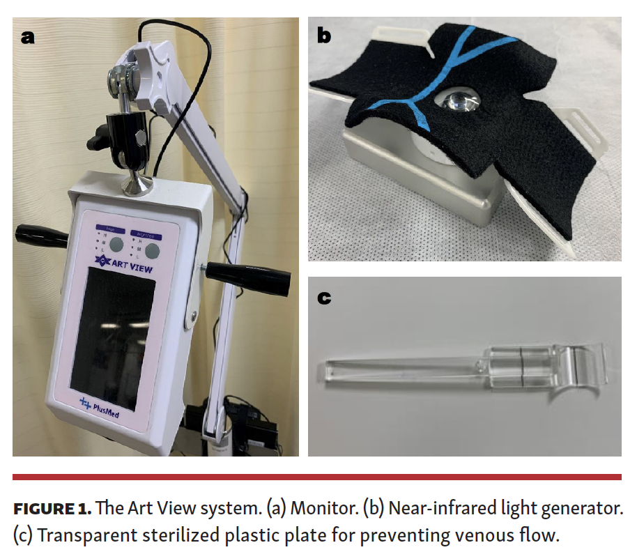
The Art View (Forte Grow Medical Company), a visualization device for blood flow using near-infrared rays, has been developed. This device consists of the generator of near-infrared rays and monitor for visualization (Figure 1). Infrared rays refer to wavelengths ranging from 0.7 µm to 1000 µm. Near-infrared rays involve wavelengths ranging from 0.7 µm to 2.5 µm, and are classified as electromagnetic waves, which have a slightly longer wavelength than visible red light. Therefore, near-infrared rays exhibit features similar to those of visible light, and the risk of biological hazard associated with their use as microwaves has not been previously reported. Based on these features, near-infrared rays are widely utilized in various medical devices.11-13 In the Art View device, the 0.85 µm center wavelength of the near-infrared ray spectrum is used. In addition, near-infrared rays exhibit approximately 10-fold higher biological transmittance compared with visible light, and they are absorbed by hemoglobin. In other words, near-infrared rays do not penetrate red blood cells, but penetrate subcutaneous tissue around the wrist. These features enable clinicians to discriminate blood flow and other tissues on the Art View monitor with contrast.
In this study, it was hypothesized that RA puncture under visual guidance by the Art View with vessel discrimination between veins and arteries may ensure an accurate and safe RA puncture during TRA.
Methods
This was a retrospective and observational study conducted in a single center from December 2019 to April 2020. Patients who underwent coronary artery angiography (CAG) by a single cardiologist via the right RA using the Art View device were included in this study. The CAG procedure with TRA was performed according to the general indication, excluding cases with non-palpable RA, patients undergoing dialysis, and those with possibility of arteriovenous shunt creation for dialysis. Following confirmation of RA pulsation, TRA was considered the first choice for the CAG procedure. The diameter of the right RA was measured using vascular ultrasound, and flow was determined through Doppler ultrasound prior to puncture. After placing the near-infrared ray generator under each patient’s right wrist, the location and pulsation of the right RA were confirmed on the Art View monitor, which was placed directly above the puncture site.
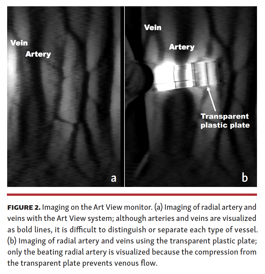
Distinction of the artery and veins is necessary for a puncture to the target artery. This discrimination is a fundamental method for RA puncture under visual guidance by the Art View device. Although the visible pulsation of the vessel on the monitor is effective in distinguishing the artery and veins, occasionally, pulsation is unclear and the network of veins disturbs the detection of the RA. To overcome this problem, a transparent, sterilized, plastic plate was used for preventing the venous flow. Pressing the puncture site and preventing the venous flow using the plate enhances the view of the artery and its pulsation on the Art View monitor (Figure 2).
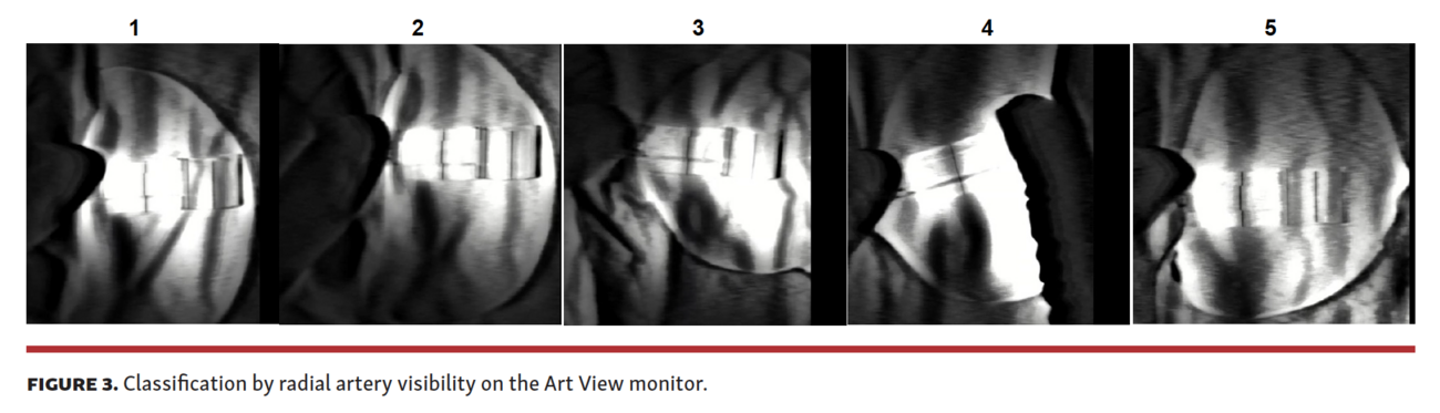
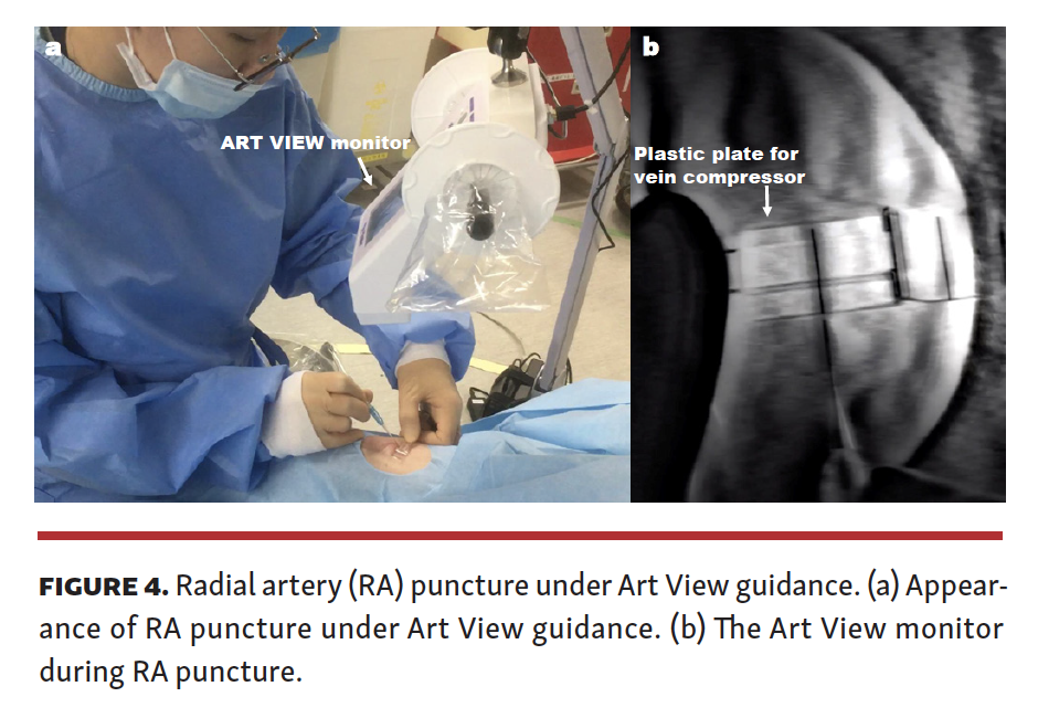
In this study, patients were divided according to the quality of RA visibility on the Art View monitor into 5 groups as follows: 5 (excellent) = constantly confirmed RA with a plastic plate; 4 (good) = confirmed RA with a plastic plate, with a quality between excellent and fair; 3 (fair) = confirmed RA with a plastic plate in the systolic phase, and slightly confirmed in the diastolic phase; 2 (not good) = confirmed RA with a plastic plate, with a quality between fair and poor; and 1 (poor) = slightly confirmed RA with a plastic plate in the systolic phase alone (Figure 3). Patients without RA confirmation on the Art View monitor and those in whom the puncture method or approach site were changed were excluded from this study. Benzethonium chloride (0.02%) was used as a colorless antiseptic solution to avoid permeability loss of near-infrared rays. The bodies of patients were covered with a surgical drape, except for the puncture site. Prior to RA puncture, 0.25-0.5 mL of 1.0% lidocaine was used as local infiltration anesthesia through a 27 G needle. The puncture site was compressed using a transparent, sterilized, plastic plate to prevent the venous flow as described above, and the RA was visualized on the Art View monitor. A black line drawn on the plate was aligned with the right RA. The right RA was punctured along the black line using a 20 G or 22 G indwelling needle with the Seldinger technique (Figure 4). Following penetration of the right RA by the indwelling needle, the inner needle was removed and the external sleeve was gradually pulled. Subsequently, the sheath-attached wire was crossed to confirm the backflow of the RA and advanced under x-ray fluoroscopic imaging to the brachial artery without any resistance. Finally, the 4 Fr sheath was inserted along its wire. RA puncture using the Art View was limited to 2 punctures. After unsuccessful RA puncture using the Art View device, the next step involved vascular ultrasound guiding, blind RA puncture, or change of approach site. Following insertion of the sheath, CAG was performed as usual. The aforementioned procedures were performed by a single cardiologist to avoid procedural bias.
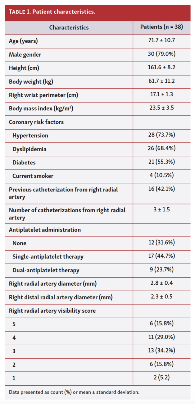
Patient information, including height, body weight, right wrist perimeter, history of catheterization procedure from the right RA and administration of antiplatelet therapy, preprocedural right RA diameter measured by vascular ultrasound, and RA visibility grouping as above, was examined in this study.
The primary endpoint of this study was procedural success of TRA using the Art View device. Secondary endpoints were procedural time (from local-infiltration anesthesia to successful crossing of the sheath-attached wire), number of right RA punctures, and change of puncture method or approach site.
All patients with available data provided written informed consent to undergo the catheterization procedure. This study conformed to the institutional ethics guidelines and those established by the American Physiological Society.
Statistical analysis. The Kruskal–Wallis test was conducted to analyze the association between RA visibility and procedural success rate or procedural time of TRA using the Art View device. Multiple-logistic regression analysis was performed to evaluate factors affecting the procedural success rate and time. P-values <.05 denoted statistically significant differences. Statistical analysis was performed with SPSS software, version 25 (IBM Corporation).
Results
Patient characteristics are shown in Table 1. Of the 38 patients surveyed in this study (30 males [79.0%] and 8 females [21.0%]; mean age, 71 ± 11 years; body mass index, 23.6 ± 3.5 kg/m2; and right wrist perimeter, 17.1 ± 1.3 cm), a total of 22 (57.9%) underwent initial CAG procedure. Preprocedural mean RA diameter, measured by vascular ultrasound, was 2.8 ± 0.4 mm. Twelve patients (31.6%), 17 patients (44.7%), and 9 patients (23.7%) received no, single, and dual-antiplatelet therapy, respectively. The RA visibility score on the Art View monitor was 6 patients (15.8%) in group 5, 11 patients (29.0%) in group 4, 13 patients (35.0%) in group 3, 6 patients (15.8%) in group 2, and 2 patients (5.2%) in group 1.
Study endpoints are shown in Table 2. Thirty patients (79.0%) achieved the primary endpoint (successful TRA using Art View) in a mean procedural time of 142 ± 85 seconds. In this study population, 23 patients (60.5%) and 7 patients (18.4%) achieved procedural success with the first and second RA puncture attempt, respectively. In contrast, 8 patients did not achieve successful RA puncture using the Art View device within the first 2 attempts. Change of puncture method for successful right RA puncture included vascular ultrasound guiding in 4 patients (10.6%) and blind puncture in 3 patients (7.9%). Change of approach site to the left RA because of unsuccessful right RA puncture was performed in only 1 patient (2.6%).
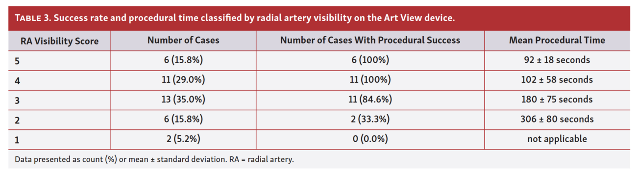
Table 3 presents the achievement rate of procedural success and mean procedural time in each group divided by RA visibility on the Art View monitor. Procedural success rate and shortness of procedural time were both significantly superior (P<.01 for both) in correlation with RA visibility on the Art View monitor. Moreover, there was a significant correlation between the preprocedural RA diameter and procedural success rate (P<.01). However, the other factors (eg, wrist perimeter, body mass index, and coronary risk factors) were not correlated with procedural success rate.
Discussion
This is the first report on the feasibility of RA puncture under visual guidance by a visualization device of blood flow using near-infrared rays. As shown in Table 3, in groups 4 and 5 (excellent and good RA visibility under the Art View, respectively), procedural success rate achieved 100%, and procedural time was significantly shorter than recorded in groups 1–3. Visualizing the location and pulsation of the RA may contribute to a safe and minimally invasive TRA procedure. Puncture in TRA is generally more difficult compared with transfemoral approach, and depends on arterial palpation and each operator’s skill with RA puncture. In addition, unsuccessful RA puncture leads to a prolonged procedure or risk of complications. Therefore, some operators hesitate to perform TRA due to the difficulty of RA puncture. Visualization of the RA may improve the procedural success rate of RA puncture. Hence, the availability of a device that can visualize the RA would be revolutionary in TRA. RA puncture under visual guidance may be easier and more obtainable with a shorter learning curve than under palpation of the radial pulse alone.14,15
Nevertheless, the development of a vessel visualization device has remained unrealized. Arterial visualization through the use of contrast agents and x-ray fluoroscopy is conflicting because it is only applied under the condition of successful arterial securing. Use of the Art View device for arterial visualization with near-infrared rays is non-invasive. Through this approach, it is possible to identify the RA using a transparent, sterilized, plastic plate for preventing the venous flow. In this study, a high success rate of RA puncture was achieved under visual guidance with the Art View device for the confirmation of the arterial location.
Generally, the RA runs straight at the puncture site. However, bending and meandering is observed in a few cases. In cases with high RA bifurcation, there are 2 pulsative arteries at the conventional RA puncture site. In such cases, it is difficult to discern a reasonable RA puncture site through arterial palpation alone. In the RA, which has numerous variations in its location and direction, the Art View enables visualization with an overview, and may contribute to an accurate puncture.
In this study population, there were some cases with RA occlusion or stenosis in a short segment because of an arterial injury during previous TRA. In such cases, it may be impossible to puncture or cannulate a sheath to the RA, despite the detection of arterial palpation at a location near the occlusion site. Puncturing under palpation of the radial pulse alone results in unsuccessful procedure or change of puncture site. However, it is possible to confirm the pulsation and direction with continuity of the RA beforehand using the Art View in those cases, and operators can change the puncture site without an initial unsuccessful puncture.
As previously mentioned, the Art View enables the visualization and identification of arteries and veins using a plastic plate to prevent the venous flow. Hence, avoidance of simultaneous artery and venous puncture may be possible. In a situation in which the RA overlaps veins, the incidence of arteriovenous fistula by puncture is high. Nevertheless, puncture under visual guidance by the Art View may contribute to the prevention of simultaneous occurrence of arterial and venous puncture and arteriovenous fistula. Similarly, unavailing venous puncture leading to hematoma during local anesthesia may be preventable by using the Art View device.
There were some issues regarding the use of the Art View for RA puncture in this study. In cases with unsuccessful RA puncture at first attempt, hematoma occurred unexpectedly despite contact with only small veins; this was particularly observed in patients who received dual-antiplatelet therapy. In 2 cases, after failure of the first RA puncture attempt, visualization on the Art View monitor became impossible due to increased subcutaneous hematoma. In those situations, change of puncture method (eg, palpation of the radial pulse or vascular ultrasound) was necessary. Moreover, the practical distance that near-infrared rays can reach is limited in contrast with its high biological transmittance. This feature offers good visibility of the RA when there is a short distance between the RA and the skin surface. Conversely, RA visibility on the Art View deteriorated in cases with thick subcutaneous fat or muscle, as near-infrared rays have to be transmitted though thicker tissues. Similarly, near-infrared rays exhibit scattering in subcutaneous fat tissue. In this study, RA puncture using the Art View failed in 1 patient with poor RA visibility and severe obesity (body mass index, 29 kg/m2) despite a large RA diameter and good palpation. In contrast, in patients with thin subcutaneous tissue, near-infrared rays were over-transmitted and produced halation, which worsens RA visibility on the Art View monitor. Therefore, maintaining an appropriate distance between the skin surface and monitor of the Art View is required for good visibility and successful puncture.
Vascular ultrasound is useful for vessel visualization in RA puncture.16,17 This approach yields high-resolution images, and arterial blood flow can be qualitatively evaluated using Doppler ultrasound.18 As opposed to the Art View’s near-infrared rays in which arterial visibility worsens in deep arteries, there is no such problem with ultrasound. Furthermore, ultrasound provides vascular images in both the short and long axis in real time, and this contributes to an accurate puncture even in the presence of subcutaneous hematoma, arterial spasm, or injury. Actually, there were 4 cases in which the RA was successfully punctured under vascular ultrasound guidance after failure of puncture under guidance by the Art View. However, acquisition of the puncture technique under vascular ultrasound guidance requires technical skill, and only 1 image in the short axis is obtained without puncture site observation during the procedure. RA puncture under guidance by the Art View allows the identification of an optimal puncture site after obtaining an overall view of the direction of arteries and veins. In addition, use of the Art View does not require a specific technique for vessel visualization. In this study, use of the Art View was limited to TRA for CAG. Nonetheless, it is also possible to utilize the Art View for securing the arterial line in an operating room or intensive care unit. This is because the Art View offers easy arterial visualization similar to vascular ultrasound. Use of the Art View may also be effective in distal RA puncture, which is a novel approach to coronary catheterization procedures. Considering the substantial bending and variation in the distal RA, confirmation of its direction using an arterial visualization device such as the Art View may contribute to a successful puncture. In addition, the Art View may be effective in posterior tibial arterial puncture for percutaneous peripheral intervention.
According to the present findings, successful RA puncture using the Art View alone was not achieved in all cases. There is considerable room for improvement in the Art View to ensure superior RA visibility, including the development of dedicated devices to maintain good visibility with ease, or a brightness adjuster to achieve high-resolution visibility. Therefore, currently, RA puncture may be performed mainly based on the conventional method (ie, palpation of the radial pulse) and balanced use of the Art View or vascular ultrasound. The RA puncture protocol described in this study is expected to provide a safe and accurate procedure even when performed by less-experienced operators.
Study limitations. First, the procedures in this study were performed by a single operator, in a small number of cases, and in a single center. A randomized, prospective study involving multiple operators is warranted to further demonstrate the usefulness of Art View guidance in RA puncture. Second, specialized devices (including a plastic plate for vein compression) and the protocol for RA puncture utilized in this study need to be improved. Third, the Art View, a newly launched vessel visualization device, requires a specific learning curve to grasp its technique. However, these limitations will eventually be overcome.
Conclusion
The Art View, a vessel visualization device using near-infrared rays, is useful for RA puncture. In the future, it is expected that the success rate of RA puncture will be further improved by the development of a puncture protocol, dedicated device for RA puncture, and performance of the Art View to facilitate arterial visualization.
Affiliations and Disclosures
From the 1Tokai University Hachioji Hospital, Division of Cardiology, Department of Internal Medicine, Hachioji, Japan; and 2Tokai University School of Medicine, Division of Cardiology, Department of Internal Medicine, Hachioji, Japan.
Disclosure: The authors have completed and returned the ICMJE Form for Disclosure of Potential Conflicts of Interest. Dr Ikari reports grant support from Boston Scientific; royalties or licenses from Terumo, Medtronics, Nipro, and Asahi Intecc. The authors report no conflicts of interest regarding the content herein.
Manuscript accepted December 17, 2021.
The authors report patient consent for the images used herein.
Address for correspondence: Yota Kawamura, MD, PhD, Division of Cardiology, Department of Internal Medicine, Tokai University Hachioji Hospital, 1838 Ishikawa-cho Hachioji City, Tokyo 192-0032, Japan. Email: youta@f4.dion.ne.jp
References
1. Garg S, Serruys PW. Coronary stents: current status. J Am Coll Cardiol. 2010;56:S1-S42.
2. Zhang Y, Farooq V, Garcia-Garcia HM, et al. Comparison of intravascular ultrasound versus angiography-guided drug-eluting stent implantation: a meta-analysis of one randomised trial and ten observational studies involving 19,619 patients. EuroIntervention. 2012;8:855-865.
3. Hong SJ, Kim BK, Shin DH, et al. Effect of intravascular ultrasound-guided vs angiography-guided everolimus-eluting stent implantation: the IVUS-XPL randomized clinical trial. JAMA. 2015;314:2155-2163.
4. Valgimigli M, Gagnor A, Calabró P, et al. Radial versus femoral access in patients with acute coronary syndromes undergoing invasive management: a randomised multicentre trial. Lancet. 2015;385:2465-2476.
5. Chase AJ, Fretz EB, Warburton WP, et al. Association of the arterial access site at angioplasty with transfusion and mortality: the M.O.R.T.A.L study (mortality benefit of reduced transfusion after percutaneous coronary intervention via the arm or leg). Heart. 2008;94:1019-1025.
6. Romagnoli E, Biondi-Zoccai G, Sciahbasi A, et al. Radial versus femoral randomized investigation in ST-segment elevation acute coronary syndrome: The RIFLE-STEACS (radial versus femoral randomized investigation in ST-elevation acute coronary syndrome) study. J Am Coll Cardiol 2012;60:2481-2489.
7. Kotowycz MA, Dẑavík V. Radial artery patency after transradial catheterization. Circ Cardiovasc Interv. 2012;5:127-133.
8. Rymer JA, Rao SV. Preventing acute radial artery occlusion: a battle on multiple fronts. JACC Cardiovasc Interv. 2018;11:2251-2253.
9. Bernat I, Aminian A, Pancholy S, et al. Best practices for the prevention of radial artery occlusion after transradial diagnostic angiography and intervention: an international consensus paper. JACC Cardiovasc Interv. 2019;12:2235-2246.
10. Barbash IM, Minha S, Gallino R, et al. Operator learning curve for transradial percutaneous coronary interventions: implications for the initiation of a transradial access program in contemporary US practice. Cardiovasc Revasc Med. 2014;15:195-199.
11. De Graaff JC, Cuper NJ, Mungra RAA, Vlaardingerbroek K, Numan SC, Kalkman CJ. Near-infrared light to aid peripheral intravenous cannulation in children: a cluster randomised clinical trial of three devices. Anaesthesia. 2013;68:835-845.
12. Waksman R, Di Mario C, Torguson R, et al. Identification of patients and plaques vulnerable to future coronary events with near-infrared spectroscopy intravascular ultrasound imaging: a prospective, cohort study. Lancet. 2019;394:1629-1637.
13. Zheng F, Sheinberg R, Yee MS, Ono M, Zheng Y, Hogue CW. Cerebral NIRS monitoring and neurological outcomes in adult cardiac surgery patients and neurological outcomes. Anesth Analg. 2013;116:1-28.
14. Stolker JM, Hadid M, Hussain ZM, et al. Training the next generation of invasive cardiologists: feasibility of implementing a trans-radial access program at an academic hospital. Cardiovasc Revasc Med. 2016;17:431-437.
15. Grandpierre RG, Bobbia X, Muller L, et al. Ultrasound guidance in difficult radial artery puncture for blood gas analysis: a prospective, randomized controlled trial. PLoS One. 2019;14:1-10.
16. Zhefeng Q, Luo C, Zhang L, Li X, He H, Chi P. Application of optimized ultrasonic localization system for radial artery puncture by intern doctors: a randomized trial. Med Sci Monit. 2019;25:1566-1571.
17. Gao YB, Yan JH, Gao FQ, Pan L, Wang XZ, Lv CJ. Effects of ultrasound-guided radial artery catheterization: an updated meta-analysis. Am J Emerg Med. 2015;33:50-55.
18. Costa F, Van Leeuwen MAH, Daemen J, et al. The Rotterdam radial access research: ultrasound-based radial artery evaluation for diagnostic and therapeutic coronary procedures. Circ Cardiovasc Interv. 2016;9:1-10.


