


Iso-Osmolar vs Low-Osmolar Contrast Agents for Coronary Optical Coherence Tomography: A Blinded Prospective Randomized Controlled Study
© 2024 HMP Global. All Rights Reserved.
Any views and opinions expressed are those of the author(s) and/or participants and do not necessarily reflect the views, policy, or position of the Journal of Invasive Cardiology or HMP Global, their employees, and affiliates.
Abstract
Background. The impact of contrast type on coronary optical coherence tomography (OCT) imaging has received limited research.
Methods. We conducted a blinded, prospective, single-center, randomized, controlled crossover study comparing iso-osmolar contrast media (IOCM) with low-osmolar contrast media (LOCM) in patients undergoing clinically indicated coronary OCT imaging. Patients were randomly assigned to undergo OCT imaging with either IOCM or LOCM as the initial contrast medium. Following a washout period, a second run of OCT imaging of the same coronary vessel was performed using the other contrast medium.
Results. A total of 62 patients were randomized to IOCM first (n = 31) or LOCM first (n = 31). Mean patient age was 65.9 ± 11.2 years and 74.2% were male, with high prevalence of dyslipidemia (82.3%) and prior myocardial infarction (41.9%). Percutaneous coronary intervention was performed in 60 cases (96.8%) and the left anterior descending artery was the most common target vessel (53.3%). The contrast volume used for OCT imaging was similar for IOCM and LOCM (8.0 [6.9, 9.0] mL vs 8.0 [6.7, 9.0] mL; P = .89), as was the length of clear OCT images (70.0 [62.8, 74.0] mm for IOCM vs 70.0 [64.0, 74.0] mm for LOCM; P = .65). Electrocardiographic changes were observed in 11 runs with IOCM (ventricular repolarization changes in 9 runs and premature ventricular contractions [PVCs] in 2 runs) vs 12 runs with LOCM (ventricular repolarization changes in 9 runs and PVCs in 3 runs).
Conclusions. The use of IOCM in coronary OCT is associated with similar contrast volume and clear imaging length when compared with LOCM.
Introduction
Coronary optical coherence tomography (OCT) provides high resolution coronary artery imaging.1,2 OCT requires red blood cell displacement from the vessel lumen during image acquisition, which is usually achieved using contrast injection, although low molecular weight dextran has also been used.3,4 Contrast agents can be broadly classified into low-osmolar contrast media (LOCM) and iso-osmolar contrast media (IOCM), with IOCM being more viscous than LOCM.5 We compared the administered contrast volume for OCT imaging, the length of clear OCT images, and the immediate electrocardiographic (ECG) changes following contrast injection between IOCM and LOCM.
Methods
Study design and participants. In this blinded, prospective, single-center, randomized, controlled crossover study, we compared IOCM with LOCM in patients undergoing clinically indicated coronary OCT imaging (Iso-Osmolar versus Low-Osmolar Contrast Agents for Optical Coherence Tomography; Clinicaltrials.gov identifier: NCT05065073). Study data were collected and managed using REDCap (Research Electronic Data Capture) electronic data capture tools hosted at the Minneapolis Heart Institute Foundation.6,7 Institutional review board approval was obtained for the study.
Patients referred to the cardiac catheterization laboratory at our center for clinically indicated coronary angiography and coronary OCT imaging were considered for enrollment. Eligible patients were at least 18 years old, were undergoing clinically indicated coronary angiography and OCT imaging, and agreed to participate in the study. Exclusion criteria were baseline ECG changes impeding interpretation (such as left bundle-branch block, and > 1-mm ST segment depression), presentation with ST-segment elevation acute myocardial infarction, inability to provide symptomatic assessment, or a history of allergic reaction to contrast agents. All patients provided written informed consent.
OCT imaging of the same target coronary artery segment was sequentially conducted using the Dragonfly OPTIS Intravascular Imaging Catheter (Abbott Vascular) during manual intracoronary administration of IOCM and LOCM at room temperature. The catheter was advanced distally to the target lesion and automated mechanical pullback was performed during contrast injection until 75 mm were imaged in the OCT survey mode. The catheter’s position remained unchanged during imaging with each contrast agent. OCT runs were performed before percutaneous coronary intervention (PCI), and balloon dilatation was routinely performed before runs.
The analysis of OCT images was conducted using the Ilumien Optis system (Abbott Vascular), with calibration preceding each measurement. OCT images were analyzed at 1-mm intervals measuring lumen area and diameter, as well as area of artifact. Co-registration of the pullbacks acquired with the 2 different contrast types was ensured using fiduciary landmarks, such as side branches and location of the target coronary lesion. Clear image segments were defined as a visible lumen border greater than 270 degrees. Post-procedure analysis of the OCT images and ECG interpretation were performed by a single reader who was blinded to the type of contrast used.
Study procedures. Enrolled patients were randomly assigned to undergo OCT image acquisition with either IOCM or LOCM first. Subsequently, patients underwent coronary angiography and OCT imaging following the clinical standard of care. Based on randomization, the initial OCT imaging run was performed with either IOCM or LOCM. After a washout period, a second run of OCT imaging of the same coronary vessel was conducted using the contrast media that was not used in the first run. The electrocardiogram was recorded during and 30 seconds after each injection and analyzed offline.
Study endpoints. The primary endpoint was contrast volume utilized for OCT imaging (compared between IOCM and LOCM). The secondary endpoints included the length of clear OCT images and the occurrence of ECG changes immediately after contrast injection compared between IOCM and LOCM. The ECG was reviewed to determine the presence of any of the following abnormalities: arrhythmias, mean axis deviation, and QRS enlargement (type A); and ventricular repolarization (ST/T) changes (type B).
Statistical analysis. Categorical variables were depicted as percentages and were compared using the Pearson’s chi-square test. Continuous variables were presented as either mean ± standard deviation or as median (interquartile range [IQR]) unless stated otherwise and were compared using the student’s t-test for normally distributed variables and the Wilcoxon rank-sum test for non-parametric variables, as appropriate. Statistical analyses were conducted using JMP, version 16.0 (SAS Institute) and R v4.0 in R-Studio 2023 environment (Posit Software PBC). A P-value of less than .05 was considered indicative of statistical significance.
Results
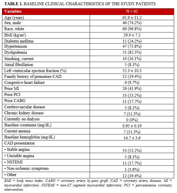
A total of 62 patients were randomized to IOCM first (n = 31) or LOCM first (n = 31). Mean patient age was 65.9 ± 11.2 years and 74.2% were men. The prevalence of dyslipidemia (82.3%), and prior myocardial infarction (41.9%) was high (Table 1).
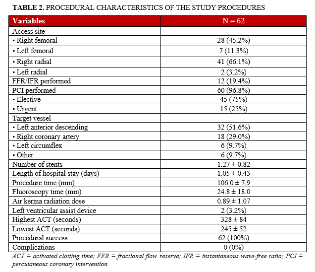
The most commonly used access site was the right radial artery (66.1%), followed by the right femoral artery (45.2%). PCI was performed in 60 of the 62 cases (96.8%). The most common target vessel was the left anterior descending artery (51.6%, n = 32), followed by the right coronary artery (29.0%, n = 18) and the left circumflex (9.7%, n = 6). A bifurcation lesion was present in 15 cases (24.2%). Balloon angioplasty was performed in all cases and a stent was placed in 80.6% of the cases (n = 50). Brachytherapy was performed in 4 cases (6.5%) and intravascular lithotripsy in 1 case (1.6%). Procedural success was achieved in all PCIs (n = 60), with no in-hospital complications (Table 2).
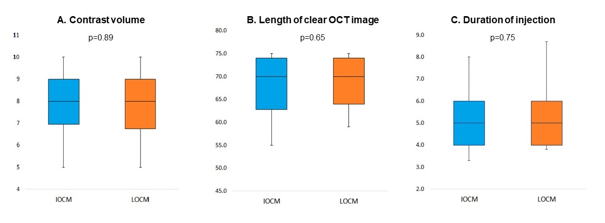
The amount of contrast used for OCT imaging with IOCM was similar to LOCM (8.0 [6.9, 9.0] mL vs 8.0 [6.7, 9.0] mL; P = .89) (Figure 1A). The length of clear OCT images was similar (70.0 [62.8, 74.0] mm for IOCM vs. 70.0 [64.0, 74.0] mm for LOCM; P = .65), as was the percentage of frames with uninterpretable data from the automated analysis (13.7% [7.3%, 20.9%] for IOCM vs 13.2% [6.7%, 20.6%] for LOCM; P = .48) (Figure 1B). There was also no difference between the 2 contrast agents during the duration of the OCT run (5.0 [4.0, 6.0] seconds for IOCM vs 5.0 [4.1, 6.0] seconds for LOCM; P = .75) (Figure 1C).
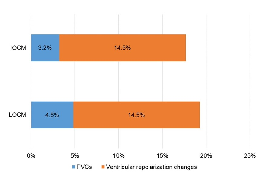
ECG changes were observed in 11 runs (17.7%) with the IOCM (ventricular repolarization [ST/T] changes in 9 runs and premature ventricular contractions [PVCs] in 2 runs) and in 12 runs (19.4%) with the LOCM (ventricular repolarization [ST/T] changes in 9 runs and PVCs in 3 runs) (Figure 2). None of the patients with ECG changes during the OCT run experienced symptoms.
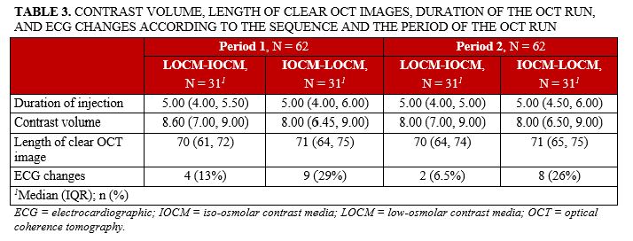
The amount of contrast, the length of clear OCT images, the duration of the OCT runs, and the ECG changes were similar when analyzed by sequence of use of the 2 contrast agents (Table 3).
Discussion
We found no significant difference between IOCM and LOCM in contrast volume used for OCT imaging, the length of clear OCT image, or the incidence of ECG changes.
OCT provides high-resolution images of the coronary lumen and wall.8 In contrast to intravascular ultrasound, OCT provides enhanced visualization and delineation of structures, but has a lower penetration depth.9 Additionally, OCT requires the displacement of red blood cells from the vessel lumen during image acquisition, which is usually achieved using radiographic contrast agents.3,9
Although the impact of contrast type on the risk of hypersensitivity reactions10,11 and contrast nephropathy12-14 has been extensively studied, the impact of various contrast agents on the quality of OCT image as well as the contrast volume used for OCT imaging has received limited study. Christakopoulos et al15 compared OCT imaging obtained using the non-ionic, iso-osmolar iodixanol with the ionic, low osmolar ioxaglate, and reported similar lumen and diameter measurements and stent strut characterization between the 2 contrast types. Similarly, in our single-center, blinded, randomized, controlled crossover study, no significant difference was observed between IOCM and LOCM in the contrast volume used for OCT imaging or the length of clear OCT image.
Metrass et al16 evaluated the impact of 2 different iodine contrast on ECG changes recorded after intra-coronary injection and reported that hyperosmolar contrast resulted in significantly more ECG changes (88%) compared with low osmolar contrast (48%). Furthermore, they reported that the incidence of both type A and type B abnormalities was similar in patients with coronary artery disease and in people with normal coronary angiography. We also found no difference between IOCM and LOCM in the incidence of ECG changes during OCT runs.
Limitations. First, our study was performed at a single center. Second, the sample size was relatively small. Third, contrast was administered with manual injection. Fourth, there was no independent blinded adjudication of the image quality and electrocardiogram interpretation.
Conclusions
The use of IOCM for coronary OCT imaging is associated with similar contrast volume and clear imaging length compared with LOCM. There was also no difference between IOCM and LOCM in the frequency of ECG changes during and after the OCT runs.
Affiliations and Disclosures
From the Minneapolis Heart Institute and Minneapolis Heart Institute Foundation, Abbott Northwestern Hospital, Minneapolis, Minnesota, USA.
Acknowledgments: Study data were collected and managed using Research Electronic Data Capture (REDCap) electronic data capture tools hosted at the Minneapolis Heart Institute Foundation (MHIF), Minneapolis, Minnesota. REDCap is a secure, web-based application designed to support data capture for research studies, providing: (1) an intuitive interface for validated data entry; (2) audit trails for tracking data manipulation and export procedures; (3) automated export procedures for seamless data downloads to common statistical packages; and (4) procedures for importing data from external sources.
Disclosures: Dr Sandoval receives consulting/speaker honoraria from Abbott Diagnostics, Roche Diagnostics, Zoll, and Philips; is an associate editor for JACC Advances; and holds patent 20210401347. Dr Allana is a consultant for Boston Scientific Corporation and Abiomed. Dr. Burke receives consulting and speaker honoraria from Abbott Vascular and Boston Scientific. Dr Brilakis receives consulting/speaker honoraria from Abbott Vascular, American Heart Association (associate editor, Circulation), Amgen, Asahi Intecc, Biotronik, Boston Scientific, Cardiovascular Innovations Foundation (Board of Directors), ControlRad, CSI, Elsevier, GE Healthcare, IMDS, InfraRedx, Medicure, Medtronic, Opsens, Siemens, and Teleflex; research support from Boston Scientific, GE Healthcare; is the owner of Hippocrates LLC; and is a shareholder of MHI Ventures, Cleerly Health, and Stallion Medical. The remaining authors report no financial relationships or conflicts of interest regarding the content herein.
Funding: This study was funded by a research grant from GE HealthCare.
Address for correspondence: Emmanouil S. Brilakis, MD, PhD, Minneapolis Heart Institute Foundation, 920 E 28th Street #300, Minneapolis, MN 55407, USA. Email: esbrilakis@gmail.com; X: @esbrilakis, @S_Kostantinis, @CCAD_MHIF
References
1. Cruz Ferreira R, Pereira-da-Silva T, Patricio L, Bezerra H, Costa M. Coronary optical coherence tomography: A practical overview of current clinical applications. Rev Port Cardiol. 2016;35(2):105-112. doi: 10.1016/j.repc.2015.09.016
2. Ali ZA, Karimi Galougahi K, Maehara A, et al. Intracoronary optical coherence tomography 2018: Current status and future directions. JACC Cardiovasc Interv. 2017;10(24):2473-2487. doi: 10.1016/j.jcin.2017.09.042
3. Kataiwa H, Tanaka A, Kitabata H, Imanishi T, Akasaka T. Safety and usefulness of non-occlusion image acquisition technique for optical coherence tomography. Circ J. 2008;72(9):1536-1537. doi: 10.1253/circj.cj-08-0406
4. Frick K, Michael TT, Alomar M, et al. Low molecular weight dextran provides similar optical coherence tomography coronary imaging compared to radiographic contrast media. Catheter Cardiovasc Interv. 2014;84(5):727-731. doi: 10.1002/ccd.25092
5. Stratta P, Quaglia M, Airoldi A, Aime S. Structure-function relationships of iodinated contrast media and risk of nephrotoxicity. Curr Med Chem. 2012;19(5):736-743. doi: 10.2174/092986712798992084
6. Harris PA, Taylor R, Minor BL, et al. The REDCap consortium: Building an international community of software platform partners. J Biomed Inform. 2019;95:103208. doi: 10.1016/j.jbi.2019.103208
7. Harris PA, Taylor R, Thielke R, Payne J, Gonzalez N, Conde JG. Research electronic data capture (REDCap)--a metadata-driven methodology and workflow process for providing translational research informatics support. J Biomed Inform. 2009;42(2):377-381. doi: 10.1016/j.jbi.2008.08.010
8. Kurogi K, Ishii M, Yamamoto N, Yamanaga K, Tsujita K. Optical coherence tomography-guided percutaneous coronary intervention: a review of current clinical applications. Cardiovasc Interv Ther. 2021;36(2):169-177. doi: 10.1007/s12928-020-00745-4
9. Kume T, Uemura S. Current clinical applications of coronary optical coherence tomography. Cardiovasc Interv Ther. 2018;33(1):1-10. doi: 10.1007/s12928-017-0483-8
10. Cha MJ, Kang DY, Lee W, et al. Hypersensitivity reactions to iodinated contrast media: a multicenter study of 196 081 patients. Radiology. 2019;293(1):117-124. doi: 10.1148/radiol.2019190485
11. Bertrand ME, Esplugas E, Piessens J, Rasch W. Influence of a nonionic, iso-osmolar contrast medium (iodixanol) versus an ionic, low-osmolar contrast medium (ioxaglate) on major adverse cardiac events in patients undergoing percutaneous transluminal coronary angioplasty: A multicenter, randomized, double-blind study. Visipaque in Percutaneous Transluminal Coronary Angioplasty [VIP] Trial Investigators. Circulation. 2000;101(2):131-136. doi: 10.1161/01.cir.101.2.131
12. Zhao F, Lei R, Yang SK, et al. Comparative effect of iso-osmolar versus low-osmolar contrast media on the incidence of contrast-induced acute kidney injury in diabetic patients: a systematic review and meta-analysis. Cancer Imaging. 2019;19(1):38. doi: 10.1186/s40644-019-0224-6
13. Lee T, Kim WK, Kim AJ, Ro H, Chang JH, Lee HH, Chung W, Jung JY. Low-osmolar vs. iso-osmolar contrast media on the risk of contrast-induced acute kidney injury: A propensity score matched atudy. Front Med (Lausanne). 2022;9:862023. doi: 10.3389/fmed.2022.862023
14. Davidson CJ, Laskey WK, Hermiller JB, et al. Randomized trial of contrast media utilization in high-risk PTCA: the COURT trial. Circulation. 2000;101(18):2172-2177. doi: 10.1161/01.cir.101.18.2172
15. Christakopoulos GE, Kotsia AP, Christopoulos G, et al. Comparison of iodixanol and ioxaglate for coronary optical coherence tomography imaging. J Invasive Cardiol. 2015;27(12):E287-E290.
16. Metrass MJ, Brito D, de Lacerda AP, da Costa BB, de Padua F, Madeira H. [Electrocardiographic changes after coronary angiography: effect of the contrast media used]. Rev Port Cardiol. 1996;15(9):639-645, 612.


