


Safety and Feasibility of a Novel Total Percutaneous Post-Closure Technique After Bedside Impella Decannulation in Patients With Cardiogenic Shock
Abstract
Objectives. The Impella (Abiomed) is a widely used percutaneous mechanical circulatory support device for high-risk percutaneous coronary intervention in patients with cardiogenic shock. This study aimed to determine the safety and feasibility of a non-angio-guided post-closure approach using the Perclose ProGlide (Abbott) to decannulate the Impella in the intensive care unit.
Methods. This retrospective study included consecutive patients who were successfully weaned from mechanical circulatory support using the Impella device between April 2019 and April 2022 at Hamamatsu University School of Medicine. Fifteen patients underwent complete post-closure of the femoral artery access sites at the bedside. Technical success of the post-closure hemostasis technique was defined as no evidence of bleeding or additional medical procedures after manual compression. The safety endpoints comprised the Valve Academic Research Consortium-3 and Bleeding Academic Research Consortium criteria.
Results. All patients achieved successful hemostasis with this novel technique without surgical conversion. There was no significant bleeding; however, procedure-related vessel occlusion was observed in 1 patient who was recanalized with balloon angioplasty.
Conclusions. Bedside post-closure using the Perclose ProGlide device is a safe and feasible alternative to manual compression and surgical removal of the Impella device with low bleeding or vascular complications rates.
Introduction
The Impella (Abiomed) is a widely used mechanical circulatory support (MCS) device for high-risk percutaneous coronary intervention in patients with cardiogenic shock.1 The Impella has been associated with access-related vascular complications of large-bore access (LBA), including inserting a 14-French (F) sheath into the common femoral artery.2 In this setting, LBA hemostasis is a critical factor in the use of the Impella device. A previous retrospective study reported that hemostasis after successful Impella weaning was achieved by manual compression, surgical femoral cutdown technique, or suture-mediated percutaneous vascular closure devices.3
Among the vascular closure devices, the Perclose ProGlide (Abbott) improves hemostasis and is a widely accepted suture-mediated vascular closure device for LBA in terms of safety and efficacy.4,5 The pre-closure technique, in which sutures are placed at the insertion site before LBA insertion without tying a knot, is a standard method for using the Perclose ProGlide device for LBA.6 However, this technique cannot be used in emergency procedures for cardiogenic shock. Therefore, the feasibility of post-closure has been increasingly reported.7,8
Although angiography-guided post-closure in the catheterization laboratory is safe for vascular complications, significant risks are associated with transferring patients with MCS.9 Critical patients who need to be transferred out of the intensive care unit (ICU) are at increased risk, and additional staffing is required for their transfer.10 Moreover, there are no reports on total bedside post-closure techniques for the Impella device. Therefore, this study aimed to determine the safety and feasibility of a complete percutaneous post-closure approach using the Perclose ProGlide device to decannulate the Impella in the ICU.
Methods
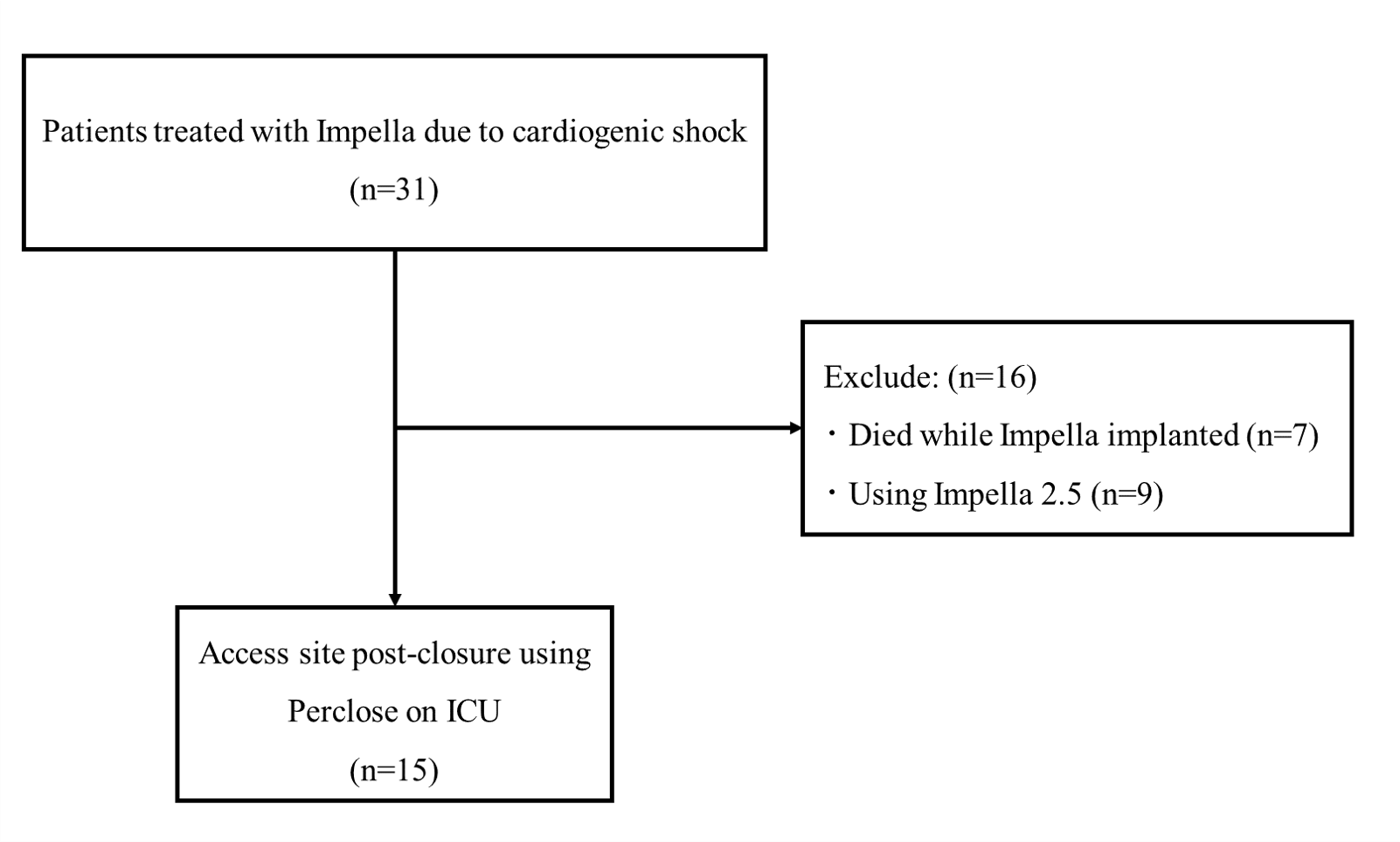
Patient population. This single-center retrospective study included 31 consecutive patients with cardiogenic shock who required Impella 2.5, CP, or CP Smart Assist between April 2019 and April 2022 at Hamamatsu University School of Medicine (Figure 1). The mechanisms and efficacy of the Perclose ProGlide device have been described in previous reports.4,11 All patients underwent echo- or angiography-guided initial percutaneous cannulation in the emergency setting, and hemostatic procedures were performed by experienced interventional cardiologists.
Description of procedure. Anesthetic administration and hemodynamic monitoring were overseen by anesthesiologists who remained ready to promptly act when the patient's condition changed. Our post-closure protocol recommendation is to withhold heparin for at least 1 hour until the activated clotting time is less than 180 seconds. The Perclose ProGlide device typically requires a standard sheath, as the Impella 2.5 device does not have a wire port for guidewire insertion. This issue has been addressed by including a wire port in the Impella CP or CP Smart Assist sheath (Figure 2A). After sterile preparation of the Impella sheath, a 0.035-inch guidewire was inserted into the wire port (Figure 2B). The sheath and the Impella were removed simultaneously (Figure 2C). During sheath removal, another cardiologist manually compressed the proximal insertion site to prevent bleeding and damage to the device foot (Figure 2C).
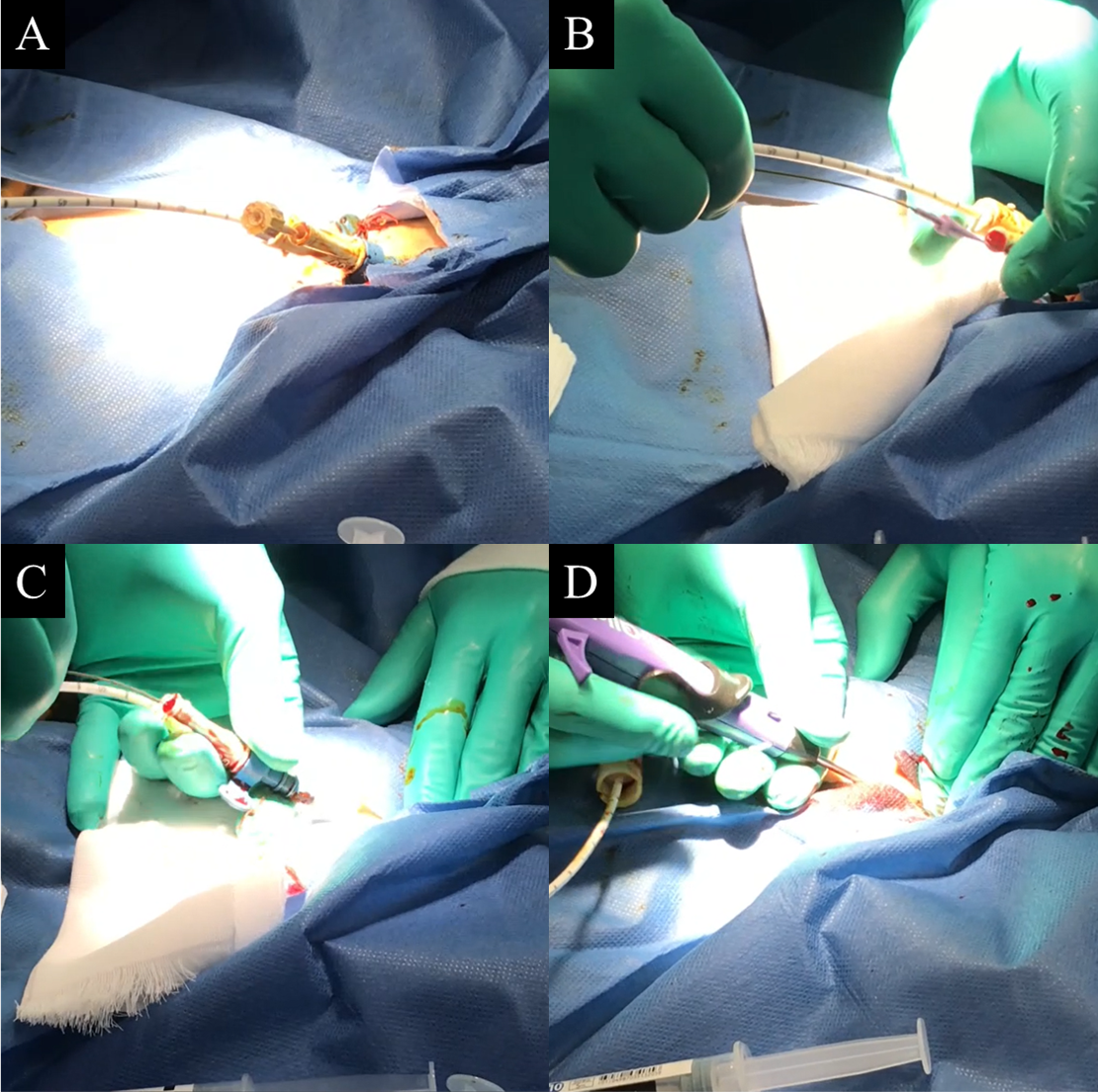
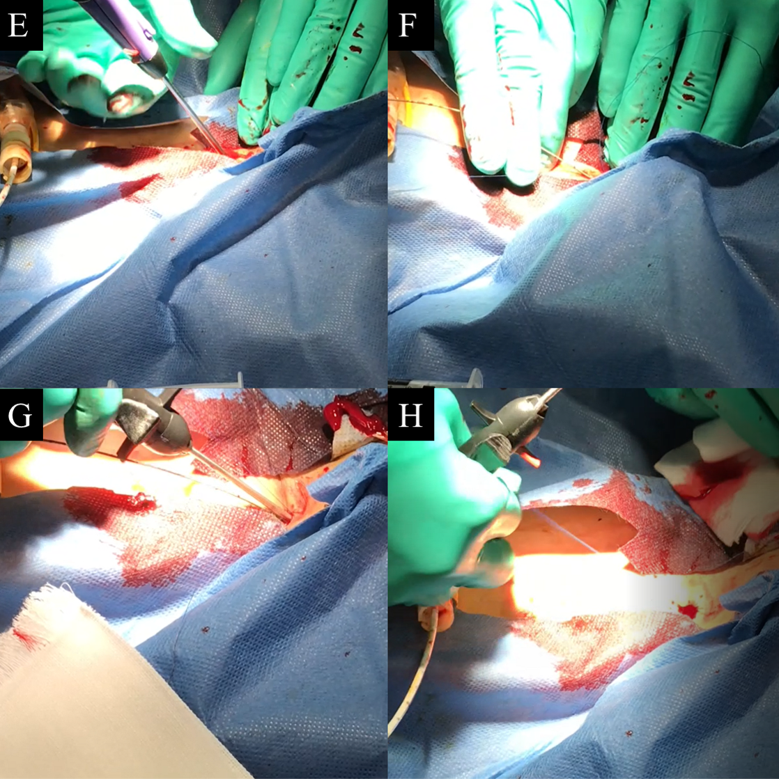
The Perclose ProGlide device was then advanced over the wire, and arterial-like back bleeding from the port was observed to confirm intraluminal placement (Figure 2D). At this point, the assistant slightly released the pressure to assist with inserting and deploying the Perclose ProGlide device. After confirming that the foot of the device had made secure contact with the vessel wall while applying a constant force, the first Perclose ProGlide sutures were deployed (Figure 2E). Subsequently, a 0.035-inch guidewire was reinserted through the Perclose ProGlide device into the femoral artery (Figure 2F). The guidewire was removed after tightening the blue knot with a pusher and confirming hemostasis (Figure 2G, H). If adequate hemostasis was not achieved, a second Perclose ProGlide device was advanced over the guidewire. Manual compression was then applied for approximately 5 to 10 minutes per patient. Finally, a compression bandage was routinely applied to the site for 4 hours, even if immediate hemostasis was achieved. The puncture sites and peripheral arterial circulation were clinically evaluated for ischemia and wound complications during the first 24 hours after hemostasis.
Study endpoints. Technical success of the total percutaneous post-closure hemostasis was defined as no evidence of bleeding or requirement of additional medical procedures after manual compression. Successful Perclose ProGlide device procedure was defined as correct device deployment. Safety endpoints comprised the Valve Academic Research Consortium-3 and Bleeding Academic Research Consortium12,13 criteria. In addition, the clinical outcomes were analyzed at a 30-day follow-up. The medical record review provided comprehensive data on clinical characteristics, laboratory parameters, and procedural parameters.
Ethics. The study was conducted in accordance with the Declaration of Helsinki, and the study protocol was approved by the Ethics Committee of Hamamatsu University School of Medicine (approval number 23-122). Informed consent was obtained in the form of an opt-out on the website. The authors have conformed to institutional guidelines and those of the American Physiological Society.
Statistical analysis. Statistical analyses were performed by an independent physician using the R statistical software (The R Foundation for Statistical Computing). Normally distributed data were presented as means and standard deviations. Categorical variables were presented as numbers with percentages.
Results
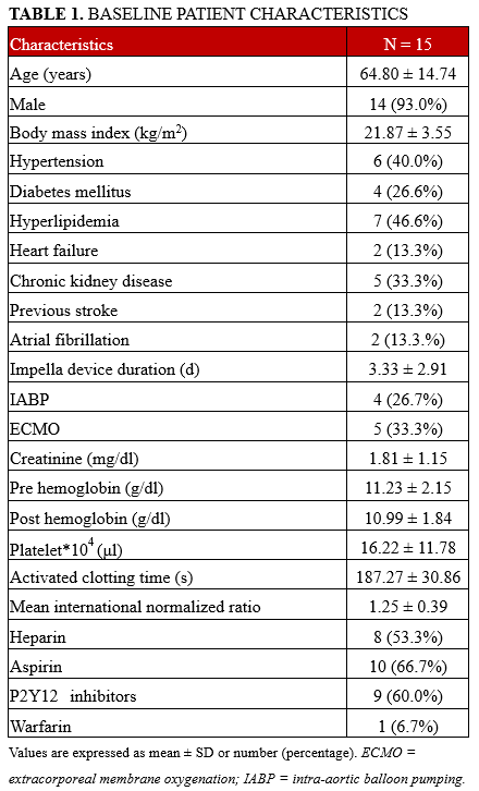
This study included 24 patients who were successfully weaned from the Impella 2.5, CP, or CP Smart Assist device between April 2019 and April 2022. Of these, 15 consecutive patients underwent removal of the Impella CP or CP Smart Assist device post-closure in the ICU (Figure 1). Baseline characteristics of the patients are shown in Table 1. Cardiogenic shock was the clinical indication for Impella use in all patients. The Impella device was withdrawn after 3.30 ± 2.91 days. Primary hemostasis and technical success were achieved in all patients. Manual compression time at the weaning site was 5.30 ± 6.92 minutes. Results of safety and efficacy endpoints are described in Table 2.
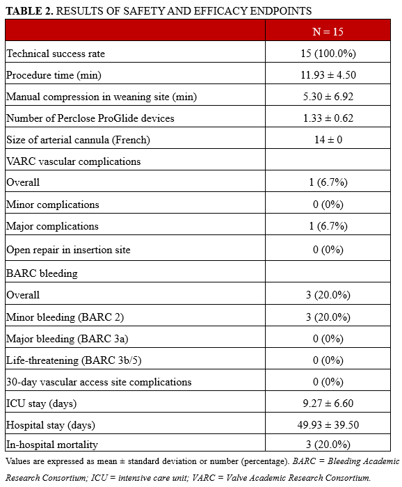
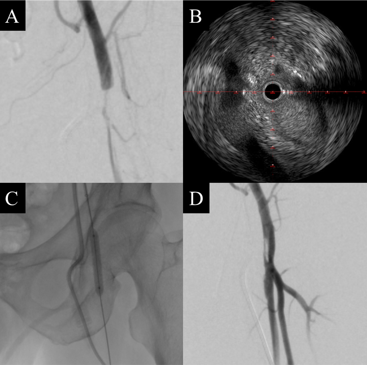
There were no major bleeding events or surgical conversions. One patient (6.7%) developed a vascular complication of limb ischemia due to access site stenosis during hospitalization. Angiography revealed total arterial occlusion due to the dissection, followed by successful percutaneous recanalization of the lesion (Supplemental Figure). There were no additional vascular complications at 30 days, especially access site infections.
Discussion
This study demonstrated the safety and feasibility of access site hemostasis using the bedside post-closure technique with Impella device decannulation after weaning in patients with cardiogenic shock. All patients achieved successful hemostasis with this novel technique without surgical conversion. Complete hemostasis was achieved in an average of 5.3 minutes using the post-closure technique and manual compression combination. One patient experienced procedure-related vessel occlusion that was recanalized using balloon angioplasty.
The Impella device requires a 14-F sheath (Impella CP or CP Smart Assist); therefore, hemostasis with manual compression is challenging and may lead to vascular complications. In this situation, the Perclose ProGlide device can reduce the time to achieve hemostasis in LBA compared with manual compression and surgery.14 Perclose ProGlide devices for LBA can be deployed using pre- and post-closure techniques. The safety and feasibility of the pre-closure technique for endovascular aortic aneurysm repair and veno-arterial extracorporeal membrane oxygenation (VA-ECMO) have been reported.4,11 Transcatheter aortic valve replacement has been established as a pre-closure technique with a single Perclose ProGlide device.15 A retrospective study on the Impella device reported that patients with LBA achieved 99% (68 of 69) hemostatic success with the Perclose ProGlide for delayed closure using 2 pre-closures wrapped in sterile towels and a Tegaderm dressing (3M).6 However, the pre-closure technique may be unsafe during cardiopulmonary resuscitation or cardiogenic shock with MCS devices because of the insufficient time to apply the Perclose ProGlide and the risk of insertion site infection.
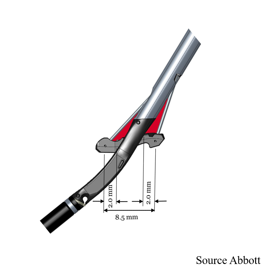
Therefore, the post-closure technique could be an effective alternative to the pre-closure technique. Post-closure reports for VA-ECMO can generally be divided into 2 groups: angiography-guided in the catheterization laboratory and non-angiography-guided at the patient's bedside.16-18 Both have been demonstrated to be safe and effective. However, there are no reports on total bedside post-closure techniques for the Impella device. One problem with post-closure is that the foot pedals of the Perclose ProGlide device may not reliably capture the anterior wall of the vessel. Therefore, extraction using two 8F sheaths has been reported.8,19 However, we assume that the Perclose ProGlide foot pedals can capture the anterior wall due to the 8.5-mm distance between them, elasticity of the vessels, and compression-induced narrowing of the entrance space (Figure 3). In angiography-guided post-closure, hemodynamic status must be maintained while transferring a patient with MCS.9 On the other hand, bedside percutaneous post-closure prevents the risk of patient transfer and reduces staffing requirements compared with conventional angiography-guided procedures.
Several techniques are required to successfully remove the Impella device at the bedside. The first technique involves a combination of moderately releasing pressure during foot deployment and puncturing the anterior vessel wall with the foot while the operator applies a pulling force. The next step is determining whether temporary hemostasis could be achieved by pulling the blue knots under tension. In addition, proper preparation for expanding the subcutaneous tissue at the insertion site using mosquito forceps is essential because the knot may not fall directly over the vessel wall. Using a J-shaped coil wire without a smooth coating allows for safe wire insertion without the requirement for angiography, and the wire should remain in the common femoral artery until hemostasis is confirmed.
There were no cases of failed hemostasis in this series; however, there was 1 case of vessel occlusion. The vessel occlusion may have been caused by vessel wall injury due to the foot of the Perclose ProGlide device. Other possible mechanisms include vessel calcification, stenosis, branch puncture, and suture overdrawing after the proper initial placement.20 Severe calcified or stenotic lesions are contraindications for the Perclose ProGlide device, and this technique should be avoided if calcium in the vessel has been confirmed by fluoroscopy, angiography, echography, or computed tomography. Significantly, a learning curve was associated with the use of the Perclose ProGlide device, and the correct use of the device was critical for achieving successful hemostasis. In our institution, all percutaneous insertions were performed by experienced operators. This technique requires experience before it can be applied routinely.
Study limitations. This study was a single-center, retrospective, observational, feasibility study without a comparison group. The learning curve and outcomes are anticipated to exhibit variability based on the expertise of the facility and operator. Further extensive investigations, incorporating a suitable control group, are necessary.
Conclusions
To the best of our knowledge, this study is the first to report the outcomes of total percutaneous bedside Perclose PrpGlide-assisted post-closure of an Impella CP device in a cohort. Bedside post-closure using the Perclose ProGlide device is a safe and feasible alternative to manual compression and surgery for Impella withdrawal from the common femoral artery.
Affiliations and Disclosures
From the Division of Cardiology, Internal Medicine Ⅲ, Hamamatsu University School of Medicine, Hamamatsu, Japan.
Disclosures: Dr Maekawa reports receipt of Scholarship funds or Donations Scholarship funds from Abbott Medical Japan LLC, and BIOTRONIK JAPAN. The funding organizations did not have any role in the study design, collection, analysis, or interpretation of data, in the writing of the manuscript, or in the decision to submit the article for publication. The remaining authors report no financial relationships or conflicts of interest regarding the content herein.
Address for correspondence: Yuichiro Maekawa, MD, PhD, Division of Cardiology, Internal Medicine Ⅲ, Hamamatsu University School of Medicine, 1-20-1 Handayama, Higashi-Ku, Hamamatsu 431-3192, Japan. Email: ymaekawa@hama-med.ac.jp
Supplemental Material
References
- Iannaccone M, Albani S, Giannini F, et al. Short term outcomes of Impella in cardiogenic shock: A review and meta-analysis of observational studies. Int J Cardiol. 2021;324:44-51. doi:10.1016/j.ijcard.2020.09.044
- Zein R, Patel C, Mercado-Alamo A, Schreiber T, Kaki A. A review of the Impella devices. Interv Cardiol. 2022;17:e05. doi:10.15420/icr.2021.11
- O'Neill WW, Kleiman NS, Moses J, et al. A prospective, randomized clinical trial of hemodynamic support with Impella 2.5 versus intra-aortic balloon pump in patients undergoing high-risk percutaneous coronary intervention: the PROTECT II study. Circulation. 2012;126(14):1717-1727. doi:10.1161/CIRCULATIONAHA.112.098194
- Nelson PR, Kracjer Z, Kansal N, et al. A multicenter, randomized, controlled trial of totally percutaneous access versus open femoral exposure for endovascular aortic aneurysm repair (the PEVAR trial). J Vasc Surg. 2014;59(5):1181-1193. doi:10.1016/j.jvs.2013.10.101
- Sohal S, Mathai SV, Nagraj S, et al. Comparison of suture-based and collagen-based vascular closure devices for large bore arteriotomies-a meta-analysis of bleeding and vascular outcomes. J Cardiovasc Dev Dis. 2022;9(10):331. doi:10.3390/jcdd9100331
- Lata K, Kaki A, Grines C, Blank N, Elder M, Schreiber T. Pre-close technique of percutaneous closure for delayed hemostasis of large-bore femoral sheaths. J Interv Cardiol. 2018;31(4):504-510. doi:10.1111/joic.12490
- Hwang JW, Yang JH, Sung K, et al. Percutaneous removal using Perclose ProGlide closure devices versus surgical removal for weaning after percutaneous cannulation for venoarterial extracorporeal membrane oxygenation. J Vasc Surg. 2016;63(4):998-1003.e1. doi:10.1016/j.jvs.2015.10.067
- Thawabi M, Cohen M, Wasty N. Post-close technique for arteriotomy hemostasis after Impella removal. J Invasive Cardiol. 2019;31(6):E159.
- Martin-Tuffreau AS, Bagate F, Boukantar M, et al. Complete percutaneous angio-guided approach using preclosing for venoarterial extracorporeal membrane oxygenation implantation and explantation in patients with refractory cardiogenic shock or cardiac arrest. Crit Care. 2021;25(1):93. doi:10.1186/s13054-021-03522-8
- Szem JW, Hydo LJ, Fischer E, Kapur S, Klemperer J, Barie PS. High-risk intrahospital transport of critically ill patients: safety and outcome of the necessary "road trip". Crit Care Med. 1995;23(10):1660-1666. doi: 10.097/00003246-199510000-00009
- Torsello GB, Kasprzak B, Klenk E, Tessarek J, Osada N, Torsello GF. Endovascular suture versus cutdown for endovascular aneurysm repair: A prospective randomized pilot study. J Vasc Surg. 2003;38(1):78-82. doi:10.1016/s0741-5214(02)75454-2
- Varc-3 Writing Committee; Généreux P, Piazza N, Alu MC, et al. Valve Academic Research Consortium 3: Updated endpoint definitions for aortic valve clinical research. J Am Coll Cardiol. 2021;77(21):2717-2746. doi:10.1016/j.jacc.2021.02.038
- Mehran R, Rao SV, Bhatt DL, et al. Standardized bleeding definitions for cardiovascular clinical trials: A consensus report from the Bleeding Academic Research Consortium. Circulation. 2011;123(23):2736-2747. doi:10.1161/CIRCULATIONAHA.110.009449
- Karatolios K, Hunziker P, Schibilsky D. Managing vascular access and closure for percutaneous mechanical circulatory support. Eur Heart J Suppl. 2021;23(Suppl A):A10-A14. doi:10.1093/eurheartj/suab002
- Hollowed J, Akhondi A, Rabbani A, et al. Single versus double Perclose techniques for vascular closure during transfemoral transcatheter aortic valve replacement. Catheter Cardiovasc Interv. 2022;99(7):2125-2130. doi:10.1002/ccd.30176
- Au SY, Chan KS, Fong KM, et al. One-year experience of bedside percutaneous VA-ECMO decannulation in a high-ECMO-volume center in Hong Kong. Perfusion. 2021;36(8):803-807. doi:10.1177/0267659120971998
- Au SY, Chan KS, Fong KM, Leung PWR, George Ng WY, Leung KHA. Bedside decannulation of peripheral VA-ECMO using percutaneous Perclose ProGlide post-close technique. J Emerg Crit Care Med. 2020;4:4. doi:10.21037/jeccm.2019.09.08
- Xu X, Liu Z, Han P, et al. Feasibility and safety of total percutaneous closure of femoral arterial access sites after veno-arterial extracorporeal membrane oxygenation. Medicine (Baltimore). 2019;98(45):e17910. doi:10.1097/MD.0000000000017910
- Choi CH, Hall JK, Malaver D, Applegate RJ, Zhao DXM. A novel technique for postclosure of large-bore sheaths using two Perclose devices. Catheter Cardiovasc Interv. 2021;97(5):905-909. doi:10.1002/ccd.29351
- Mousa AY, Campbell JE, Broce M, et al. Predictors of percutaneous access failure requiring open femoral surgical conversion during endovascular aortic aneurysm repair. J Vasc Surg. 2013;58(5):1213-1219. doi:10.1016/j.jvs.2013.04.065


