

Serial Assessment of Coronary Artery Healing of a Biodegradable Polymer Drug-Eluting Stent at 1, 2, and 3 Months by Optical Coherence Tomography (OCT)—The REPAIR Trial
Watch the accompanying author interview here.
Abstract
Background. Although first-generation drug-eluting stent (DES) devices have effectively achieved their main goal of reducing restenosis, their safety has been limited by suboptimal polymer biocompatibility, delayed stent endothelialization, and local drug toxicity, which ultimately prompted the development of new-generation DES options carrying biocompatible or even biodegradable polymers. Aims. We sought to assess the vessel-healing pattern of the novel sirolimus-eluting Inspiron DES (Scitech Medical) using serial optical coherence tomography (OCT) and assuming the hypothesis that this thin-strut (75-µm), biodegradable-polymer DES promotes a faster healing, with very early strut coverage. Methods. This is a prospective, multicenter, open-label, single-arm study enrolling 68 patients who underwent percutaneous coronary intervention guided by OCT. These patients were consecutively assigned into 3 groups. The first group had its OCT imaging follow-up performed at 3 months, the second group at 2 months, and the third group at 1 month. Results. Mean age was 59.5 years, 70.6% were male, 41.2% had type 2 diabetes, and 29.4% presented with acute coronary syndrome. A total of 72 lesions were treated and 1.06 stents were implanted per patient. OCT assessment of the stents at 1, 2, and 3 months showed a strut coverage of 90.41%, 93.96%, and 97.21%, respectively (P=.04). Conclusion. The Inspiron DES showed an early strut healing pattern, with >90% of the struts covered by neointima within the first month and with almost all struts covered by the third month.
J INVASIVE CARDIOL 2023;35(5):E225-E233. Epub March 13.
Although first-generation drug-eluting stent (DES) devices have effectively achieved their main goal, reducing restenosis across virtually all lesions and patient subsets, their safety has been limited by suboptimal polymer biocompatibility, delayed stent endothelialization leading to late/very late stent thrombosis (ST), and local drug toxicity, which ultimately prompted the development of new-generation DESs carrying biocompatible or even biodegradable polymers. The cause of late ST after DES implantation is multifactorial, with delayed healing in combination with other clinical and/or procedural risk factors, such as withdrawal of antiplatelet therapy, malapposition/incomplete apposition, and bifurcation stenting playing important roles.1
Previous analyses comparing first- and second-generation DESs have demonstrated lower rates of ST over time with second-generation stents. However, it is not possible to determine whether all DES devices could benefit from short-term regimens of dual-antiplatelet therapy (DAPT) based on previous reports.2-4 The prevention of major bleeding may represent an important step in improving outcomes by balancing safety and efficacy in the contemporary treatment of coronary lesions.
The presence of a durable, less-biocompatible polymer, the amount of polymer used in the DES system, strut thickness, and the type and amount of antiproliferative drug delivered might also play central roles in the safety profile of these novel devices. We sought to assess the vessel-healing pattern of the Inspiron DES (Scitech Medical) using serial optical coherence tomography (OCT) evaluation, assuming the hypothesis that this thin-strut, biodegradable DES would promote faster healing, with very early strut coverage.
Methods
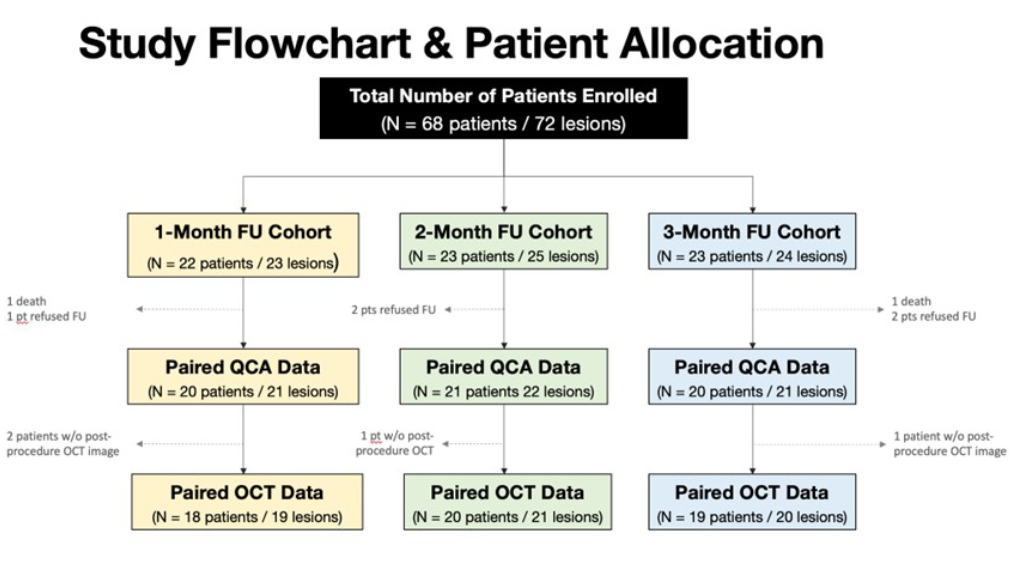
Study design. The REPAIR (serial assessment of coronary artery healing of a biodegradable polymer drug-eluting stent at 1, 2 and 3 months by optical coherence tomography) trial is a prospective, multicenter, open-label, single-arm, mechanistic study conducted at 4 local sites (Figure 1).
The study protocol was developed by the steering committee and approved by the sponsoring company, Scitech Medical, which had no role in the analysis/interpretation of the data. All data were collected prospectively and recorded.
The study protocol and related procedures were approved by the institutional review board and ethical committee of each center and followed the principles of the Declaration of Helsinki. All patients signed informed consent approved by the ethics committees. The study was registered in the ClinicalTrials.gov database (unique identifier: NCT0326946).
Study stent. The Inspiron’s sirolimus-eluting stent (SES) characteristics have been detailed described in previous reports.5 In brief, it is a second-generation DES with an open-cell, thin (75 µm), cobalt-chromium platform, eluting sirolimus (4.4 µg/mm of stent, 80% of the dose being released in the first month and 100% up to 3 months) from an abluminal bioabsorbable polymer comprised of a blend of polylactic acid and poly(lactic-co-glycolic acid) of 5-µm thickness, fully absorbed within 6-9 months.
Study population. The REPAIR trial included 68 patients who underwent percutaneous coronary intervention (PCI) guided by OCT and agreed to undergo an invasive follow-up. These patients were consecutively assigned into 3 groups. The first group had its OCT imaging follow-up performed at 3 months, the second group at 2 months, and the third group at 1 month.
The main inclusion criteria were age ≥18 years and ≤80 years, submitted to cineangiocoronariography due to symptomatic chronic coronary artery disease with invasive or non-invasive evidence of myocardial ischemia in the territory supplied by the target vessel(s) (native epicardial vessels only) or acute coronary syndrome, presented with up to 2 de novo lesions, <30-mm extension each, reference diameter visually estimated between 2.5 and 3.5 mm, and 70%-99% coronary artery stenosis.
Patients who were pregnant or planning to be pregnant within the next 12 months, as well as those with history of coagulopathy, bleeding <6 months, ST segment-elevation myocardial infarction within the first 72 hours of presentation, cardiogenic shock, life expectancy <1 year, renal insufficiency (glomerular filtration rate <60 mL/min/1.73 m2, left main stenosis ≥50%, heavily calcified lesions, excessive tortuosity, total occlusions, thrombus-containing lesions, bifurcations requiring 2 stents, saphenous vein grafts, and aorto-ostial lesions were excluded.
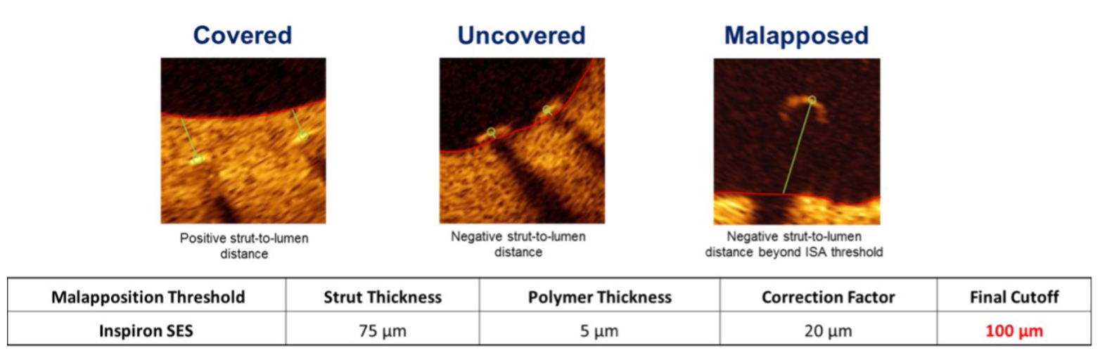
Quantitative coronary angiography (QCA) and OCT methodology. QCA was performed by a commercially validated software for offline analysis (QAngio 7.3; Medis Medical Imaging). OCT images were acquired with commercially available fourier-domain OCT systems (C7 XR or Ilumien Optis; St Jude Medical) and analyzed by a commercially validated software for offline analysis (QIVUS, version 3.0; Medis Medical Imaging). Quantitative analyses were performed at 0.6-mm longitudinal intervals. Stented segments were analyzed by an independent analyst blinded to timepoint after SES implantation. The neointimal hyperplasia (NIH) thickness was determined based on automated measurements performed from the center of the luminal surface of each strut blooming and its distance to the lumen contour. An uncovered strut was defined as having NIH thickness of 0 mm (Figure 2).6 A malapposed strut was defined as a distance between the center reflection of the strut and the vessel wall greater than strut thickness (bioresorbable polymer sirolimus-eluting stent [BP-SES], 0.80 µm).7 All QCA and OCT images were analyzed by an independent imaging core laboratory (Cardiovascular Research Center, Sao Paulo, Brazil) blinded to the timepoints of the study.
Study endpoints. The primary endpoint was the percentage of stent strut coverage by OCT assessment at 3 different timepoints (1, 2, and 3 months) after the procedure. The hypothesis was that we would find <15% uncovered stent struts at the earliest follow-up (1 month) and <5% at the last assessment (3 months).
Secondary endpoints included NIH area, NIH thickness, and NIH obstruction by OCT at 1, 2, or 3 months and 2-year clinical outcomes, performed by office appointment. DAPT was recommended for a minimum of 6 months in stable patients and a minimum of 12 months after acute coronary syndrome.
Clinical endpoints were defined according to the Academic Research Consortium.8 Analyses of clinical endpoints were based on events adjudicated by an independent clinical event committee.
Statistical analysis. Study sample size was not formally calculated, since this is an exploratory, mechanistic trial. Continuous data are reported as mean ± standard deviation, and categorical variables are reported as number and percentage of patients. A sample t test with 95% upper confidence limit was performed to evaluate whether the primary endpoint was <10%.
Intergroup comparison for continuous normal variables was assessed by 1-way analysis of variance (ANOVA) with Bonferroni for posthoc testing. The Kruskal-Wallis test was used for non-normal variables. Non-parametric tests (Wilcoxon signed-rank test) were used for the comparisons of continuous variables with dependent time relationships. The assumption of homogeneity of variances was evaluated by the Levène test and the normality by the Kolmogorov-Smirnov test.
For OCT analysis, to adjust the ranking of the measures evaluated, mixed generalized linear models with random intercept were adjusted according to the level of the ranking at the lowest possible level, considering normal or gamma distributions (with logarithmic connection) for continuous variables, or binomial for binary variables. Data from this analysis have a cluster distribution. We used generalized linear mixed models for variables derived at the cross-section and stem levels, with variable intercept (random intercept) per lesion, assuming the distribution that best fit the data. Assuming these 2 models, data were presented regarding the trend in time (basal, 1 month, 2 months, 3 months), having 20 cases monthly and as paired comparisons, ie, separating the values of the index procedure for each subgroup of patients reassessed at 1 month, 2 months, and 3 months.
The hypothesis tests were interpreted considering a significance level of 5%. All analyses were performed with R, version 3.6.0 (R Core Team, 2019).
Results
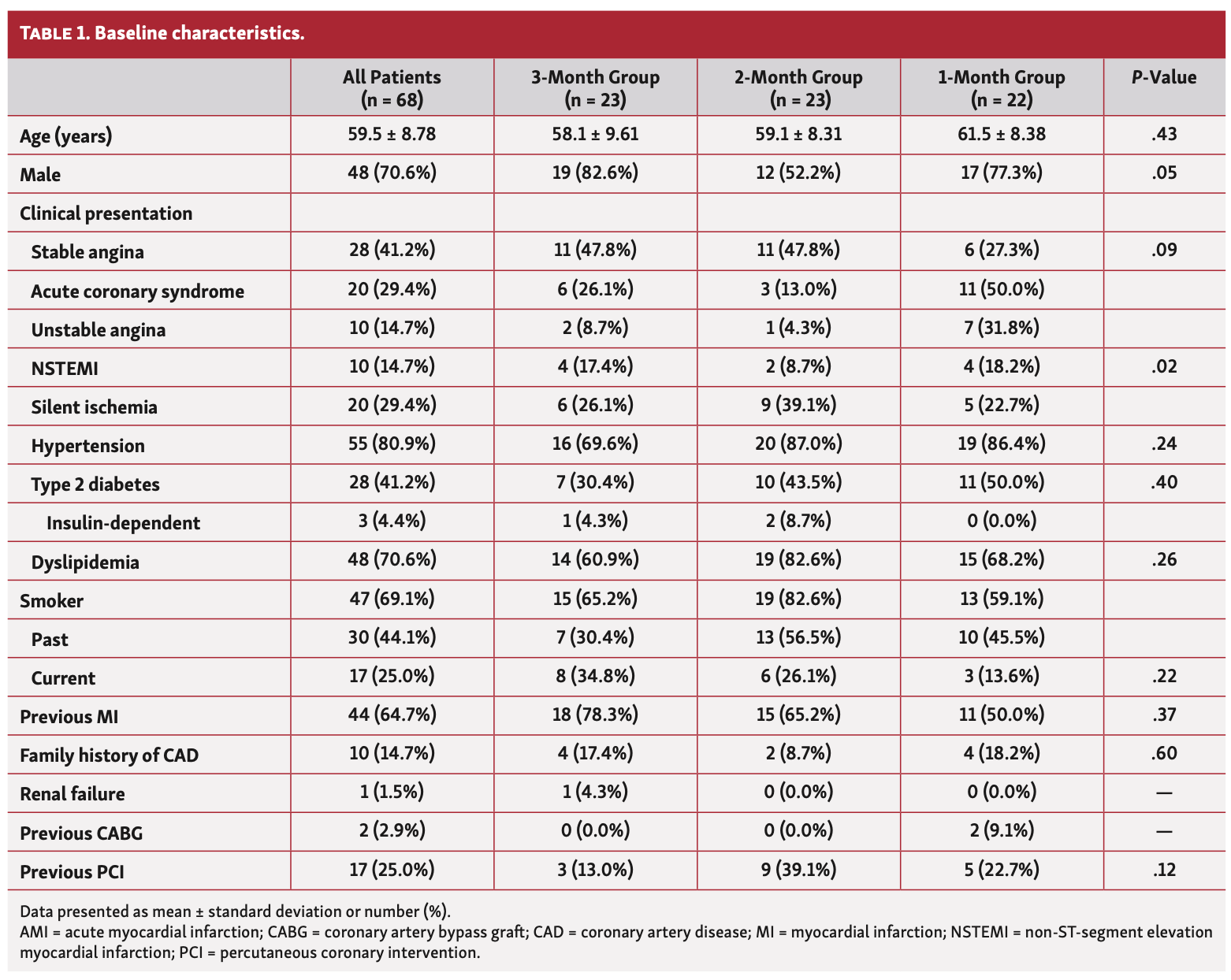
Between October 2017 and August 2018, a total of 68 patients were submitted to OCT-guided PCI at 4 participant centers. Baseline patient and angiographic characteristics are summarized in Table 1 and Table 2, respectively. Demographic analysis showed homogeneity between the groups and 70.6% of the patients recruited were male. Mean age was 59.5 ± 8.78 years, 41.2% had type 2 diabetes, and 29.4% presented with acute coronary syndrome.
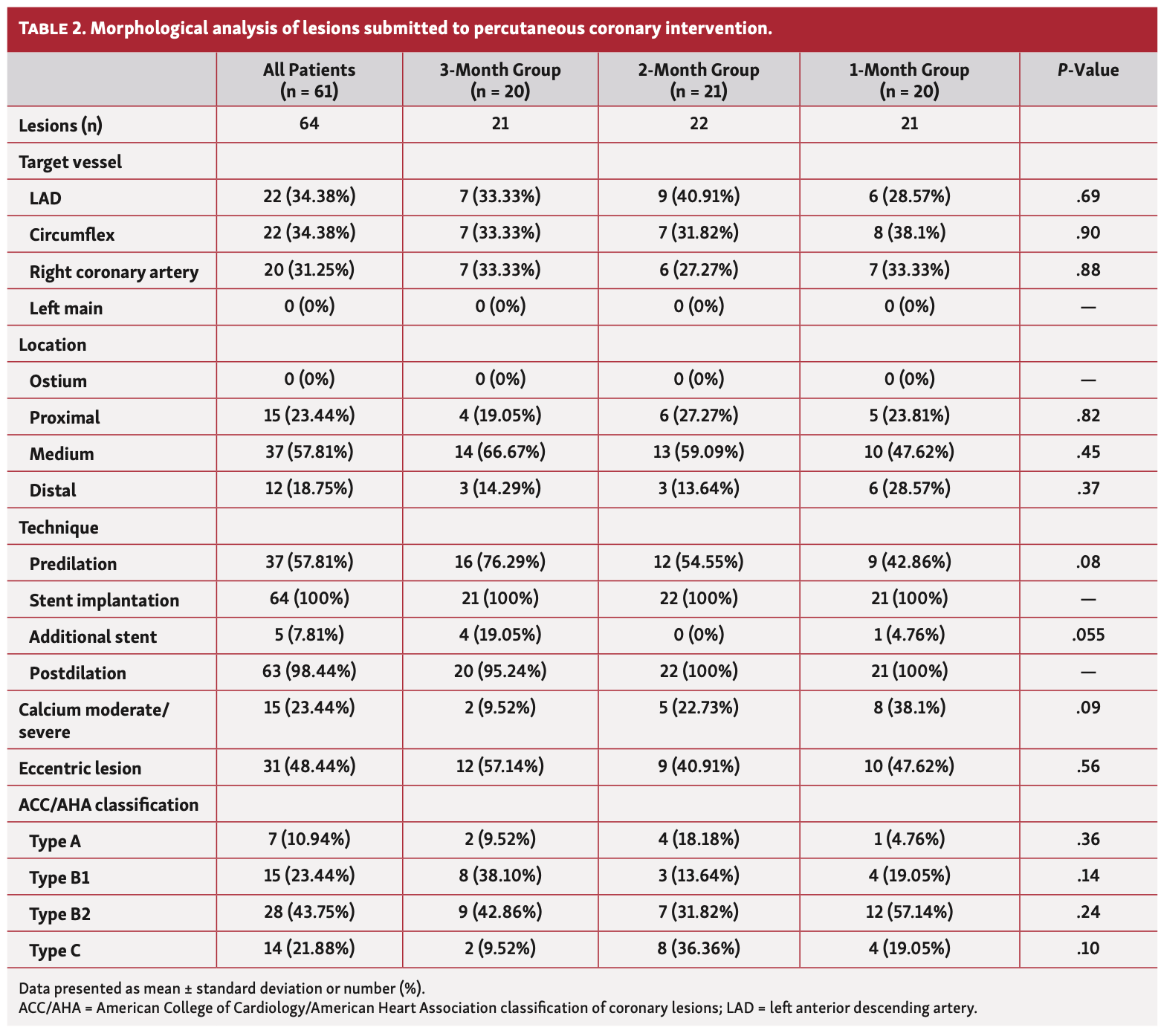
A total of 72 lesions were treated and 1.06 stents were implanted per patient. Morphological analysis of lesions submitted to PCI are presented in Table 2. Lesions were equally distributed along all 3 major epicardial vessels and postdilation was performed in >95% of all cases.
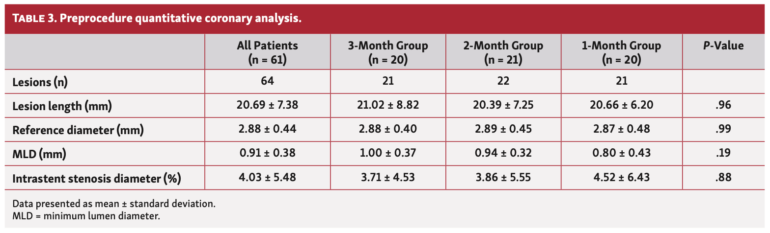
QCA results. A total of 64 lesions in 61 patients were analyzed using QCA (20 patients and 21 lesions in the 1-month group, 21 patients and 22 lesions in the 2-month group, and 20 patients and 21 lesions in the 3-month group).
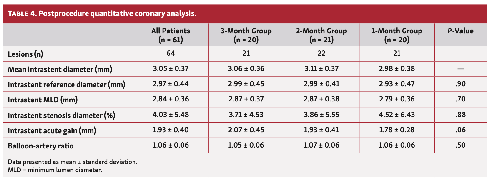
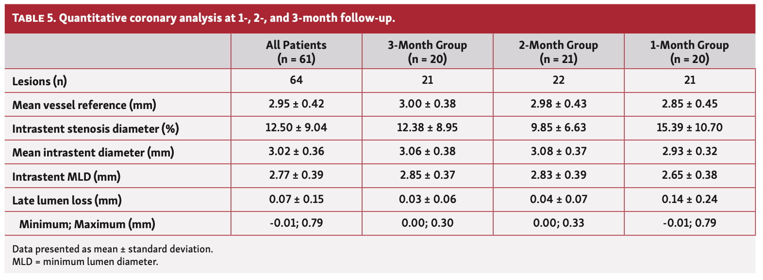
Preprocedure QCA data (Table 3) showed mean vessel diameter and lesion length of 2.88 ± 0.44 mm and 12.86 ± 6.72 mm, respectively. Minimum luminal diameter was 0.91 ± 0.38 mm and mean percentage of stenosis was 68.62 ± 11.48%. Angiographic characteristics were also well balanced among the 3 cohorts. Table 4 summarizes postprocedure QCA data. QCA at 1-, 2-, and 3-month follow-up showed that late lumen loss (LLL) was 0.03 ± 0.06 mm in the first month, 0.04 ± 0.07 mm in the second month, and 0.14 ± 0.24 mm in the third month (Table 5).
OCT results. OCT assessment was performed in 83% of the lesions (60 lesions in 57 patients), encompassing >10,000 struts evaluated. Figure 1 displays the study flowchart.
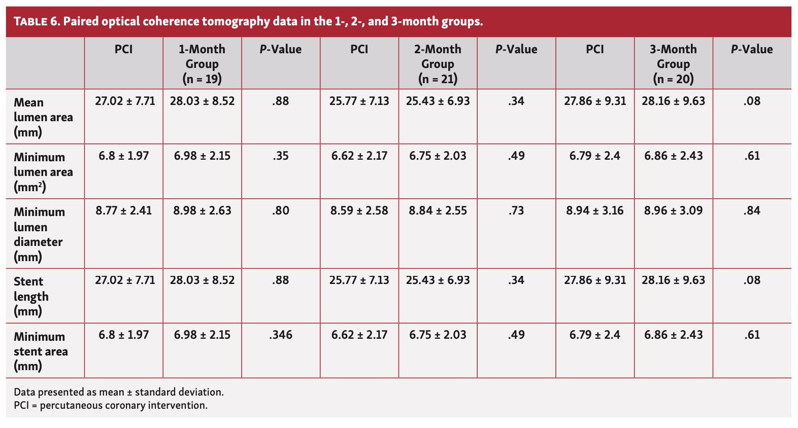
Stent length and minimum stent area did not vary significantly across the 3 cohorts. The index procedure malapposition rates were extremely low in all 3 groups (2.53 ± 2.54% in the 1-month group, 2.6 ± 2.87 in the 2-month group, and 1.72 ± 2.24 in the 3-month group). In all groups, there was a significant reduction in malapposition throughout the different timepoints (1, 2, and 3 months), as detailed in Table 6.
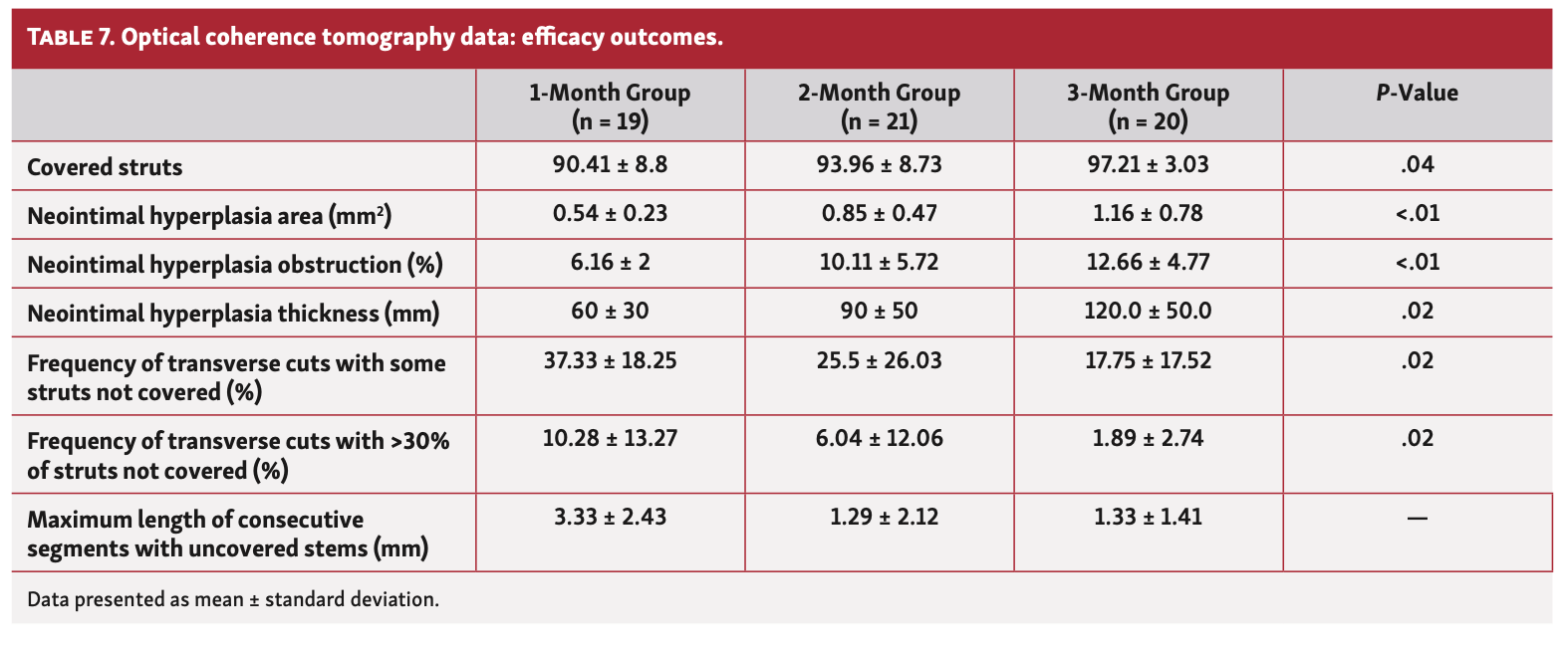
Tissue coverage, the primary outcome of this trial, was observed in 90.8 ± 8.8% of all stent struts analyzed at 1 month, in 93.9 ± 8.7% of the struts analyzed at the 2 months, and in 96.9 ± 3.3% of the struts analyzed at 3 months (P=.04) (Figure 3).
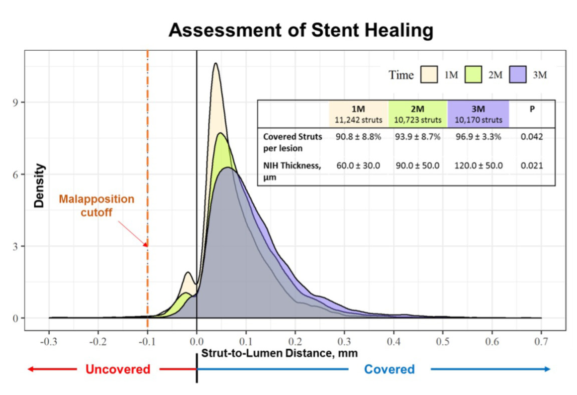
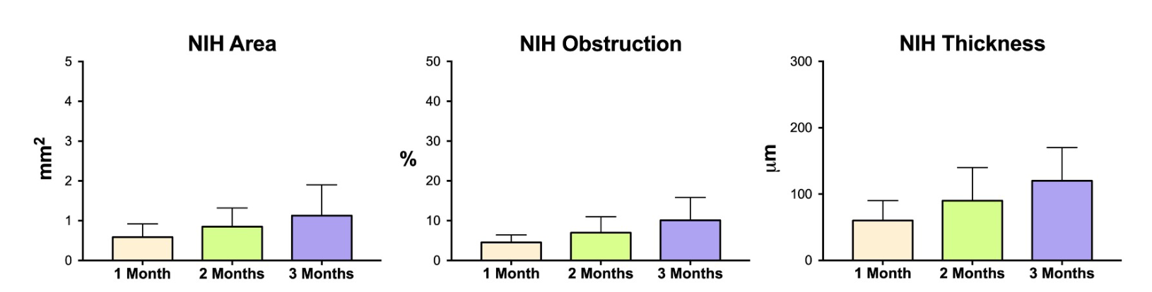
Median NIH thickness over covered struts was 0.06 mm, 0.09 mm, and 0.12 mm, mean NIH obstruction was 4.45 ± 1.85%, 6.95 ± 4.04%, and 10.11 ± 5.72%, and NIH area was 0.59 ± 0.33%, 0.85 ± 0.47%, and 1.13 ± 0.77% at 1, 2, and 3 months, respectively (Figure 4).
The frequency of transverse cuts that demonstrated >30% uncovered struts showed a progressive reduction over the study period, from 10.28 ± 13.27% at 1 month, to 6.04 ± 12.06 % at 2 months, and finally 1.89 ± 2.74 % at 3 months (P=.02) (Table 7).
Since almost 30% of the total samples consisted of ACS patients, we compared the main OCT parameters between them with patients with stable coronary disease. Of note, OCT findings (% of strut coverage, % of struts apposed, NIH thickness, NIH area, and % of NIH obstruction) did not significantly vary between the different clinical presentations.
Clinical follow-up. During the 2-year follow-up period, there were 2 deaths, comprising 1 secondary to congestive heart failure and cardiogenic shock and 1 classified as possible subacute ST in a patient who prematurely discontinued DAPT. No other adverse events, including death, non-fatal myocardial infarction, stroke, or target-lesion revascularization were observed during this period.
Discussion
This is the first study to assess in vivo, with OCT, the early healing profile of the thin-strut, cobalt-chromium Inspiron stent, eluting a low dose of sirolimus from a bioabsorbable abluminal polymer. The main findings include a high level of strut coverage (>90%) at 1 month and almost full coverage at only 3 months of follow-up. Additionally, the rates of acute malposition, in procedures guided by OCT, were overall very low and continuously decreased over the study period, with virtually all struts fully apposed at 3 months.
These additional data support the potential clinical impact of the novel generation of DES as well as the importance of adequate stent implantation (perhaps using intravascular imaging guidance) in facilitating faster stent healing, improving safety, and possibility offering, in specific scenarios (eg, high bleeding risk population), the likelihood of abbreviating DAPT.
To understand the improvement in the safety profile of the new generation of DES, it is important to comprehend the modifications done in all 3 key elements of these systems, including platform, polymer, and antiproliferative drug. While first-generation DES devices (eg, Cypher and Taxus) had their platforms made of stainless-steel, with closed-cell design and thick struts (≥130 µm), the new DES platform incorporates cobalt-chromium and platinum-chromium alloys, which allows maintenance or improvement of radial force and x-ray visibility despite the reduction in the amount of nickel, an element that has been related to local inflammatory/allergic reactions, including eosinophilic type-4 reactions.9
Moreover, most of the current platforms have tubular, open-cell design, with thin (<100 µm) or ultrathin (<80 µm) struts, such as the Inspiron platform (75 µm). Kastrati et al have previously demonstrated that strut thickness is directly related to the development of NIH and increased thrombogenicity.10 In addition, DES strut thickness influences strut malapposition and neointimal coverage.11 A recent meta-analysis comparing stents of similar composition and thickness demonstrated a significant reduction in ST and acute myocardial infarction in patients after PCI with ultrathin struts (<80 µm) when compared with thicker strut stents (>120 µm). Of note, the thinner the strut, the greater the benefit.12
Regarding the antiproliferative drug, most of the current systems discontinued the use of taxol/paclitaxel, present in the first-generation Taxus DES. Presently, almost all DES options use sirolimus or a similar drug (eg, biolimus A9, zotarolimus, everolimus, etc). Furthermore, most of the systems use abluminal polymer coverage and have an open-cell design, which reduces the metal to surface ratio and therefore the amount of drug delivered. While the first-generation Cypher stent, which has a circumferential polymer distribution and closed-cell design, carried a dose of sirolimus of 9.7 µg/mm of stent, the Inspiron carries the same drug at a dose of 4.4 µg/mm of stent (a decrease of 45% in the sirolimus dose). Importantly, most of the drug is released within the 1st month of the procedure, while there is still a small amount being released up to 2 months, covering the critical period of vessel repair, but not lasting too long to delay strut re-endothelialization.
Finally, the polymer technology changed a lot in the recent years. The recent generations of DES use more biocompatible durable polymers, including elements present in our body composition such as fluorine, which might potentially attenuate local inflammatory reactions. However, more recently, most new DES systems moved from durable to biodegradable polymers. Polymers have a central role in storing and local delivery the antiproliferative agent. After the drug is gone, polymers might be fully eliminated, which is the concept behind the bioabsorbable technology. It is important to highlight that not all polymers consist of the same material and even the ones with the same composition might have different resorption times, which might impact the occurrence of local toxicity, especially when it occurs too early. The abluminal polymer present in the Inspiron stent was conceived to fully resorb within 6 to 9 months, which is considered a reasonable period to avoid marked local inflammation.
Similar in concept to the present trial, DISCOVERY 1TO3 was a prospective, single-arm, multicenter trial with a total of 60 patients with multivessel disease treated with the Ultimaster DES (Terumo) who were submitted to OCT evaluation at baseline and at 1, 2, and 3 months.13 A total of 132 lesions were treated and the strut coverage of a single implanted stent at 3 months was 95.2 ± 5.2%. The median NIH thickness was 0.04 mm, 0.05 mm, and 0.06 mm and mean NIH obstruction was 4.5 ± 2.4%, 5.2 ± 3.4%, and 6.6 ± 3.3% at 1, 2, and 3 months, respectively. The in-stent LLL for the Ultimaster stent was 0.04 ± 0.3 mm at 3 months of follow-up. The results were consistent with our study, showing clear signs of very early strut coverage after OCT-guided PCI.13 Similarly, the stent used in the DISCOVERY 1TO3 trial also has thin struts, an open-cell design, and elutes sirolimus from a bioabsorbable polymer with comparable resorption characteristics.
The concept of faster healing of the Ultimaster stent demonstrated in the DISCOVERY 1TO3 trial was recently tested in the larger-scale MASTER DAPT clinical study.14 One month after patients had undergone Ultimaster implantation, investigators randomly assigned those at high bleeding risk to discontinue DAPT immediately (abbreviated therapy) or to continue it for at least 2 additional months (standard therapy). The 3 ranked primary outcomes were net adverse clinical events (a composite of death from any cause, myocardial infarction, stroke, or major bleeding), major adverse cardiac or cerebral events (a composite of death from any cause, myocardial infarction, or stroke), and major or clinically relevant non-major bleeding. Among the 4434 patients in the per-protocol population, net adverse clinical events occurred in 165 patients (7.5%) in the abbreviated-therapy group and in 172 patients (7.7%) in the standard-therapy group (difference, -0.23 percentage points; 95% confidence interval [CI], -1.80 to 1.33; P<.001 for non-inferiority). Notably, Among the 4579 patients in the intention-to-treat population, major or clinically relevant non-major bleeding occurred in 148 patients (6.5%) in the abbreviated-therapy group and in 211 patients (9.4%) in the standard-therapy group (difference, -2.82 percentage points; 95% CI, -4.40 to -1.24; P<.001 for superiority), confirming the safety of shortened DAPT among high bleeding risk populations.14
The results of the REPAIR trial are encouraging regarding the prospect of a larger clinical trial with the Inspiron DES, since it is not clear whether there is a class effect for all current DES options in terms of the safe use of abbreviated DAPT.
Study limitations. There are some limitations in the present study. First, this is a mechanistic trial, with insufficient sample size to address clinical endpoints. Second, it is a single-arm study, with no stent as a comparator. Comparisons and assumptions are based on the extrapolation of results from other trials with similar designs that have been published. Third, it is important to note that the 3 different invasive timepoints were not obtained in the same cohort of patients since it would be unethical to submit the same patient to 3 invasive follow-ups in such a short period without a clear clinical motivation. However, similarities in baseline clinical and angiographic characteristics as well as procedure results among the 3 cohorts might have attenuated this possible bias. Fourth, OCT resolution allows a detailed assessment of strut coverage, almost at a microscopic level. However, despite the resolution of this intravascular method, it is unable to evaluate the type and quality of tissue covering the struts, which might represent normal repair endothelium or other entities, including fibrin, dysfunctional tissue, thrombus, etc. Finally, since there is a paucity of evidence that allows the titration of the duration of DAPT based on strut coverage by OCT, this type of study should work as a hypothesis generator for a larger study with clinical endpoints.
Conclusion
The present study confirms in vivo, in a non-complex population, the fast degree of strut coverage achieved with the Inspiron DES. More than 90% of all struts were covered at 1 month and almost all were covered at 3 months by OCT evaluation. These findings, combined with the rest of the clinical program, pave the way for a large trial assessing the possibility of shortening DAPT regimen among patients treated with this device.
Affiliations and Disclosures
From the 1Department of Interventional Cardiology, Instituto Dante Pazzanese de Cardiologia, Sao Paulo, Brazil; 2Department of Medicine, Universidade Federal de Sergipe, Aracaju, Brazil; 3Department of Interventional Cardiology, Mount Sinai Medical Center, New York, New York; 4Instituto de Cardiologia—Fundação Universitária de Cardiologia, Porto Alegre, Brazil; and 5Associação Evangélica Beneficente Espírito Santo, Vitoria, Brazil.
Funding: This study was partially sponsored by Scitech Medical (institutional grant).
Disclosure: The authors have completed and returned the ICMJE Form for Disclosure of Potential Conflicts of Interest. The authors report no conflicts of interest regarding the content herein.
Manuscript accepted December 8, 2022.
Address for correspondence: J. Ribamar Costa Jr, MD, PhD, Av. Dante Pazzanese, n. 500, 04012-909, Sao Paulo, Brazil. Email: rmvcosta@uol.com.br
References
1. Joner M, Finn AV, Farb A, et al. Pathology of drug-eluting stents in humans: delayed healing and late thrombotic risk. J Am Coll Cardiol. 2006;48(1):193-202. Epub 2006 May 5. doi:10.1016/j.jacc.2006.03.042
2. Stone GW, Rizvi A, Newman W, et al; SPIRIT IV Investigators. Everolimus-eluting versus paclitaxel-eluting stents in coronary artery disease. N Engl J Med. 2010;362(18):1663-1674. doi:10.1056/NEJMoa0910496
3. Kirtane AJ, Leon MB, Ball MW, et al; ENDEAVOR IV Investigators. The “final” 5-year follow-up from the ENDEAVOR IV trial comparing a zotarolimus-eluting stent with a paclitaxel-eluting stent. JACC Cardiovasc Interv. 2013;6(4):325-333. Epub 2013 Mar 20. doi:10.1016/j.jcin.2012.12.123
4. Bhatt DL. EXAMINATION of new drug-eluting stents-top of the class! Lancet. 2012;380(9852):1453-1455. Epub 2012 Sep 3. doi:10.1016/S0140-6736(12)61021-6
5. Ribeiro EE, Campos CM, Ribeiro HB, et al. First-in-man randomised comparison of a novel sirolimus-eluting stent with abluminal biodegradable polymer and thin-strut cobalt-chromium alloy: INSPIRON-I trial. EuroIntervention. 2014;9(12):1380-1384. doi:10.4244/EIJV9I12A234
6. Tanigawa J, Barlis P, Di Mario C. Intravascular optical coherence tomography: optimisation of image acquisition and quantitative assessment of stent strut apposition. EuroIntervention. 2007;3(1):128-136.
7. Gutiérrez-Chico JL, van Geuns RJ, Regar E, et al. Tissue coverage of a hydrophilic polymer-coated zotarolimus-eluting stent vs. a fluoropolymer-coated everolimus-eluting stent at 13-month follow-up: an optical coherence tomography substudy from the RESOLUTE All Comers trial. Eur Heart J. 2011;32(19):2454-2463. Epub 2011 Jun 9. doi:10.1093/eurheartj/ehr182
8. Cutlip DE, Windecker S, Mehran R, et al; Academic Research Consortium. Clinical end points in coronary stent trials: a case for standardized definitions. Circulation. 2007;115(17):2344-2351. doi:10.1161/CIRCULATIONAHA.106.685313
9. Kounis NG, Giannopoulos S, Tsigkas GG, Goudevenos J. Eosinophilic responses to stent implantation and the risk of Kounis hypersensitivity associated coronary syndrome. Int J Cardiol. 2012;156(2):125-132. Epub 2011 Jun 22. doi:10.1016/j.ijcard.2011.05.052
10. Kastrati A, Mehilli J, Dirschinger J, et al. Intracoronary stenting and angiographic results: strut thickness effect on restenosis outcome (ISAR-STEREO) trial. Circulation. 2001;103(23):2816-2821. doi:10.1161/01.cir.103.23.2816
11. Tanigawa J, Barlis P, Dimopoulos K, Dalby M, Moore P, Di Mario C. The influence of strut thickness and cell design on immediate apposition of drug-eluting stents assessed by optical coherence tomography. Int J Cardiol. 2009;134(2):180-188. Epub 2008 Sep 4. doi:10.1016/j.ijcard.2008.05.069
12. Iantorno M, Lipinski MJ, Garcia-Garcia HM, et al. Meta-analysis of the impact of strut thickness on outcomes in patients with drug-eluting stents in a coronary artery. Am J Cardiol. 2018;122(10):1652-1660. Epub 2018 Sep 8. doi:10.1016/j.amjcard.2018.07.040
13. Chevalier B, Smits PC, Carrié D, et al. Serial assessment of strut coverage of biodegradable polymer drug-eluting stent at 1, 2, and 3 months after stent implantation by optical frequency domain imaging: the DISCOVERY 1TO3 study (Evaluation With OFDI of Strut Coverage of Terumo New Drug Eluting Stent With Biodegradable Polymer at 1, 2, and 3 Months). Circ Cardiovasc Interv. 2017;10(12):e004801. doi:10.1161/CIRCINTERVENTIONS.116.004801
14. Valgimigli M, Frigoli E, Heg D, et al; MASTER DAPT Investigators. Dual antiplatelet therapy after PCI in patients at high bleeding risk. N Engl J Med. 2021;385(18):1643-1655. Epub 2021 Aug 28. doi:10.1056/NEJMoa2108749


