


Current Insights On Bracing For Hindfoot Osteoarthritis
Foot orthoses (FOs) have been a standard treatment in podiatric clinics for decades. Until a decade ago, however, it was rare for American podiatrists to dispense ankle foot orthoses (AFOs) of any kind. In 1996, the Richie Brace was introduced and it was the first ankle brace to incorporate a custom functional foot orthosis (FFO). Two years later, the Arizona Brace, the first gauntlet AFO to incorporate a polypropylene shell, arrived on the market and was soon widely used within the podiatric profession. In the last decade, foot orthoses and AFOs of many types have become far more prevalent modalities in the podiatric office as podiatrists have used them for the treatment of pathologies ranging from posterior tibial tendon dysfunction (PTTD) to lateral ankle instability to Charcot arthropathy. With this in mind, let us take a closer look at the use of orthoses for the treatment of hindfoot osteoarthritis (OA).
What Orthoses Can One Use To Treat Hindfoot Arthridites?
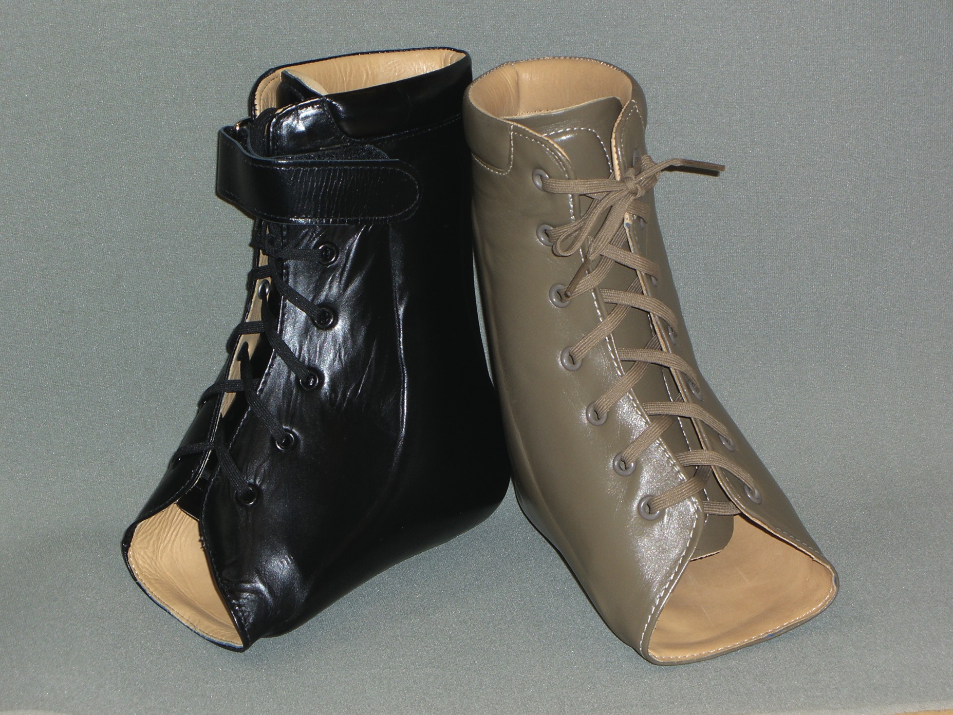
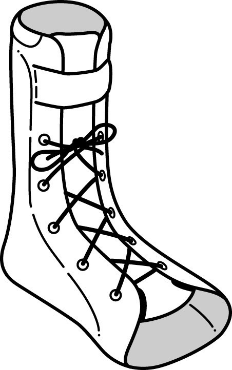
There are many types of FOs and AFOs, and it is easy to become overwhelmed with the many choices involved in prescribing each type. However, when it comes to routine use, podiatrists tend to only use the following five types. • Functional foot orthoses. There is evidence that balanced custom functional foot orthoses can provide effective symptomatic relief in some patients with subtalar joint OA and, to a lesser extent, those with ankle OA. • Solid AFO. Solid AFOs are molded of plastic and conform to the posterior leg and foot. They include medial and lateral flanges, and act to limit or eliminate ankle joint motion. • Podiatric AFO (Richie Brace™). This podiatric AFO is a custom balanced functional foot orthosis attached to medial and lateral uprights. The uprights can be attached via an articulated or fixed pivot (hinge). One would align the pivots with the ankle joint axis to encourage normal ankle joint motion in the articulated model. Both articulated and fixed devices will limit subtalar joint motion while the podiatric AFO with a fixed pivot will also limit ankle joint motion.1 • Gauntlet AFO (Arizona Brace™). Introduced by Ernestro Castro, CPed in 1998, the gauntlet AFO is a custom-made AFO with a plastic shell sandwiched between layers of leather. The device wraps around the circumference of the foot and ankle, and thus provides significant strength with a relatively thin profile. It is designed to stabilize the ankle area and the talocalcaneal, midtarsal and subtalar joints. It provides medial and lateral stability to minimize sinus tarsi impingement, and can reduce forefoot abduction or adduction.2
What The Studies Reveal About Treatment Goals With Lower Extremity OA
The primary goal of bracing in treating osteoarthritis of the hindfoot joints is to limit motion of the involved joint. Literature directly relating to bracing of the arthritic hindfoot is scarce. However, several articles provide direction on how to achieve optimal clinical outcomes. In a 2006 study, Kitakao, et. al., compared the effects on gait of differing types of custom-made polypropylene orthoses. Researchers evaluated the ankle-foot orthosis, rigid hindfoot orthosis (HFO-R) and the articulated hindfoot orthosis (HFO-A).3 The solid AFO supports the back of the calf down to the base of the toes. The HFO-R starts midcalf and includes the ankle, but not the foot or toes. The HFO-A starts midcalf, has a hinge joint at the ankle and goes to the base of the toes. The researchers concluded that gait alterations were affected by orthosis design. Orthoses with a rigid component crossing a joint restricted motion at that joint but caused changes in typical gait kinetics. When it comes to immobilizing the hindfoot, the researchers concluded that the HFO-A may be more comfortable and still provide more stability than the HFO-R or solid AFO. In a 2006 Mayo Clinic study, Huang, et. al., looked to determine whether different foot orthoses have a similar effect on foot kinematics in patients with subtalar osteoarthritis (OA) when they are walking on various ground conditions. This study measured the effects of differing brace designs on triplane range of motion of the calcaneus relative to the tibia and the metatarsal relative to the calcaneus with patients walking on level ground, an ascending ramp, a descending ramp and side slope conditions. Researchers concluded that the HFO-R provides significant subtalar joint motion restriction for a greater variety of ground conditions than did the ankle foot orthosis or the HFO-A. Researchers also suggested considering the HFO-R as an optimal orthosis for patients with subtalar osteoarthritis pain arising from subtalar motion.4 This group of researchers subsequently performed the same study on patients with osteoarthritis of the ankle joint. In this study, they also concluded that the HFO-R not only provides selective restriction to the ankle-hindfoot motion but also allows sufficient forefoot motion in comparison with the AFO. They maintain that the HFO-R is the best option of all tested orthoses for treating patients with ankle OA pain arising from ankle motion.5
When RA And Hemophilia A Affect The Lower Extremity: Can Custom Foot Orthoses Have An Impact?
Also bear in mind that researchers have found custom foot orthoses useful in the treatment of rheumatoid arthritis (RA) patients with pain due to rearfoot valgus. In a study published in 2003, researchers evaluated 3D kinematics of the ankle joint complex. In comparison with healthy control patients, all patients with RA demonstrated excessive subtalar joint pronation. Custom foot orthoses significantly reduced eversion through stance. After 12 months, researchers noted that internal leg rotation started to decrease and this suggested re-coupling of motion. According to the study authors, the study results support the continuous use of custom foot orthoses to correct deformity and optimize function of the ankle joint complex in RA patients with early painful deformity of the rearfoot.6 In 2003, Woodburn, et. al., investigated the clinical effectiveness of early custom rigid foot orthosis intervention for painful correctable valgus deformity of the rearfoot in patients with RA. Patients wearing foot orthoses demonstrated an immediate clinical improvement with the effect peaking at 12 months. After 30 months, the continuous use of custom designed foot orthoses over the treatment period resulted in a reduction in foot pain by 19.1 percent, foot disability by 30.8 percent and functional limitation by 13.5 percent.7 Slattery, et. al., studied patients with ankle joint damage due to hemophilia A and evaluated the efficacy of functional foot orthoses on pain, disability and activity levels. All patients reported a reduction in level of pain. However, there was no improvement in the disability and activity index scores.8
Summarizing The Study Results
Both foot orthoses and AFOs offer some relief in patients with hindfoot arthridities. The AFO studies offer conflicting results and would seem to indicate that one can achieve positive clinical outcomes with both rigid and articulated AFOs. However, given that a basic tenet of reducing pain in the arthritic joint is to limit motion at the joint, it is critical to consider whether the hindfoot arthritis is affecting the ankle or subtalar joints. Accordingly, should the physician focus treatment on eliminating motion at the involved joint while otherwise maintaining normal joint kinematics and gait? If only the subtalar joint is involved, physicians should choose a brace that will allow normal ankle joint motion. If the ankle joint or both the subtalar and ankle joints are involved, then one should use a brace that limits ankle joint motion.
Key Insights On Orthotic Prescriptions
When patients only have subtalar joint OA. If the ankle joint is free of OA, then an orthosis that does not limit ankle joint motion will offer the patient a more normal gait. Subtalar joint motion can be limited and one can maintain ankle joint motion with the use of a functional foot orthosis or an articulated podiatric AFO. Those patients who have lost mechanical coupling between the leg and the foot are less likely to achieve adequate control from foot orthoses only and are more likely to require an AFO. Several tests can help evaluate whether a patient is likely to achieve adequate relief with a foot orthosis or will require an AFO. A simple test is to apply a standard low-Dye taping and evaluate the patient’s response. Those patients who report complete or significant reduction of their hindfoot pain with the low-Dye taping would likely see similar reduction in symptoms with a foot orthosis. If the low-Dye taping does not provide relief, then additional control might be necessary and it would be logical to consider the use of a podiatric AFO. Other tests that can help determine whether the patient has lost mechanical coupling include the Hubscher maneuver (Jack test). Perform the test with the patient weightbearing while you dorsiflex the hallux. Watch for the formation of an arch. In a positive test, dorsiflexion of the hallux results in first ray plantarflexion, subtalar joint (STJ) supination and external rotation of the tibia. When coupling has been lost between the leg and the foot, the tibia will not externally rotate.9 If the patient responds positively to the low-Dye taping and the Hubscher maneuver is positive, then one should start treatment with a functional foot orthosis. Physicians should prescribe this device in such a manner to limit subtalar joint pronation. While there is not a single “correct” prescription, a possible prescription might be as follows: Material: Semi-rigid polypropylene orthosis shell Heel cup: 20 mm Width: Wide or medial flange Cast fill: Minimum Cast correction: Medial heel skive External posts: 4/4 rearfoot post If the patient does not respond to the low-Dye taping and/or the Hubscher maneuver is negative, start treatment with a podiatric AFO. When prescribing a podiatric AFO, the clinician will prescribe the components of the functional foot orthosis section and also choose whether the pivot should be flexible or fixed. A prescription for the podiatric AFO for a patient with subtalar OA could be as follows: Pivot: Flexible Heel cup: 35 mm Width: Wide or medial flange Cast fill: Minimum Cast correction: Medial heel skive
Utilizing Orthotics For Ankle OA: What You Should Know
When patients have ankle OA. Although there is some indication in the literature that foot orthoses can reduce pain in patients with ankle OA, most evidence points toward the need to limit ankle joint motion in order to most significantly reduce pain. This requires the use of an AFO that crosses the ankle joint and allows little or no ankle joint motion. One could use a solid AFO but the standard solid AFO has limitations that make either a fixed-pivot podiatric AFO or the gauntlet AFO superior choices. Both the solid AFO and the fixed-pivot podiatric AFO limit ankle joint motion. However, unlike podiatric AFOs, solid AFOs do not incorporate a functional foot orthoses. As several studies have indicated that FFOs can be effective in limiting ankle pain in patients with ankle arthritis, it would seem prudent to include this control. Since the gauntlet AFO wraps the foot and ankle, rather than primarily providing control posterior and plantar as per the solid AFO, it offers superior control while utilizing a thinner plastic. In my clinical experience, both the podiatric AFO and the gauntlet AFO are easier to fit into shoes than are solid AFOs. The choice for these patients comes down to prescribing a podiatric AFO or a gauntlet AFO. Literature is limited in helping to make the distinction as to which brace individual patients should receive. Overall, the gauntlet provides superior limitation of motion both at the subtalar and the ankle joint. In cases in which patients have severe deformity and involvement of both the STJ and the ankle joint, the gauntlet AFO is recommended. In situations in which ankle joint OA is present but the patient has relatively normal subtalar joint function, then the podiatric AFO would be recommended. However, both AFOs will provide adequate control for most patients. Podatric AFO prescription for patients with ankle joint OA and normal STJ. This prescription is essentially the same as the one for the patient with STJ arthritis but has a fixed pivot. Pivot: Fixed Heel cup: 35 mm Width: Wide or medial flange Cast fill: Minimum Cast correction: Medial heel skive Gauntlet AFO prescription for patients with ankle joint and STJ OA. The gauntlet AFO is prescribed somewhat differently as a functional orthosis is not a component of this AFO. When prescribing a gauntlet, the practitioner will indicate the shell shape, the calf height, length of the foot plate and a closure technique. All of the gauntlets will function in a similar manner regarding the ankle and subtalar function. These prescription items have to do more with shoe fit, patient size and patient convenience. Shell: Cut-out heel Height: 7 inches Foot plate: To toes Closure: Lace bottom with two Velcro straps on top
In Summary
The podiatric practitioner has numerous choices available when using orthoses to treat patients with arthritic changes affecting the subtalar joint and/or the ankle joint. It is imperative that clinicians treating these patients be familiar with the use of and indications for a variety of available orthoses in order to offer their patients the best possible clinical outcomes. Dr. Huppin is an Adjunct Associate Professor in the Department of Applied Biomechanics at the California School of Podiatric Medicine. He is also the Medical Director for ProLab Orthotics/USA and Shoes-n-Feet shoe stores.
References:
1. Richie DH. Clearing Up the Confusion about Posterior Tibial Tendon Dysfunction. Podiatry Today 14(12): 38-44, 2001.
2. Augustin JF, Lin SS, Berberian WS, Johnson JE. Nonoperative treatment of adult acquired flat foot with the Arizona brace. Foot Ankle Clin. 2003 Sep;8(3):491-502.
3. Kitaoka HB, Crevoisier XM, Harbst K, Hansen D, Kotajarvi B, Kaufman K. Arch. The effect of custom-made braces for the ankle and hindfoot on ankle and foot kinematics and ground reaction forces. Phys Med Rehabil. 2006 Jan;87(1):130-5.
4. Huang YC, Harbst K, Kotajarvi B, Hansen D, Koff MF, Kitaoka HB, Kaufman KR. Effects of ankle-foot orthoses on ankle and foot kinematics in patients with subtalar osteoarthritis. Arch Phys Med Rehabil. 2006 Aug;87(8):1131-6.
5. Huang YC, Harbst K, Kotajarvi B, Hansen D, Koff MF, Kitaoka HB, Kaufman KR. Effects of ankle-foot orthoses on ankle and foot kinematics in patient with ankle osteoarthritis. Arch Phys Med Rehabil. 2006 May;87(5):710-6.
6. Woodburn J, Helliwell PS, Barker S. Changes in 3D joint kinematics support the continuous use of orthoses in the management of painful rearfoot deformity in rheumatoid arthritis. J Rheumatol. 2003 Nov;30(11):2356-64.
7. Woodburn J, Barker S, Helliwell PS. A randomized controlled trial of foot orthoses in rheumatoid arthritis. J Rheumatol. 2002 Jul;29(7):1377-83.
8. Slattery M, Tinley P.The efficacy of functional foot orthoses in the control of pain in ankle joint disintegration in hemophilia. J Am Podiatr Med Assoc. 2001 May;91(5):240-4.
9. Richie DH Jr. Biomechanics and clinical analysis of the adult acquired flatfoot. Clin Podiatr Med Surg. 2007 Oct;24(4):617-44, vii.

