How Do Minimally Invasive Bunionectomies Heal?
© 2024 HMP Global. All Rights Reserved.
Any views and opinions expressed are those of the author(s) and/or participants and do not necessarily reflect the views, policy, or position of Podiatry Today or HMP Global, their employees, and affiliates.
In training, I was in an environment where it seemed like minimally invasive surgery (MIS) was all the rage, but many of my mentors and attendings disagreed with the idea. For bunions in particular, we are now on the fourth generation of MIS.1,2 The more recent resurgence thus far seems to be meeting with better success than its predecessors, but it is not without debate. I heard my mentor question the bone healing potential of these recent approaches. Despite these musings, surgeons worldwide are reporting excellent outcomes with MIS procedures, prompting me to reconsider our understanding of bone healing.
Traditional bone healing relies on the fixation of cancellous bone to cancellous bone, either through internal or external splints, with relative or absolute stability being crucial for complete union.3 Absolute stability is achieved when the strain at the fracture site is less than 2% and secondary stability occurs when the strain is 2–10%. Beyond this, increased strain can result in delayed or nonunion.3
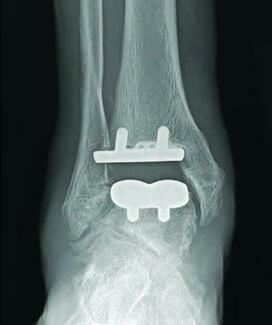
In a standard MIS bunionectomy, only cortical bone interfaces with cortical bone. There are several different fixation methods to secure the osteotomy, including: 2 screws, an intramedullary plate, or Steinmann pin which create a stable construct. There is a degree of micromotion after fixation; however, there is no cancellous bone apposition, and yet these still heal and develop significant bone callus. This challenges our conventional understanding of bone healing, suggesting there is much we have yet to grasp.1
In my experience, in these procedures, there is typically a large amount of bone callus that forms.1,2 Therefore, it is important to understand that this heals with endochondral ossification, also known as secondary bone healing.4-6 This is achieved primarily by the periosteum and periosteally derived cells. After the fracture (or in this case, osteotomy), a hematoma forms, which is then stabilized by surrounding soft tissues and reconstituted by the fibrous layer of the periosteum. Closer to the periphery of the osteotomy, the inner periosteal layer begins membranous ossification and closer to the osteotomy site a mass of cartilage forms, which undergoes endochondral ossification.4–6
However, the periosteum is likely not the full explanation for bone healing, as when people age, the periosteum thins considerably and has significantly less osteoblastic potential than in children.7 That being said, in experiments where bone is stripped of periosteum after a fracture, healing is delayed, but stripping does not obviate healing completely.4,8 Thanks to the micromotion that occurs after these osteotomies with patients bearing weight immediately, it seems to me that there is ample opportunity for callus formation and endochondral ossification. It is important to understand that while radiographs may appear unconventional, they are indicative of appropriate bone healing.
The endosteum can also be a source of healing after an MIS case with 100% shift of the capital fragment. The endosteum lines all the inner surfaces of the cortex, trabeculae, and Haversian systems, and consists of cells with osteoblastic potential. The endosteum is a potent source of osteogenic cells in a rich environment.
While this blog refrains from endorsing MIS as the definitive solution for bunionectomies, it underscores that unconventional radiographs do not negate the procedure’s efficacy. The ability of the periosteum to provide bone healing to even complete shifts of the capital fragment is remarkable. It’s worth noting that first-generation ankle replacements had very poor outcomes, similar to the initial MIS for bunions, yet persistence has transformed ankle replacement into a gold standard for ankle arthritis.9
Dr. Ehlers is in private practice in Arvada, CO, and is an attending at the Highlands-Presbyterian/St. Luke’s Podiatric Residency Program. He finds interest in debunking medical myths and dogma.
References
1. Neufeld SK, Dean D, Hussaini S. Outcomes and surgical strategies of minimally invasive chevron/Akin procedures. Foot Ankle Int. 2021 Jun;42(6):676-688. doi: 10.1177/1071100720982967. Epub 2021 Jan 27. PMID: 33501844.
2. Lewis TL, Lau B, Alkhalfan Y, et al. Fourth-generation minimally invasive hallux valgus surgery with Metaphyseal Extra-Articular Transverse and Akin Osteotomy (META): 12 month clinical and radiologic results. Foot Ankle Int. 2023 Mar;44(3):178-191. doi: 10.1177/10711007231152491. Epub 2023 Feb 14. PMID: 36788732.
3. Sheen JR, Mabrouk A, Garla VV. Fracture healing overview. [Updated 2023 Apr 8]. In: StatPearls [Internet]. Treasure Island (FL): StatPearls Publishing; 2023 Jan-.
4. Dwek JR. The periosteum: what is it, where is it, and what mimics it in its absence? Skeletal Radiol. 2010 Apr;39(4):319-23. doi: 10.1007/s00256-009-0849-9. PMID: 20049593; PMCID: PMC2826636.
5. Newman H, Shih YV, Varghese S. Resolution of inflammation in bone regeneration: From understandings to therapeutic applications. Biomaterials. 2021 Oct;277:121114. doi: 10.1016/j.biomaterials.2021.121114. Epub 2021 Sep 1. PMID: 34488119; PMCID: PMC8545578.
6. Bahney CS, Zondervan RL, Allison P, et al. Cellular biology of fracture healing. J Orthop Res. 2019 Jan;37(1):35-50. doi: 10.1002/jor.24170. Epub 2018 Nov 30. PMID: 30370699; PMCID: PMC6542569.
7. Allen MR, Hock JM, Burr DB. Periosteum: biology, regulation, and response to osteoporosis therapies. Bone. 2004 Nov;35(5):1003-12. doi: 10.1016/j.bone.2004.07.014. PMID: 15542024.
8. Schulze S, Rothe R, Neuber C, et al. Men who stare at bone: multimodal monitoring of bone healing. Biol Chem. 2021 Jul 26;402(11):1397-1413. doi: 10.1515/hsz-2021-0170. PMID: 34313084.
9. McKenna BJ, Cook J, Cook EA, Crafton et al. Total ankle arthroplasty survivorship: a meta-analysis. J Foot Ankle Surg. 2020 Sep-Oct;59(5):1040-1048. doi: 10.1053/j.jfas.2019.10.011. Epub 2020 Jun 26. PMID: 32600863.
Disclaimer: The views and opinions expressed are those of the author(s) and do not necessarily reflect the official policy or position of Podiatry Today or HMP Global, their employees and affiliates. Any content provided by our bloggers or authors are of their opinion and are not intended to malign any religion, ethnic group, club, association, organization, company, individual, anyone or anything.