Blue Bone Discoloration: Incidental Finding During Foot Surgery Associated With Minocycline Intake
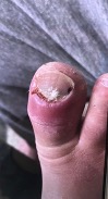
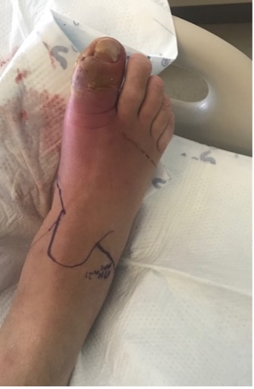
A 57-year-old male presented to his primary care clinic for evaluation of a painful, red right hallux. He reported a contusion to the toe two weeks prior, due to which he experienced a partial toenail avulsion, causing a laceration of the eponychium and nail matrix area.
His past medical history included type 2 diabetes, gout, psoriasis, hypertension, and cardiomyopathy. He is a lifelong nonsmoker. Past surgical history included only an L5-S1 micro-decompression in 2017.
His primary care provider described edema to the foot and ankle with a sausage appearance of the hallux upon examination. The report also included notation of normal pedal pulses, along with laceration and purulent drainage under the hallux nail. Erythema tracked from the hallux to the medial aspect of the thigh. As a result of these findings, he underwent admission to the medical floor with a diagnosis of diabetic foot infection. His orders included a request for podiatry consultation.
The podiatric evaluation revealed intact pedal pulses and peripheral neuropathy with loss of protective sensation. Erythema extended at this time from the right hallux to the ankle. The hallux skin color was purple and cyanotic. Also of note was a laceration/open wound on the eponychium and nail matrix area of right hallux with purulent drainage. Temperature was 36.8 degrees Celsius with pain level of 0/10 on a visual analog scale.
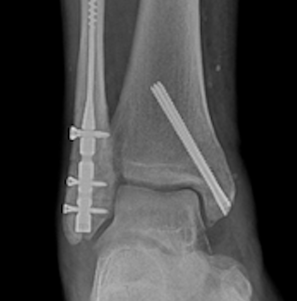
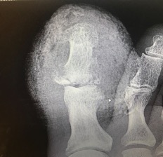
X-Rays on admission revealed a fracture of the medial base of the first distal phalanx. Erosive changes at the lateral distal phalanx and the distal aspect of the proximal phalanx suggested potential osteomyelitis. Blood and deep tissue cultures showed methicillin-susceptible Staphylococcus aureus (MSSA).
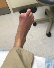
Initial treatment prior to podiatric consultation consisted of IV antibiotic therapy. The podiatry team assessed the case and performed initial wound debridement one day after admission. The patient refused surgical intervention, so the treatment plan included IV antibiotics for six weeks and wound care. After two weeks of treatment with IV antibiotics and little sign of improvement, the patient consented to amputation of the hallux.
| Laboratories | Result |
| WBC | 15.2 |
| Neutrophils | 11.3 |
| ESR | 127 |
| AST | 112 |
| ALT | 89 |
| BUN | 24 |
| Creatinine | 1.27 |
When Surgeons Encounter A Surprising Intraoperative Finding
During surgery, the distal aspect of the hallux had a typical presentation for cellulitis and osteomyelitis. After disarticulating the metatarsophalangeal joint, the remaining soft tissue was healthy and viable in appearance. The head of the first metatarsal and the base of the proximal phalanx, however had signs of osteoarthritis and a blue/gray discoloration. Morphology was otherwise normal.
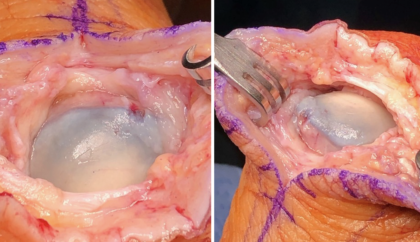
The team considered resecting the distal first metatarsal but ultimately decided against it due to the normal consistency of the bone and surrounding soft tissue. We sent a sample of the proximal phalanx that was also discolored to pathology along with the distal hallux for analysis. The patient followed up at the podiatry clinic and his surgical site completely healed in four weeks.
The pathology report described sections of bone that were gray-blue-tan with smooth articular surfaces. The distal phalanx and interphalangeal joint had devitalized bone with marrow space fibrosis and scattered lymphocytes consistent with osteomyelitis.
After the incidental finding of blue discoloration of bone, we reviewed the patient's past medical history in greater detail, revealing long-term use of minocycline to treat cystic acne. He began taking minocycline in 1995 while on active duty and continued to take the medication until discontinuing it in 2019, over 25 years.
Review of the surgery report from his prior L5-S1 micro-decompression in 2017 revealed similar bony findings. The orthopedic surgeon described discolored ligamentum flavum covering the thecal sac with brown/gray/black bone discoloration during that surgery. Pathology described benign fibrocartilaginous tissue and bone without evidence of inflammation.
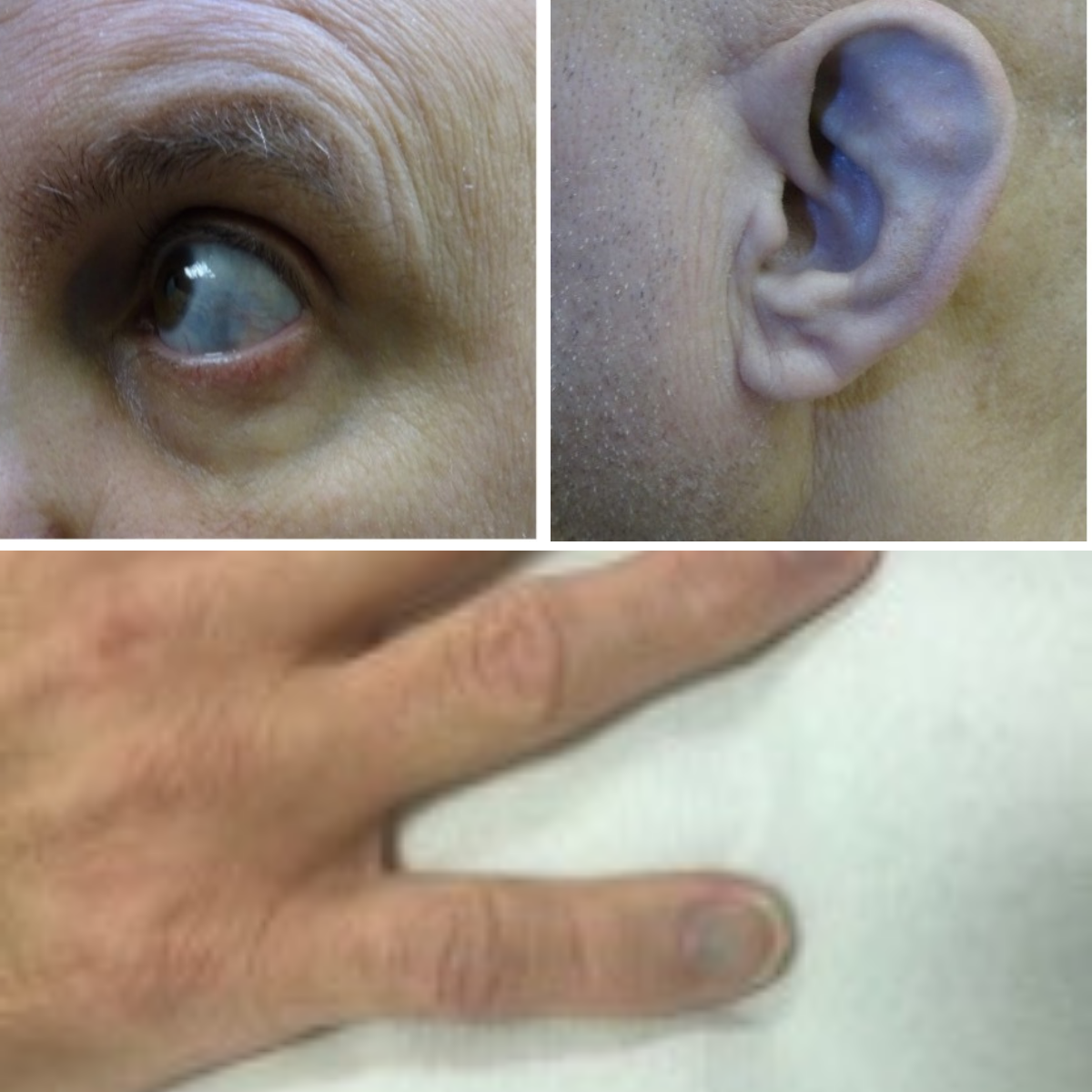
The patient agreed to be evaluated by the staff dermatologist during subsequent follow-up, who described bluish, well-demarcated patches on the sclera bilaterally. They noted a bluish dermal hue on the cartilage of the ears bilaterally. Brown-to-bluish irregular asymmetric patches on lower legs were also present with a bluish dermal hue on the fingernails. He also agreed to evaluation at the dental clinic, which found gray-blue anterior teeth and a gray-blue tinge on maxillary anterior alveolar mucosa.
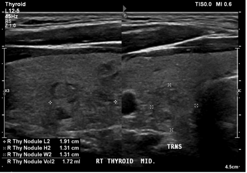
Due to thyroid cancer being a complication of longer-term minocycline treatment, an otolaryngologist evaluated the patient next. After performing a thyroid sonogram, this evaluation revealed multinodular goiter with two concerning nodules. The provider recommended observation and a repeat thyroid sonogram in six months.

What You Should Know About Minocycline and Bone Discoloration
Minocycline is widely used to treat several conditions, including acne and rosacea.1-6 Some also use this drug off-label for rheumatoid arthritis.2,4,5,7-9 Long-term use of minocycline increases the probability of developing adverse effects related to pigmentation.1,3,9-14
Minocycline hydrochloride attaches easily to plasma proteins, is lipid soluble, and has great tissue and fluid absorption.3,10 Minocycline degrades to a black product that can deposit in various tissues. The dark pigmentation in bones usually contains iron and calcium.2,3,5,8,10,13-15
The US Food and Drug Administration (FDA) documents the the following adverse reactions associated with minocycline: hyperpigmentation of skin, nails, mucous membrane, photosensitivity, erythema multiforme and Steven-Johnson syndrome. Also included in this list are: arthritis, arthralgia, bone discoloration, myalgia, joint stiffness, joint swelling, thyroid cancer, and thyroid discoloration.1,8-14,16-20
Minocycline cutaneous hyperpigmentation of the skin classically divides into three types, however, there is a more recent proposal of a fourth category.3,16
Type I: blue-black discoloration in areas of scarring or inflammation of the face
Type II: blue-gray pigmentation of normal skin of the extremities
Type III: brown hyperpigmentation, symmetrically, in sun-exposed skin
Type IV: blue/gray discoloration in areas with prior inflammation of the back
While there is a classification for cutaneous manifestations there is no universally accepted classification for bone discoloration due to minocycline. Minocycline-induced bone discoloration is usually referred to as black bone disease, but it can present in a variety of different colorations or shades.9 The differential diagnosis includes metal deposits, malignancy, bone necrosis, sequestrum, alkaptonuria, and metabolic bone disease.1,2,4,5,11,12,21
Diagnosis of minocycline-induced bone discoloration takes place via a thorough review of the medical history to verify long-term use of minocycline.2 Histological examination by detecting fluorescence of the tissue in question under ultraviolet light can provide confirmation of the diagnosis.2,5,8,10,11,17 Most pathology labs do not have ultraviolet microscopes and thus cannot provide fluorescence examination. Failure to diagnose minocycline-induced pigmentation of skin, bone, and other body parts may lead to unnecessary procedures and testing.
The treatment for minocycline discoloration is discontinuation of the medication.3,9 The hyperpigmentation of the skin and nails may fade over time, but the brown pigmentation may persist. Laser and tretinoin creams may be beneficial.3,16 Tooth pigmentation is usually permanent.3,14,18 Oral mucosa hyperpigmentation usually resolves over time. The bone pigmentation is irreversible, but there are no reports of reduced bone healing capacity due to said pigmentation.3-5,10,11,13 Ocular pigmentation resolves occasionally, but there are no reported harmful effects.3 Bone discoloration is rare and often seen in the oral cavity, accounting for about 20 percent of documented cases.3,10,13,15
Taking An International Perspective
We performed a literature review on this condition, examining reports from Australia, Japan, Singapore, England, Ireland, Scotland, South Africa, and the United States. We did not include bone discoloration of the oral cavity. There is a variety of bone coloration //reported associated with minocycline//, but it is most frequently described as black bone discoloration. Other color variations included blue, green, brown, violet and gray.1,2,4,5,8,10-13,15,17,19,21,22,23
|
Location |
Bone Coloration |
Year of Publication |
Author |
|
Ankle |
Black |
2012 |
Yang, et al. |
|
Ankle (fibula) |
Green |
2018 |
Judge, et al. |
|
Clavicle |
Green |
1984 |
Wolfe, et al. |
|
Clavicle (acromion) |
Black |
2004 |
Pandit, et al. |
|
Costal Cartilages |
Dark |
1976 |
Attwood, et al. |
|
Femoral bone |
Brown/Gray/Black |
2005 |
Hepburn, et al. |
|
Femoral shaft |
Brown/Black |
2016 |
Thiam, et al. |
|
Foot (bilateral 1st Ray) |
Black |
2011 |
Middleton, et al. |
|
Foot (first metatarsal head) |
Green/Blue |
2018 |
Judge, et al. |
|
Foot (first metatarsal head) |
Black |
2019 |
Nirenberg, et al. |
|
Foot (first metatarsal head and base of proximal phalanx) |
Brown/Green/Blue/Gray |
2013 |
Kerbleski, et al. |
|
Foot (first metatarsal head, base proximal phalanx and Head second proximal phalanx) |
Discolored (Described only as discolored, but pictures show Blue/Gray/Black coloration.) |
2018 |
Cornejo, et al. |
|
Hip (2 cases) |
Black |
2012 |
Yang, et al. |
|
Hip (acetabulum and femoral head) |
Brown/Black |
2012 |
Chan, et al. |
|
Knee (femur and tibia) |
Blue/Green/Gray |
2004 |
McCleskey, et al |
|
Knee (femur and tibia) |
Violet/Black |
2012 |
Reed, et al |
|
Knee (bilaterally) |
Black |
2014 |
Chauhan, et al |
|
Knee (2 cases) |
Black |
2012 |
Yang, et al |
|
Metacarpals |
Brown |
2010 |
Somayazula, et al |
|
Shoulder |
Gray/Black |
2016 |
Ashukem, et al |
|
Thoracic Vertebra |
Black |
1991 |
Rumbak, et al |
Incidence of bone pigmentation associated with long term minocycline therapy
In Conclusion
Minocycline-induced bone pigmentation is usually an incidental finding, and as such, its incidence is unknown, but it appears to have no impact on healing capability. Minocycline-related bone changes outside of the oral cavity are not commonly found in the literature. We recommend surgeons publish their findings to familiarize other clinicians and surgeons of this condition and its proper management.
Dr. Serrano and Dr. Dahlenburg practice with the Alaska VA Health Care System in Anchorage, AK.
The authors would like to acknowledge the VA librarians' network and Mrs. Susan Knecht for their assistance related to this piece.
References
1. McCleskey,PE, Littleton KH. Minocycline-induced blue-green discoloration of bone. J Bone Joint Surg. 2004;86(1):146-148.
2. Reed DN, Gregg FO, Corpe RC. Minocycline induced black bone disease encountered during a total knee arthroplasty. Orthopedics. 2012;35(5):e737-9.
3. Eisen D, Hakim MD,. Minocycline-induced pigmentation. Incidence, prevention and management. Drug Safety. 1998;18(6):431-440.
4. Yang S, Takakubo Y, Kobayashi S, et al. Minocycline-induced periarticular black bones in inflamed joints which underwent arthroplastic reconstruction. Clin Orthop Surg. 2012;4(3):181-187.
5. Ashukem M, Levy J, Formaini N. Minocycline induced black bone disease: an incidental finding during total shoulder arthroplasty. Curr Orthop Pract. 2016;27(6):608-701.
6. Somayazula R, Rogers GF. Metacarpal darkening associated with minocycline therapy. J Hand Surg Eu. 2010;35(9):760-1.
7. Vidaurri VA, Cochrane ZA. Minocycline for rheumatoid arthritis: does it work?. Healthline.com. 2018; https://www.medicalnewstoday.com/articles/325710 . Published July 10, 2019. Accessed March 21, 2022.
8. Judge MS, Miller M, Lyons M. Green bone: Minocycline-induced discoloration of bone rarely reported in foot and ankle. J Foot Ankle Surg. 2018;57(4):801-807.
9. Cornejo A, Kerbleski GJ. Black bone disease of the foot: A two-year follow-up case study. J Foot Ankle Surg. 2018;57(6):1259-1262.
10. Chan CM, Hicks DG, Giordano BD, Minocycline-induced discoloration. JBJB Case Connector. 2012;2(3):p e47.
11. Thiam D, Teo TY, Malhotra R, Tan KB, Chee YH. Black bone disease in a healing fracture. BMJ. 2016;ber2015211915.
12. Nirenberg M. Black bone disease of the foot: a case study of minocycline-induced bone pigmentation. Curr Orthop Pract. 2019;30(5):487-489.
13. Middleton SD, Anakwe RE, McKinley JC. Black Bone disease of the foot. Minocycline related pigmentation. J Foot Ankle Surg. 2011;17(2):e34-e36.
14. Wolfe ID, Reichmister J. Minocycline hyperpigmentation: skin, tooth, nail, and bone involvement. Cutis. 1984;33(5):457-8.
15. Kerbleski GJ, Hampton TT, Cornejo A. Black bone disease of the foot. A case study and review of literature demonstrating a correlation of long term minocycline therapy and bone hyperpigmentation. J Foot Ankle Surg. 2013;52(2):239-241.
16. Wetter DA, Minocycline hyperpigmentation. Mayo Clinic Proc. 2012;87(5):e33.
17. Pandit S, Hadden W. Black pigmentation of bone due to long-term minocycline use. Surgeon. 2004;2(4):236-237.
18. Odell EW, Hodgson RP. Oral presentation of minocycline-induced black bone disease. J Oral Surg Oral Med Oral Path Oral Radiol Endod. 1995;79:459-461.
19. Rumbak MJ, Pitcock JA, Palmieri GMA, Robertson JT. Black bones following long-term minocycline treatment. Arch Pathol Lab Med. 1991;115(9):939-41.
20. Bann DV, Goyal N, Crist H, Goldenberg D. Black Thyroid. J Ear Nose Throat. 2014;93(0):E54-E55.
21. Chauhan V, McDougall C. Black bones: minocycline-induced bone pigmentation. Med J Aust. 2014;201(2):114.
22. Hepburn MJ, Dooley DP, Hayda RA. Minocycline induce black bone disease. Orthopedics. 2005;28(5):501-2.
23. Attwood HD, Dennett X. A black thyroid and minocycline treatment. J Br Med. 1976;2(6044):1109-1110.