


A Rare Case of Forefoot Melorheostosis Complicated by Infiltrating Angiolipoma
Melorheostosis is a rare skeletal abnormality that affects cortical bone as well as the surrounding soft tissue. It has hyperostotic features and typically presents with pain to the affected site.1 This abnormality classically has a “dripping candle wax” characteristic on radiographs. Melorheostosis can present similarly to tumors and other types of bone diseases and affects both females and males equally. The onset may develop at any age; however, in 50% of cases, it occurs in adolescence.2 Melorheostosis is a benign process; however, due to the disease’s progression, it may lead to limb shortening, bone deformity, and joint stiffness, which are all factors leading to increases in pain and potential incapacity of the affected limb.2
Many times, melorheostosis and other bony diseases are accompanied by adjacent soft tissue growth that has developed around the affected bone. In this report, we present such a case: a large multilobular infiltrating angiolipoma at the plantar surface of the forefoot surrounding the region of the hyperostotic bone. Angiolipomas are mature adipose tissue composed of vascular tissue that are subdivided into 2 types: infiltrating and noninfiltrating.3 Angiolipomas are a benign process. However, they can lead to neuropathies and discomfort. Preferred treatment is wide local surgical excision due to the high rate of recurrence.3 In our patient, the patient underwent surgical excision using a longitudinal incision, and after 6 months of follow-up, the patient exhibited no signs of recurrence.
When a Patient Presents With a Longstanding Pedal Mass
A 64-year-old male presented to our outpatient clinic complaining of a progressively slow growing mass to the dorsal aspect of the right fourth digit and fourth metatarsal shaft. He stated he first noticed the mass 25 years before and reported that the mass had significantly increased in size in the past 4 years. He reported dorsal skin irritation in shoewear due to the prominent mass. He also experienced difficulty ambulating in shoe gear and stiffness to the fourth digit.
The patient denied seeing a health care provider for the right foot or any history of trauma or previous surgical interventions. He had a history of hypertension, taking metoprolol and amlodipine. The patient had no known drug allergies, denied any smoking, and rarely drank alcohol. All labs, including complete blood count, erythrocyte sedimentation rate, alkaline phosphatase, calcium, uric acid, phosphorus, parathyroidhormone, and vitamin D were all within normal levels.
Pertinent Findings from the Physical Exam
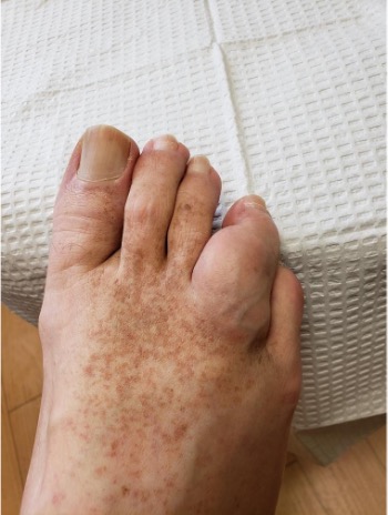
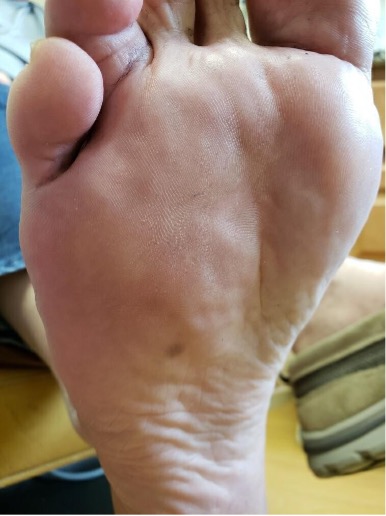
There were no open skin tears or lesions noted to the right foot. There was a palpable, mobile soft tissue mass evident on the dorsum of the right fourth digit, extending just proximal to the dorsal interphalangeal joint and extending from the dorsal digit to the fourth metatarsophalangeal joint, infiltrating the third and fourth interspaces. The mass measured 3.0 cm x 3.5 cm. There was no overlying erythema. The right fourth toe appeared significantly larger than the other lesser toes. One could also palpate a firm subcutaneous mass plantar to the fourth metatarsal shaft. There was no fluctuance or crepitus present. Range of motion was limited to the 4th digit due to the large localized mass. The areas of the mass were not tender to palpation. The patient had diminished sensation to the fourth digit of the right foot; otherwise neurological status was intact. Pedal pulses were palpable with capillary fill time intact to the digits. Skin temperature was normal and symmetrical bilaterally.
What the Imaging Revealed
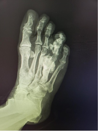
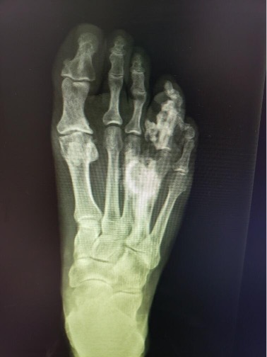
On plain films, one could note radiodensity at the right fourth metatarsal shaft and right fourth proximal and middle phalanges with significant hypertrophic osseous formation of the right fourth metatarsal shaft and right fourth proximal phalanx. The fourth digit appeared pedunculated from the medial aspect of the base of the proximal phalanx with possible synostosis to the right third metatarsal shaft.
Magnetic resonance imaging (MRI) showed abnormal cortical thickening, expansion, and irregularity of the fourth ray, including involvement of the metatarsal and all the phalanges. Based on radiography there was involvement of the calcaneus (seen on lateral view). Plain film radiography revealed involvement of the calcaneus, seen on the lateral view. There was a soft tissue mass noted at the distal aspect of the fourth ray. There was ossification in the plantar soft tissues contiguous with the fourth ray hyperostosis.
These findings were felt to be most consistent with a dysplastic bone disease; likely melorheostosis. The treatment team then decided to pursue surgical intervention.
Salient Surgical Details
The patient received intravenous sedation from the anesthesia team and a local anesthetic fourth ray block by podiatry. An ankle tourniquet at 250 mmHg maintained hemostasis. We made a 6 cm linear incision overlying the fourth metatarsal extending from the metatarsal midshaft to the proximal phalanx midshaft. During careful dissection down to bone, one could see exuberant abnormal, beefy, friable fibrous soft tissue with no signs of infection. A synostosis existed between the third and fourth metatarsals from midshaft and to the metatarsal head. Fluoroscopy confirmed this observation. We decided to keep the integrity of the fourth metatarsal intact to prevent a stress fracture.
Redirecting attention to the fourth digit, we noted an abnormal excess of subcutaneous fat resembling a lipoma, which we excised and sent to pathology. A sagittal saw then assisted in resecting the exostosis at the medial aspect of the fourth digit, which we also sent to pathology. A handheld rasp then smoothed the dorsal and medial aspects of the fourth proximal phalanx. Skin closure was uneventful, and the patient was permitted to bear weight as tolerated.
Diagnostic Data and Follow-Up
The soft tissue taken from the right fourth toe revealed highly vascular adipose tissue.
Diagnostic possibilities included angiolipoma, hemangioma, or vascular malformation. The resected bone from the fourth toe was reported as benign bone and adjacent connective tissue with patchy reactive changes.
The patient followed up with oncology, therefore limiting opportunity for postop radiographs. In a telehealth interview follow-up (due to challenges for the patient with facility distance) he expressed satisfaction with ambulation.
Concluding Thoughts
Melorheostosis is rare, with only 400 cases reported worldwide.5 However, its prevalence, undetected, is likely much more common. The age of peak presentation is from 5-20 years.5 Diagnosis relies on radiologic and histologic findings. On radiographs, hyperostosis of bone is typical and soft tissue changes may not be evident. Melorheostosis is thought to arise from a defect in intramembranous bone formation that can then cause abnormal bone thickening. Common symptoms include swelling, stiffness, and reduced joint range of motion. In addition, soft tissue changes can also occur that involve contractures, associated soft tissue masses, and/or hemangiomas.4 In this case. an associated infiltrating angiolipoma made this case unique. Due to the extensive bone and soft tissue involvement, this made it difficult for our patient to ambulate. Treatment should be individualized to the patient’s lifestyle. One should consider if there is an impedance in patient’s ambulatory status and conservative treatments should be exhausted prior to proceeding with surgical intervention. We believe this was the best approach for our patient, as there was reported satisfaction in ambulation postoperatively. Each case warrants individual consideration of the best steps to obtain a definitive diagnosis and appropriate treatment.
Sandra Haider, DPM, practices in Rancho Santa Margarita, CA.
Niki Iranmanesh, DPM, practices in Southfield, MI.
Alan Bloch, DPM, practices in Northville, MI.
References
1. Fick C, Fratzl-Zelman N, Roschger P, et al. Melorheostosis: a clinical, pathologic, and radiologic case series. Am J Surg Pathol. 2019;43(11):1554-1559.
2. Franca PM, Ferrreira CS, Figueiredo R, Matushita JP. Melorheostosis. Radiol Bras. 2015 Jan-Feb;48(1):60-61.
3. Grivas TB, Savvidou O, Psarakis SA, et al. Forefoot plantar multilobular noninfiltrating angiolipoma: a case report and review of the literature. World J Surg Oncol. 2008;6:11.
4. Dhillon MS, Saibaba B. Melorheostosis—foot and ankle perspective. The Foot. 2017;31:44-48.
5. Woolridge B, Stone NC, Denic N. Melorheostosis isolated to the calcaneus: A case report and review of the literature. Foot Ankle Int. 2005;26(8):660-663.

