


Diagnosing and Treating Common Pediatric Running Injuries
Foot and ankle injuries are common in pediatric athletes. This population has seen an increased level of participation and competitiveness in organized sports. Many patients are 3-sport athletes and participate in activities 6 to 7 days a week. With an increase in athletic participation there has also been an increase in the incidence of acute and overuse injuries in the pediatric athlete.1
Most overuse injuries are caused by overtraining. Overuse injuries are related to repetitive stress on the musculoskeletal system without sufficient recovery time, thus overwhelming the reparative process. These injuries occur often during sudden increases in training. For example, increasing total mileage, intensity of pace, or both.2 This increase in training commonly occurs during the start of a new season or during sport camps.3
Risk factors associated with injury include biomechanical factors such as pes planus, limb length discrepancy, lower extremity malalignment, muscular imbalance, and muscular tightness. Other factors include hard training surfaces, training errors, worn-out running shoes, and/or inappropriate running shoes.2 Educating patients on proper shoe size is an important part of injury prevention. In my experience, patients in this population typically do not get their feet checked regularly, if at all. Discussing frequent size checks by a knowledgeable shoe store or by a medical professional may help prevent injury.
Unique to the pediatric athlete, because of the growing musculoskeletal system, this population is at a higher risk for injuries at tendon attachment sites, joint surfaces, and at the growth plate.2 It is believed that there is an increased risk of injury during the pediatric growth spurt. This is caused by increased muscle tightness and decreased physeal strength.2 The bone is more prone to injury during this time because bone mineralization may fall behind linear bone growth.4
Apophyseal injuries occur in the growing athlete and are mainly caused by overtraining. Until the apophysis reaches skeletal maturity and fuses, the connection between the underlying bone and the apophysis is weaker than its associated tendon. Due to this, in children the apophysis, rather than the tendon, become inflamed with overuse.5 Children’s bones tend to grow at a faster rate than their adjacent tendons and muscles, which could cause muscle tightness and inflexibility after a growth spurt. Common sites of injury include the heel (Sever’s disease) and the knee (Osgood-Schlatter disease). A less common site of injury is the fifth metatarsal base (Iselin’s disease).
When Patients Present with Calcaneal Apophysitis
Sever’s disease, also known as calcaneal apophysitis, is a traction apophysitis located at the insertion of the Achilles tendon on the posterior calcaneus.6 Sever’s disease is more common in boys than girls. The age of presentation is around 10–12 years of age in boys and 8–10 in girls. The child typically presents with heel pain, which is worse when running. Pain is often bilateral and typically occurs at the beginning of a new season or sport. This is very common among soccer players. Symptoms are relieved with rest.
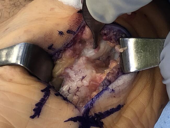
On physical exam the calcaneal apophysis is tender to palpation specifically with transverse compression of the heel (Figure 1). Swelling in the Achilles can be present. The patient may demonstrate an antalgic gait and may limp. Ankle dorsiflexion will be limited and painful. Diagnosis is based on clinical findings.
Imaging of the calcaneal apophysis is seen best on a lateral view. Findings may show sclerosis and fragmentation of the calcaneal apophysis. It is best to take bilateral views to compare the asymptomatic limb. Radiographic imaging is not usually necessary but may help with the diagnosis (Figure 2).
Treatment of calcaneal apophysitis depends on the severity of the child’s symptoms. Often it is challenging to control a child’s activity level especially in the competitive athlete. For mild symptoms a heel lift, arch support, stretching, ice, rest, nonsteroidal anti-inflammatory drugs (NSAIDs) or acetaminophen are appropriate. For more severe cases it is important to have a conversation with the patient and family to explain the importance of rest. Orthotics are an excellent choice to manage this condition with a built-in heel lift that lifts the strain off the Achilles tendon. As symptoms continue to improve, low impact exercises can be used to recondition the athlete, including stationary biking or swimming. A gradual return to higher impact activities can be implemented in severe cases at the 6-to-8-week mark. Corticosteroids are contraindicated.7 Complete resolution does not occur until closure of the growth plate.
What You Should Know About Iselin’s Disease
Apophysitis at the base of the fifth metatarsal (Iselin’s disease) can occur as an overuse injury in runners but is much less common than calcaneal apophysitis and is possibly underdiagnosed by many physicians due to lack of suspicion. The apophysis does not ossify until about 15 or 16 years of age. Iselin’s disease is seen in children with sporting activities that require running and jumping. The patient typically presents with lateral foot pain and difficulty wearing shoes. Symptoms are aggravated with activity and alleviated with rest. Swelling may be present at the base of the fifth metatarsal and there is tenderness with palpation. The etiology is due to repetitive traction of tendons inserted to the base of the fifth metatarsal.2,8
Imaging is helpful in the diagnosis of Iselin’s disease. The apophysis will show fragmentation and physeal line will show irregularities different from a normal apophysis, it is important to take bilateral films to compare. Imaging is valuable to rule out a fracture or a painful accessory bone. In avulsion, stress, and Jones fractures the orientation of the fracture line and the apophyseal line is different and is almost always perpendicular to each other.9
Symptoms respond to rest and ice. NSAIDs can be prescribed. It is important to determine the biomechanics of the patient in treating Iselin’s disease. A custom foot orthotic could benefit the patient.
Insights on Treating Osgood-Schlatter Disease
Osgood-Schlatter disease is a traction apophysitis of the tibial tuberosity and a common cause of knee pain in young athletes. This is the most common apophyseal disorder. It is seen in sports that have repetitive running, jumping, or quick turns such as soccer, basketball and football. Onset is around 14–16 in boys and 12–14 in girls and commonly occurs around a growth spurt.10 Pain is elicited with motions that exacerbate the quadriceps muscle such as jumping, running, and squatting.10
The patient presents with pain and swelling over the tibial tubercle. Pain is present with extension of the knee against resistance. Tightness of the lower extremity is also present at the hamstrings and quadriceps. The tibial tubercle may appear enlarged due to swelling or could have bony irregularly. The diagnosis can be made clinically.11
X-rays can be used to rule out an avulsion fracture of the tibial tubercle, but most of the time they are not required. A lateral knee radiograph is the best view to outline the tibial tubercle. This may show soft tissue swelling anterior to the tibial tuberosity or may show separation of the apophysis.12
Treatment involves rest, ice, NSAIDs or acetaminophen, and activity modification. Low-impact exercises can be utilized while recovering. Physical therapy or a stretching program should be initiated targeting the quadriceps and hamstring muscles. Ice before an activity can be helpful. An infrapatellar strap for 6–8 weeks can provide relief during activity. Steroids are contraindicated. Symptoms improve over 4–6 weeks.
How to Treat Stress Fractures in Children
Stress fractures are a common overuse injury in pediatric athletes. Stress fractures can involve the lower one-third of the tibia, and can also occur in the metatarsals, tarsal bones, femur, and fibula. Risk factors include increase in physical activity, osteoporosis, poor footwear, hard running surface, overtraining, prior stress fracture, and females if there is a history of an eating disorder or menstrual issue.3
When athletes load excess stress on their bones such as overtraining, or a sudden increase in their exercise regimen, this produces an imbalance between bone formation and bone resorption. This leads to microfractures and with continued loading of stress leads to stress fractures. The patient will present with local, sharp pain, exacerbated by weight-bearing activities. Patients with tibial stress fractures may have a prior diagnosis of shin splints.
Radiographs are typically normal the first two to three weeks after onset of symptoms. X-rays will show periosteal elevation, cortical thickening, sclerosis, or they may show a true fracture line. More recently MRI has become the study of choice over nuclear bone scans to rule out stress fractures. MRIs are highly sensitive and specific for diagnosing stress fracture and are important to be used to get a definitive diagnosis.1
When Young Runners Develop Tendonitis
Tendonitis is seen less frequently in young athletes in comparison to adults. The apophysis is weaker than the tendon itself and is more prone to injury. The tendon may be tender to palpation. Pain is reproduced with strength training of the involved muscle. For example, a patient with peroneal tendonitis will have pain with foot eversion against resistance. Treatment of tendonitis such as Achilles, peroneal or posterior tibial requires activity modification and physical therapy.
Soft Tissue Injuries in Runners
The most common soft tissue running injuries are blisters and lacerations. Blisters can be caused by a shoe that is too tight or too loose, parents buy shoes that “kids can grow into.” It is important for parents to periodically examine their children’s feet for the correct shoe size and for blisters. Second skin and moleskin are great modalities that may help protect children who develop blisters frequently. Education to parents is important for preseason screening.
In Conclusion
Proper stretching and training can prevent injuries as well as enhance sport performance. The role of the pediatric sports medicine foot and ankle specialist in youth sports is extremely important to educate the patient using preventative medicine. Most patients come to our offices after injury; at that time it is important to properly diagnose and treat the condition. After the patient has completed their recovery, education of patients and parents on injury prevention is pivotal. Most children are flexible, but some are not mainly during periods of rapid growth. A flexibility conditioning program should be implemented prior and during athletic activities. Physical therapy is an excellent treatment for both conditions combined with arch support, taping, and strapping.
With the increasing amount of participation of children in organized and recreational activities, podiatrists can expect to see an increase in sports related overuse injuries. It is important to understand the unique aspects of the growing child/adolescent. Many of the injuries discussed, such as tendonitis, apophysitis, shin splints, and stress fractures respond well to the treatments discussed.
Dr. Kaufman practices in Westchester, NY. She is board certified with the American Board of Podiatric Medicine and board qualified with the American Board of Foot and Ankle Surgery.
References
1. Soprano JV. Musculoskeletal injuries in the pediatric and adolescent athlete. Curr Sports Med Rep. 2005 Dec;4(6):329-34. doi: 10.1097/01.csmr.0000306295.49707.1f. PMID: 16282035.
2. Seto CK, Statuta SM, Solari IL. Pediatric running injuries. Clin Sports Med. 2010 Jul;29(3):499-511. doi: 10.1016/j.csm.2010.03.005. PMID: 20610035.
3. Brenner JS; American Academy of Pediatrics Council on Sports Medicine and Fitness. Overuse injuries, overtraining, and burnout in child and adolescent athletes. Pediatrics. 2007 Jun;119(6):1242-5. Doi: 10.1542/peds.2007-0887. PMID: 17545398.
4. Bailey DA, Wedge JH, McCulloch RG, Martin AD, Bernhardson SC. Epidemiology of fractures of the distal end of the radius in children as associated with growth. J Bone Joint Surg Am. 1989 Sep;71(8):1225-31. PMID: 2777851.
5. Adirim TA, Cheng TL. Overview of injuries in the young athlete. Sports Med. 2003;33(1):75-81. Doi: 10.2165/00007256-200333010-00006. PMID: 12477379.
6. Fares MY, Salhab HA, Khachfe HH, Fares J, Haidar R, Musharrafieh U. Sever’s disease of the pediatric population: clinical, pathologic, and therapeutic considerations. Clin Med Res. 2021 Sep;19(3):132-137. doi: 10.3121/cmr.2021.1639. PMID: 34531270; PMCID: PMC8445662.
7. Maffulli N, Wong J, Almekinders LC. Types and epidemiology of tendinopathy. Clin Sports Med. 2003 Oct;22(4):675-92. Doi: 10.1016/s0278-5919(03)00004-8. PMID: 14560540.
8. Pell RF 4th, Khanuja HS, Cooley GR. Leg pain in the running athlete. J Am Acad Orthop Surg. 2004 Nov-Dec;12(6):396-404. doi: 10.5435/00124635-200411000-00004. PMID: 15615505.
9. Deniz G, Kose O, Guneri B, Duygun F. Traction apophysitis of the fifth metatarsal base in a child: Iselin’s disease. BMJ Case Rep. 2014 May 15;2014:bcr2014204687. Doi: 10.1136/bcr-2014-204687. PMID: 24832713; PMCID: PMC4025211.
10. Gholve PA, Scher DM, Khakharia S, Widmann RF, Green DW. Osgood Schlatter syndrome. Curr Opin Pediatr. 2007 Feb;19(1):44-50. Doi: 10.1097/MOP.0b013e328013dbea. PMID: 17224661.
11. Martin TJ, Martin JS. Special issues and concerns for the high school- and college-aged athletes. Pediatr Clin North Am. 2002 Jun;49(3):533-52. doi: 10.1016/s0031-3955(02)00004-4. PMID: 12119864.
12. Duri ZA, Patel DV, Aichroth PM. The immature athlete. Clin Sports Med. 2002 Jul;21(3):461-82, ix. Doi: 10.1016/s0278-5919(01)00008-4. PMID: 12365238.
13. Moen MH, Tol JL, Weir A, Steunebrink M, De Winter TC. Medial tibial stress syndrome: a critical review. Sports Med. 2009;39(7):523-46. doi: 10.2165/00007256-200939070-00002. PMID: 19530750.
14. Caine D, Maffulli N, Caine C. Epidemiology of injury in child and adolescent sports: injury rates, risk factors, and prevention. Clin Sports Med. 2008 Jan;27(1):19-50, vii. Doi: 10.1016/j.csm.2007.10.008. PMID: 18206567.


