Emerging Insights In The Surgical Management Of Talocalcaneal Coalitions
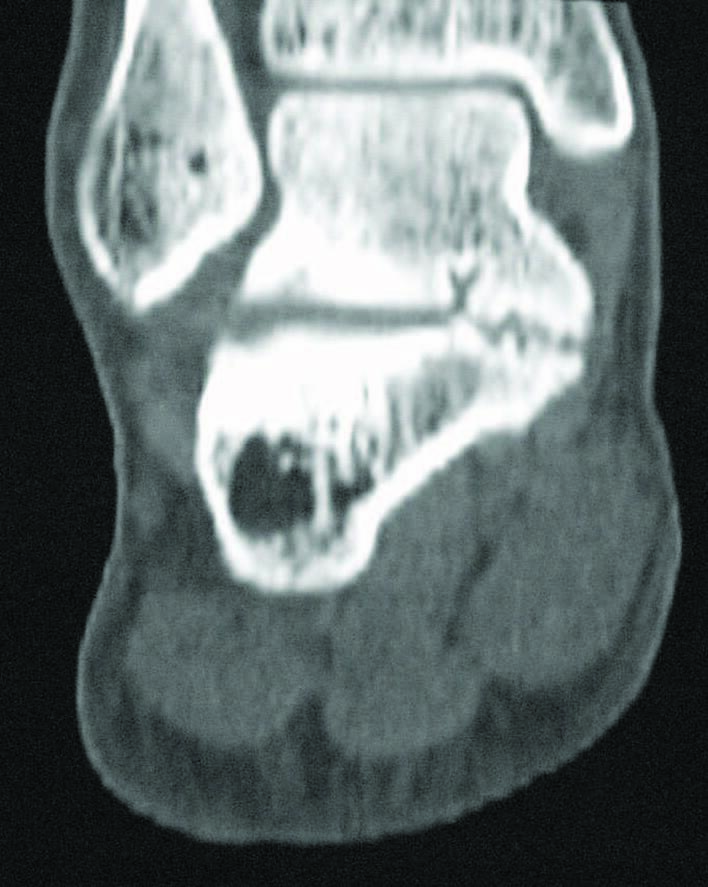
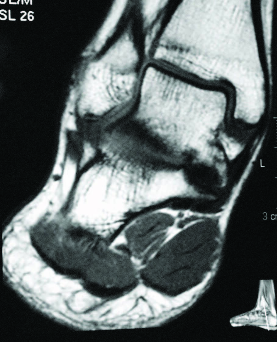
A tarsal coalition exists when there is a congenital, complete or incomplete union between two or more tarsal bones causing restricted motion or absence of motion. Tarsal coalitions remain challenging for the foot and ankle physician and surgeon. Their clinical hallmarks are primarily pain, rigid pes valgus and/or rigid pes planus deformity, and often tonic peroneal muscle spasm. Although the diagnosis of a tarsal coalition is often readily apparent upon clinical examination, diagnostic imaging is confirmatory and often aids in planning any necessary surgical intervention. Sequentially, the author always obtains radiographs of the involved foot first followed as necessary by advanced imaging with either computerized tomography (CT) or magnetic resonance imaging (MRI) scans, or in some instances of incomplete tarsal coalition, both CT and MRI.1
One should recognize that tarsal coalitions vary tremendously in presentation, configuration, and type. Although there are many similarities, one must always anticipate individual differences and appreciate them when possible. By far, the most common tarsal coalitions are the middle facet talocalcaneal coalition and the calcaneonavicular coalition.1 The former presents in many different ways, and in this paper the author will discuss some of the more recent clinical insights and debates regarding surgical management of talocalcaneal coalition.
Identifying The Most Appropriate Treatment Pathway
In managing a talocalcaneal coalition, one must consider both conservative and surgical treatment options. The mainstays of conservative treatment center on immobilization, off-loading, rest and anti-inflammatory modalities.1 Immobilization can include the use of a short-leg cast, a controlled ankle motion (CAM) walker, a brace such as a Richie Brace® or Arizona Brace,® a maximum-control foot orthotic (such as a University of California-Berkley Lab (UCBL) orthotic) or a similar orthotic with a neutral rearfoot post and a deep heel seat.1 Anti-inflammatory modalities can include corticosteroid injections, non-steroidal anti-inflammatory medications and a variety of physical therapy modalities focusing on relieving the inflamed areas.1
When conservative treatment fails, is likely to fail, or considered inappropriate, surgery is frequently indicated. Traditionally, surgical approaches for talocalcaneal coalition have been divided between resection (arthroplasty) and fusion (arthrodesis) procedures. More recently, a number of researchers attempted to provide predictable criteria for assisting in decision-making between these two diverse approaches. In 1991, this author attempted to provide a surgical classification for tarsal coalitions, including the talocalcaneal coalition.2 The criteria espoused at that time for development of a surgical treatment plan included the patient’s age (general premise: younger patients do better with resection; older patients do better with fusion), articular involvement (general premise: extraarticular coalitions do better with resection; intraarticular coalitions do better with fusion) and the extent of secondary arthritic changes (general premise: patients without arthritic changes do better with resection; patients with arthritic changes do better with fusion).2 Clearly then, the talocalcaneal coalition presents a dichotomy of options. The talocalcaneal coalition is intraarticular, and its resection by all accounts necessitates at least partial destruction of the subtalar joint. Conversely, calcaneonavicular coalitions are extraarticular and resection does not typically destroy the joint.1,2
So, do all talocalcaneal coalitions require fusion? Today, the consensus is a firm, “certainly not.” Particularly in the younger patient who has not yet reached osseous maturity, resection of a talocalcaneal coalition is an often successful consideration. Conversely, in many older patients (i.e. those who have reached developmental maturity) and/or who have secondary adaptive or arthritic changes of the surrounding joints, the surgical decision most often centers on how many joints to fuse: the subtalar joint alone; a medial double arthrodesis; or a triple arthrodesis.1 There is a “gray zone” of patients with talocalcaneal coalitions between these two extremes, where one may consider and attempt either option. Contemporary research based upon surgical treatments and clinical outcomes attempts to provide further guidance to the surgeon addressing a particular patient with a talocalcaneal coalition.
Additional Factors To Consider When Determining Surgical Intervention
Recently, the literature presents arguments for the inclusion of the size and type of talocalcaneal coalition, and the degree of heel valgus as factors to consider when deciding between joint resection and joint fusion.3-11 Some hypothesize that the size of a talocalcaneal coalition inversely correlates with the success rate of resection, and some researchers used preoperative advanced imaging studies, particularly CT, to investigate.3,4 Arbitrarily, based only on their personal experience, these investigators concluded that talocalcaneal coalitions involving greater than 50 percent of the talocalcaneal joint are less amenable to surgical resection.3,4 More recently though, several authors challenged this subjective assumption and instead determined that no definitive talocalcaneal coalition size precludes an attempt at resection in every patient.5-7
Additionally, different described shapes and morphologies of talocalcaneal coalitions may be a factor to consider when debating arthroplasty or arthrodesis. In 2010, Rozansky and colleagues used thin-cut 2D and 3D CT scans to divide talocalcaneal coalitions into five different types based on their cartilaginous or bony nature, location and facet joint orientation.8 In 2016, Bixby and associates used CT scans to assess 97 patients with a total of 138 talocalcaneal coalitions.9 They found that 97 (70 percent) of the coalitions involved the middle facet, 39 (28 percent) the posteromedial aspect of the joint and two (1.4 percent) involved the posterior facet.9 They described the posteromedial talocalcaneal coalition as being associated with an intact, but shorter middle facet with a long sustentaculum tali.9
In a subsequent study, these authors compared the clinical outcomes of resection in 36 patients (51 feet) with middle facet talocalcaneal coalitions (36 feet) and posteromedial talocalcaneal coalitions (15 feet).10 They found that patients with posteromedial talocalcaneal coalitions had significantly better clinical outcomes compared to middle facet talocalcaneal coalitions after resection.10 These researchers concluded that the differences in the patient-based outcomes were likely due to not only the typically smaller size of the posteromedial variant, but also due to its simpler and more superficial anatomic location.10 Whether preoperative consideration of joint morphology can improve patient outcomes and expectations following talocalcaneal coalition resection certainly merits further study.
The degree of hindfoot valgus present is also a proposed possible variant in the debate between when to resect and when to fuse. Wilde and team found that more than 16 degrees of heel valgus correlated with a poor outcome after talocalcaneal coalition resection.4 Conversely, Luhmann and Schoenecker found no problems with 16 degrees of heel valgus, but did find worse outcomes with resection of talocalcaneal coalitions in patients with more than 21 degrees of heel valgus.11These investigators recommended either a medializing calcaneal osteotomy or a lateral column lengthening procedure when performing resection of a talocalcaneal coalition associated with severe hindfoot valgus.11 However, more recent research calls the negative relationship between heel valgus and the success of talocalcaneal coalition resection into question. Other investigators failed to find increased hindfoot valgus predictive of clinical outcome in their series of tarsal coalition resections.6,7
Pertinent Pearls In Localization And Resection Of Talocalcaneal Coalitions
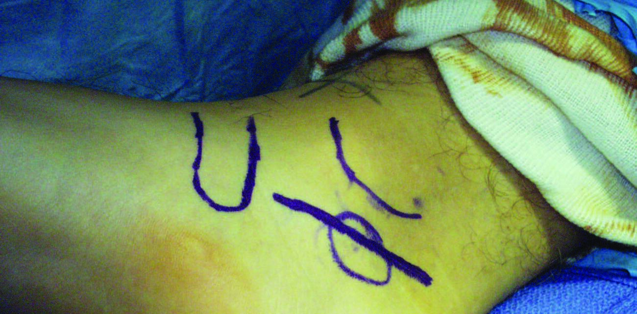
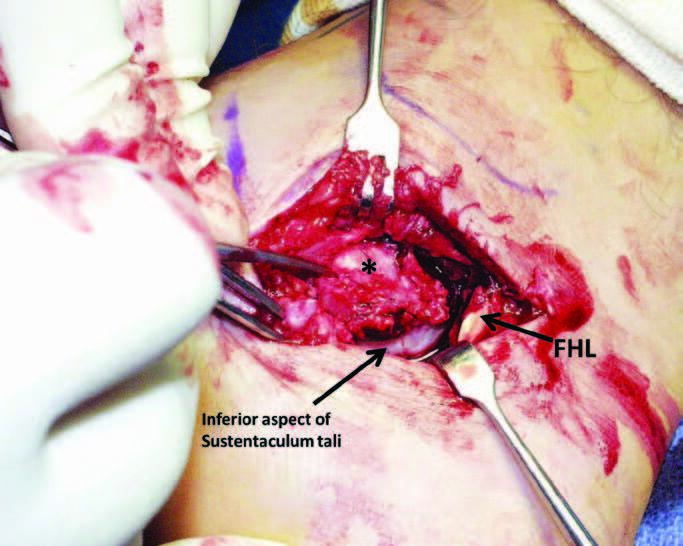
The current surgical approach to resection of a middle facet or medial talocalcaneal coalition typically involves a direct medial incisional approach, between the flexor digitorum longus tendon and the neurovascular bundle, with the tibialis posterior tendon and flexor digitorum longus tendon retracted dorsally and the neurovascular bundle and flexor hallucis longus tendon retracted plantarly. With this approach, in most instances, there is no readily visible middle facet joint space, and localization of the talocalcaneal coalition can be a challenge. Therefore, a needle, K-wire, small osteotome or other flat instrument can identify the area of the middle facet with or without the aid of intraoperative fluoroscopy.1
Humbyrd and Myerson described a technique using a laterally inserted guide wire at the level of the sinus tarsi and subsequent use of a cannulated arthroereisis sizing or dilation probe to identify the middle facet coalition and/or create a fracture through the coalition to assist in its localization.12 Alternatively, Field and Ng advocated for subtalar joint arthroscopy and a K-wire to assist in localizing the coalition.13 In 2019, Edmonds and colleagues described a K-wire technique to demarcate the coalition.14 These surgeons used the traditional medial incision over the sustentaculum tali and exposed the general area of the talocalcaneal coalition. Next, they used a second, ancillary, lateral incision extending from just distal to the fibular malleolus, over the sinus tarsi, towards the fourth metatarsal base. After incising the extensor digitorum brevis muscle and exposing the sinus tarsi, they inserted 0.045-inch K-wires from lateral to medial, outlining and defining the coalition. Through the medial incision, they visualized the K-wires and used them to directly expose and resect the talocalcaneal coalition.14
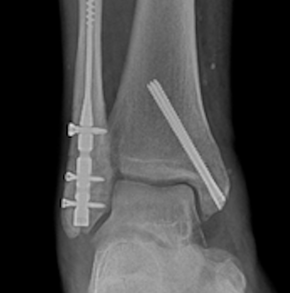
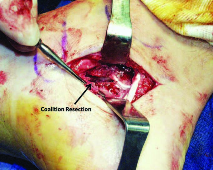
Regardless of the method used to delineate the coalition, once one identifies the extent of the talocalcaneal coalition, one can generously resect it with any combination of rongeurs, osteotomes, curettes, a power burr and/or a power saw. Care is necessary to preserve the structure, or at the very least the inferior bony margin, of the sustentaculum tali. One will typically observe an intraoperative increase in subtalar joint motion after resection of the coalition.1
Once one resects the talocalcaneal coalition, the best method to manage the defect created is also subject to debate. In the author’s opinion, the key to a successful tarsal coalition resection is a full and complete resection and not the material placed into the resulting defect. In the author’s opinion, if one generously resects a talocalcaneal coalition, there is no mandate to place anything in the defect. If desired, one can use bone wax on the resected bone margins. Many surgeons harvest the autogenous fat pad from Kager’s triangle (the area anterior to the Achilles tendon and posterior to the deep flexor compartment) or from the local soft tissues, and place the fat obtained into the resection void.1 Alternatively, some investigators advocate splitting the flexor hallucis longus longitudinally and rerouting the superior one-half of the split tendon through the resection site.15,16 Whether the insertion of these organic or nonorganic materials into the resection defect is necessary in an attempt to maintain the resection space and decrease the likelihood of recurrence remains unknown. The diversity of surgical alternatives, small size of the case series reported and the criteria for grading the results at follow up make concise conclusions difficult. Once one addresses the defect, layered closure concludes the procedure.
Can CT Scans Play A Role Intraoperatively?
Recently, CT scans have been used to assist not only in preoperative planning but also intraoperatively. In 2014, de Wouters and colleagues preoperatively created patient-specific instrument guides by 3D CT scan modeling of the coalition and then used the guides to resect a series of seven talocalcaneal coalitions.17 The guides were made-to-measure and designed to fit the coalition, enabling potentially more precise resection. They found that the guides resulted in easier and more predictable resection of the talocalcaneal coalitions in their series.17 Since this original description, other surgeons also advocated the use of customized 3D-printed surgical guides based on 3D CT reconstructions for talocalcaneal coalition resection.18
Kemppainen and associates retrospectively looked at 14 feet where intraoperative CT assessed talocalcaneal resection.19They found that intraoperative CT scans altered their operative technique in three (21 percent) of the 14 feet. On blinded review, use of intraoperative CT resulted in an “excellent” resection of the coalition in eight (57 percent) of the 14 feet compared to three (25 percent) of the 12 feet in their control group that underwent treatment without intraoperative CT scan assistance.19
In 2017, Aibinder and colleagues introduced an interesting technique of intraoperative CT-guided navigation for resection of a talocalcaneal coalition.20 Their technique required placement of a reference frame and performance of two intraoperative CT scans. The first scan delineated the coalition, and the second scan took place after a navigated burr resected the coalition. The authors hypothesized that their technique allowed for efficient, more thorough, and more controlled resection of the coalition along with decreased risk of incomplete resection. The noted disadvantages of this technique are radiation exposure and increased cost of using intraoperative CT scans.20
In 2018, Stokman and team also described the use of intraoperative CT navigation to assist in immediate localization and guided resection of talocalcaneal coalitions.21 They used a high-speed burr to resect the coalition that, unlike the technique used by Aibinder and colleagues, was not registered to the navigation system.21 Although these preoperative and intraoperative techniques utilizing 3D-CT scanning and 3D-modeling/printing technologies are not yet in widespread use, they clearly merit further investigation and comparison of clinical outcomes achieved with these methods to those achieved with more traditional surgical approaches.
A Closer Look At Endoscopic/ Arthroscopic Approaches, Postop Course And Complex Deformities
Another recent surgical advance for talocalcaneal coalitions is the endoscopic/ arthroscopic approach to resection. Arguably, the open approach to talocalcaneal coalition resection has several inherent disadvantages, including wound dehiscence or wound healing problems, additional operative time and potential damage to surrounding structures.22-26 In 2011, Bonasia and colleagues first described the resection of a talocalcaneal coalition through a posterior arthroscopic approach.22 The authors performed their resection with the patient under spinal or general anesthesia and in the prone position. They used posterolateral and posteromedial portals on either side of the Achilles tendon with the arthroscopic instrument interchangeably switched between the portals until achieving full identification and and resection of the coalition. Since that time, several other studies revealed good-to-excellent results with endoscopic/arthroscopic resection of a talocalcaneal coalition either through two lateral arthroscopic portals over the sinus tarsi23 or through two posterior portals.24,25 In a recently published systematic review, Malik-Tabassum and associates concluded that arthroscopic resection of symptomatic talocalcaneal coalitions is a feasible and effective treatment approach.26 Appropriately, the authors recommended future studies compare clinical outcomes of arthroscopic resection with open resection.26
Postoperatively, this author prefers to keep patients undergoing talocalcaneal resection non-weight bearing for anywhere from two to six weeks, depending upon the extent of dissection required for the resection. During this time, I place the patient either in a CAM-walker boot or a short leg cast. Active and passive range of motion exercises, focusing on inversion and eversion of the subtalar joint, begin as soon as possible postoperatively considering the patient’s comfort. If needed, physical therapy may further assist in motion restoration after about three to six weeks. I typically recommend long-term use of functional foot orthotics, especially if residual pes plano valgus deformity remains.
In that regard, following resection of any talocalcaneal coalition associated with a pre-existing pes plano valgus deformity, the pes plano valgus deformity may potentially become more problematic and/ or symptomatic after the coalition resection surgery. Some authors advocate that a single-stage approach is best to concomitantly perform both talocalcaneal coalition resection and flatfoot reconstruction.27,28 Kernbach and associates reported good results in three patients (six feet) using this approach.27 Lisella and team reported on a retrospective study of seven consecutive patients (eight feet) who underwent talocalcaneal coalition resection with simultaneous pes valgoplanus reconstruction.28 The authors stated that the clinical and radiographic hindfoot misalignment reliably corrected in their series. When a tarsal coalition is associated with severe hindfoot valgus, one must address the valgus either conservatively or surgically. When surgery is done for the heel valgus deformity, dispute remains as to whether this correction should occur in one surgical stage with the coalition resection or in a second surgical operation.
In Summary
It is the author’s present opinion, from personal experience and the consensus of current outcome studies, that surgical resection of a symptomatic middle facet talocalcaneal coalition is a reasonable and viable procedure. However, not all talocalcaneal coalitions are the same and, in fact, they vary significantly in their subjective and objective presentations. Current consensus findings supported by the literature regarding talocalcaneal coalition resection include the following:
1. Most studies are in younger patient populations, and although older age does not appear to be a contraindication to talocalcaneal coalition resection, younger patients are generally considered more amenable to resection;
2. The presence of compensatory or adaptive changes, such as talonavicular joint beaking, does not appear to be a contraindication to resection. However, narrowing and/or degenerative changes of the talonavicular joint do appear to be a contraindication to talocalcaneal coalition resection, and arthrodesis may be more appropriate;
3. The tissue type (fibrous tissue, cartilage, bone) of the talocalcaneal coalition does not seem to be a limiting factor regarding whether indication for resection or arthrodesis. However, incomplete coalitions are generally more amenable to resection than complete coalitions;
4. Debate continues as to whether coalition size may or may not be a contraindication to resection. Patients with coalitions involving less than 50 percent of the talocalcaneal joint are considered better candidates for resection. Coalitions involving more than 50 percent of the talocalcaneal joint may or may not be amenable to resection. The 50 percent threshold has been arbritrarily set. Further studies involving resections in coalitions involving more than 50 percent of the talocalcaneal joint, and specifically looking at the architectural shape and composition of the coalition are necessary to determine what composition, shape, and percentage of joint involvement preclude a reasonable chance of a good long-term functional outcome with resection;
5. The material interposed in the defect after coalition resection does not appear to influence results. Full and complete resection of a talocalcaneal coalition is the most important determinant of a successful clinical outcome;
6. The degree of hindfoot valgus and/ or pes plano valgus deformity present may affect the postoperative result. Hindfoot valgus deformity greater than 16 degrees may negatively impact the clinical outcome of talocalcaneal coalition resection. Again, this amount of valgus deformity has been subjectively set, and debate continues regarding whether or not any specific degree of hindfoot valgus affects clinical outcome. Concomitant correction of associated hindfoot valgus or pes plano valgus deformity may be appropriate for tarsal coalitions associated with significant and/ or painful hindfoot deformities. Alternatively, one may also address these deformities with conservative measures or at a second surgical session;
7. Preoperative and intraoperative 3D-CT scans need further investigation but appear to offer significant potential for improving clinical outcomes following talocalcaneal coalition resection. In particular, both preoperative 3D-modeled/printed templates and intraoperative CT-guided resection navigation are ripe for additional study as they incorporate some of the latest orthopedic technologies available;
8. Additional research to determine whether or not endoscopic/arthroscopic approaches for talocalcaneal coalition resection offer improved clinical outcomes and lower risks than traditional open approaches is warranted. Endoscopic/arthroscopic approaches currently appear feasible but clearly have an associated steep learning curve; and
9. When talocalcaneal coalition resection fails or is likely to fail, arthrodesis of the involved hindfoot joints remains an established and practical surgical option. If arthrodesis is the best option, the surgeon should carefully select the optimal approach: isolated subtalar arthrodesis; medial double arthrodesis; or triple arthrodesis.1
Dr. Downey is the Chief of the Division of Podiatric Surgery at Penn Presbyterian Medical Center and a Clinical Professor in the Department of Surgery at Temple University School of Podiatric Medicine in Philadelphia. He is a Fellow of the American College of Foot and Ankle Surgeons and a Senior Faculty member of The Podiatry Institute in Decatur, Ga. He practices with Penn Podiatry in Philadelphia, Pa. and Mount Laurel, NJ.
1. Downey MS, DeWaters AM. Tarsal coalition. In: Southerland JT, Boberg JS, Downey MS, Nakra A, Rabjohn LV (eds). McGlamry’s Comprehensive Textbook of Foot and Ankle Surgery, 4th ed. Philadelphia:Wolters Kluwer/Lippincott Williams & Wilkins;2013:598-635.
2. Downey MS. Tarsal coalitions: a surgical classification. J Am Podiatr Med Assoc. 1991;81(4):187–197.
3. Comfort TK, Johnson LO. Resection for symptomatic talocalcaneal coalition. J Pediatr Orthop. 1998;18(3):283–288.
4. Wilde PH, Torode IP, Dickens DR, Cole WG. Resection for symptomatic talocalcaneal coalition. J Bone Joint Surg Br. 1994;76(5):797–801.
5. Gantsoudes GD, Roocroft JH, Mubarak SJ. Treatment of talocalcaneal coalitions. J Pediatr Orthop. 2012;32(3):301-307.
6. Khoshbin A, Bouchard M, Wasserstein D, et al. Reoperations after tarsal coalition resection: a population-based study. J Foot Ankle Surg. 2015;54(3):306-310.
7. Mahan ST, Spencer SA, Vezeridis PS, Kasser JR. Patient-reported outcomes of tarsal coalitions treated with surgical excision. J Pediatr Orthop. 2015;35(6):583-588.
8. Rozansky A, Varley E, Moor M, Wenger DR, Mubarak SJ. A radiologic classification of talocalcaneal coalitions based on 3D reconstruction. J Child Orthop. 2010;4(2):129-135.
9. Bixby SD, Jarrett DY, Johnston P, Mahan ST, Kleinman PK. Posteromedial subtalar coalitions: prevalence and associated morphological alterations of the sustentaculum tali. Pediatr Radiol. 2016;46(8):1142-1149.
10. Mahan ST, Prete VI, Spencer SA, Kasser JR, Bixby SD. Subtalar coalitions: does the morphology of the subtalar joint involvement influence outcomes after coalition excision? J Foot Ankle Surg. 2017;56(4):797-801.
11. Luhmann SJ, Schoenecker PL. Symptomatic talocalcaneal coalition resection: indications and results. J Pediatr Orthop. 1998;18(6):748-754.
12. Humbyrd CJ, Myerson MS. Use of a cannulated guide in talocalcaneal coalition resection: technique tip. Foot Ankle Int. 2015;36(2):225-228.
13. Field C, Ng A. Resection of middle facet coalition with arthroscopic guidance. J Foot Ankle Surg. 2009;48(2):273-276.
14. Edmonds WB, Wiley K, Panas K. Technique article: tarsal coalition resection using Kirschner
wires across the subtalar Joint in a two-incision approach. J Foot Ankle Surg. 2019;58(2):337-340.
15. Blakemore LC, Cooperman DR, Thompson GH. The rigid flatfoot: tarsal coalitions. Foot Ankle Clin. 1998;3:609–631.
16. Raikin S, Cooperman DR, Thompson GH. Interposition of the split flexor hallucis longus tendon after resection of a coalition of the middle facet of the talocalcaneal joint. J Bone Joint Surg. 1999;81A:11-19.
17. de Wouters S, Tran Duy K, Docquier PL. Patient-specific instruments for surgical resection of painful tarsal coalition in adolescents. Orthop Traumatol Surg Res. 2014;100(4):423-427.
18. Sobron FB, Benjumea A, Alonso MB, Parra G, Perez-Mananes R, Vaquero J. 3D printing surgical guide for talocalcaneal coalition resection: technique tip. Foot Ankle Int. 2019;40(6):727- 732.
19. Kemppainen J, Pennock AT, Roocroft JH, Bastrom TP, Mubarek SJ. The use of a portable CT scanner for the intraoperative assessment of talocalcaneal coalition resections. J Pediatr Orthop. 2014;34(5):559-564.
20. Aibinder WR, Young EY, Milbrandt TA. Intraoperative three-dimensional navigation for talocalcaneal coalition resection. J Foot Ankle Surg. 2017;56(5):1091-1094.
21. Stokman JJ, Mitchell J, Noonan K. Subtalar coalition resection utilizing live navigation: a technique tip. J Child Orthop. 2018;12(1):42-46.
22. Bonasia DE, Phisitkul P, Saltzman CL, Barg A, Amendola A. Arthroscopic resection of talocalcaneal coalitions. Arthroscopy. 2011;27(3):430-435.
23. Jagodzinski NA, Hughes A, Davis NP, Butler M, Winson IG, Parsons SW. Arthroscopic resection of talocalcaneal coalitions – a bicentre case series of a new technique. Foot Ankle Surg. 2013;19:125-130.
24. Knorr J, Soldado F, Menendez ME, Domenech P, Sanchez M, Sales de Gauzy J. Arthroscopic talocalcaneal coalition resection in children. Arthroscopy. 2015;31(12):2417-2423.
25. Aldashan W, Hamed A, Eisherief F, Abdelaziz AM. Endoscopic resection of different types of talocalcaneal coalition. Foot Ankle Int. 2018;39(9):1082-1088.
26. Malik-Tabassum K, Wahed K, To C, Maling L, Rose B. Post-operative outcomes of arthroscopic tarsal coalition resection: a systematic review. J Orthop. 2020;21:537-543.
27. Kernbach KJ, Blitz NM, Rush SM. Bilateral single-stage middle facet talocalcaneal coalition resection combined with flatfoot reconstruction: a report of 3 cases and review of the literature. Investigations involving middle facet coalitions- -part 1. J Foot Ankle Surg. 2008;47(3):180-190.
28. Lisella JM, Bellapianta JM, Manoli A 2nd. Tarsal coalition resection with pes planovalgus hindfoot reconstruction. J Surg Orthop Adv. 2011;20(2):102-105.