Managing Wound Pain: International Perspectives on Assessments, Challenges and Treatment Options
The International Association of the Study of Pain (IASP) defines pain as “an unpleasant sensory and emotional experience associated with or resembling that association with, actual or potential tissue damage.”1 This definition highlights the multidimensional nature of pain.
The IASP further categorizes pain as acute or chronic. Acute pain “lasts from a few minutes to less than 6 months” and chronic pain is pain that persists for 3 months or more “despite successful management of the condition that initially caused it, or because the underlying medical condition cannot be treated successfully.”1 Chronic pain can be continuous and persistent or intermittent. Regardless of the definition used, pain remains a basic bodily sensation that we have all experienced in one way or another. Generally, painful stimuli elicit a reaction causing avoidance, escape, or destruction of the causative factor. The medical community continues to debate if pain is a pathologic entity in its own right, with some clinicians still adhering to the idea that pain is simply a symptom of a greater disease state.2
Pain is frequently associated with certain types of chronic wounds. Wound-related pain (WRP) is complex and can vary in severity. Researchers have found the prevalence of pain with chronic wounds to be 48–81%, with 19–46% of patients reporting moderate-to-severe pain.3 Understanding and effectively treating wound pain represents a significant challenge to health care providers practicing wound management. WRP is a topic of great interest as we continue to gain knowledge on the effects of pain on quality of life (QoL) and wound care outcomes. There remains a gap between our understanding of WRP and successful integration of pain management pathways in chronic wounds. This article attempts to highlight the latest evidence on WRP assessments, treatment challenges, and non-pharmacological therapies from an international perspective.
A Guide to the Types of Pain
Pain is often divided into two distinct categories: nociceptive pain and neuropathic pain. Nociceptive pain is considered the body’s typical response to noxious stimuli (mechanical, thermal, chemical), often acting as a warning to impending or frank injury. This pain can be perceived as sharp, stabbing, tender, throbbing, or aching. Nociceptors are nerve endings commonly found in cutaneous muscles and tissues such as skin. Tissue injury causes the release of histamine and serotonin via the inflammatory cascade, leading to cellular changes in these nerve endings, precipitating an electrical impulse that travels up the nerve root to the spinal cord with transmission to the brain.4 Nociceptive pain, also known as acute pain, is typically localized and constant, and is a temporary experience. The nociceptive pain resolves when the tissue damage ends and the inflammation subsides. The intensity of the pain reduces or disappears after appropriate assessment and resolution or mitigation of the underlying cause. In chronic wounds, nociceptive pain occurs during wound management procedures such as wound hygiene or dressing changes, where noxious stimulus causes brief trauma to the wound and the periwound.5
In contrast, neuropathic pain is triggered by damage to or dysfunction of the nervous system, causing signals to move through atypical pathways.6 Neuropathic pain therefore results from responses generated by damaged nerves, intensifying signals and resulting in hyperalgesia characterized by burning, stinging, shooting, and even numbness.6
Additionally, chronic wounds are influenced by endogenous inflammatory mechanisms that lower the threshold of peripheral nociceptor stimulation, which in turn often intensify pain levels.6 Thus, chronic wound pain is multifactorial and often relates to a combination of tissue damage, inflammation, and nerve injury. Wound-related factors such as blood vessel dysfunction, tissue acidosis, ischemia, bacterial contamination, and infection can further contribute to WRP.7
Persistent pain in chronic wounds may also be termed as background pain that is frequently present even when the wound is undisturbed or at rest. Background pain usually relates to the underlying cause of the wound, local wound factors, and other related pathologies such as skin irritation.8 This persistent chronic pain may have features of acute and chronic pain and can be nociceptive, neuropathic, or both, which makes wound pain challenging to diagnose and treat.5,9
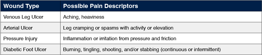
Differences in the characteristics, pattern, and quality of pain can relate to various wound etiologies (Table 1). Diabetic foot ulcers often exhibit neuropathic pain characterized by burning, shooting, and stinging sensations.10 Arterial leg ulcers often result in ischemic pain exacerbated during walking or during leg elevation,11 or in contrast, venous leg ulcers often have deep aching or muscle pain occurring after periods of dependency.12
Wound Pain, Stress, and Delayed Healing
Chronic WRP and associated stress can have a detrimental effect on wound healing through various physiologic mechanisms. During times of stress, the sympathetic nervous system activates the adrenal glands, leading to the release of adrenaline and cortisol. These hormones increase respiration, heart rate, and blood pressure.14 When WRP becomes chronic, increased levels of cortisol contribute to immune suppression by inhibiting cellular migration, differentiation, and proliferation.15 Subsequently, dysfunctions in neutrophil and macrophage activity contribute to uninhibited accumulation of wound debris and contaminants. Vasoconstriction of small arterioles occurs in the presence of cortisol.14 When prolonged, this impedes peripheral blood flow, nutrient transport, and oxygen delivery to wounded tissues. Effective pain management and stress reduction may potentially decrease wound inflammation, decrease chances for wound infection, and support wound healing.
Cortisol can also decrease fibroblast proliferation.16 Fibroblasts play a crucial role in wound healing. These “worker bee” cells help degrade dysfunctional extracellular matrix (ECM), support the creation of a functional ECM through collagen formation and aid in wound contracture. When fibroblast function is arrested, wound healing stalls and the strength of new tissues is diminished. Studies show that psychological stress impairs the wound’s inflammatory response and matrix degradation processes immediately postop.17 These findings suggest that pain reduction interventions may improve wound repair and recovery following surgery.
How Wound Pain Affects Patients
Wound care regimens often dictate patients’ daily activities. Clinic appointments, tests, and bandage changes can take many hours over several weeks or months of therapy. The indirect consequences of living with chronic wound pain often contribute to social isolation. Chronic wound patients may limit fraternization with others and experience impairments in all aspects of social interactions.18 Subsequently, this confinement and solitude can lead to feelings of depression, social detachment, withdrawal, anxiety, and sleep disturbances, lessening one’s overall quality of life (QoL). The World Health Organization Quality of Life Group defines quality of life as “an individual’s perception of their position in life in the context of the culture and value systems in which they live and in relation to their goals, expectations, standards and concerns.”15 This definition encompasses health, social, and economic aspects of overall wellbeing.
Chronic WRP can decrease patients’ QoL by preventing participation in enjoyable activities. Often these are activities that are important to maintaining daily functioning, including walking, standing, and climbing stairs. These essential actions often exacerbate wound-related pain. A limited ability to engage in physical activities limits patients’ ability to cope with stress, thus potentially escalating pain.19 As many as 80% of patients reported experience substantial levels of pain even while at rest.20 Studies have reported that pain associated with chronic wounds is one of the symptoms that patients find particularly distressing.13
A growing number of investigations aim to shine a light on the real burden of pain in the chronic wound patient population. Estimates state that chronic wounds cause decreased functional ability and QoL for up to 3% of individuals 60 years and older.3 In the US alone, that translates to 16.8% of the population, equating to roughly 75 million residents.21 QoL is a strong predictor of major amputation and death for patients who experienced deficits such as mobility restrictions, self-care deficits, inability to perform usual activities, and discomfort.22
Assessing Wound Pain
The assessment of WRP is an essential component to a complete patient evaluation. Pain cannot be treated if it cannot be assessed.23 Documenting and tracking WRP will provide important information necessary for establishing an effective, holistic patient treatment plan. To date, there are a variety of assessment tools available to measure WRP. Health care practitioners should use the most appropriate tool for their clinical setting based on the patient population served, ease of use, and staff abilities. Tailoring the selection to the type of pain and patient history ensures effective care. The same tool should be used for all subsequent patient assessments for the sake of continuity.
The time-point for the assessment pain is also important, as pain may be reported as higher during wound management procedures such as debridement and dressing changes, or during movement compared to when the patient is at rest. A lack of awareness of variation in pain intensity at different time points can have a negative consequence for ulcer treatment and management.9
Another important factor is acknowledging that patients with diabetic foot ulcers can experience WRP in the presence of peripheral neuropathy.9,24,25 Despite the common belief that pain is masked in patients with peripheral neuropathy, the presence of pain is a flag for further investigations to determine the cause of pain—whether it is symptoms of neuropathy, ischemia or infection, it is important to differentiate the pain in order to apply the appropriate treatment.
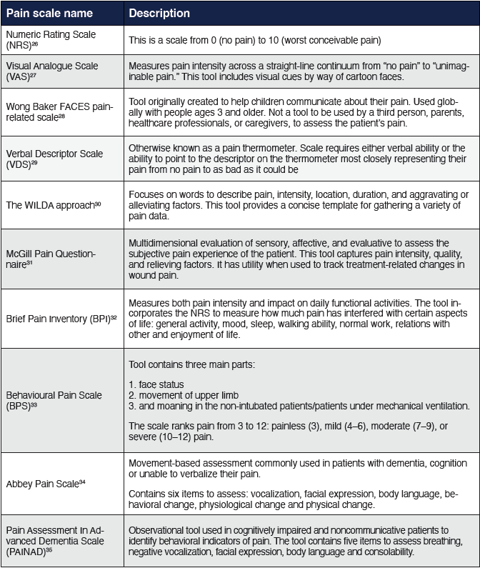
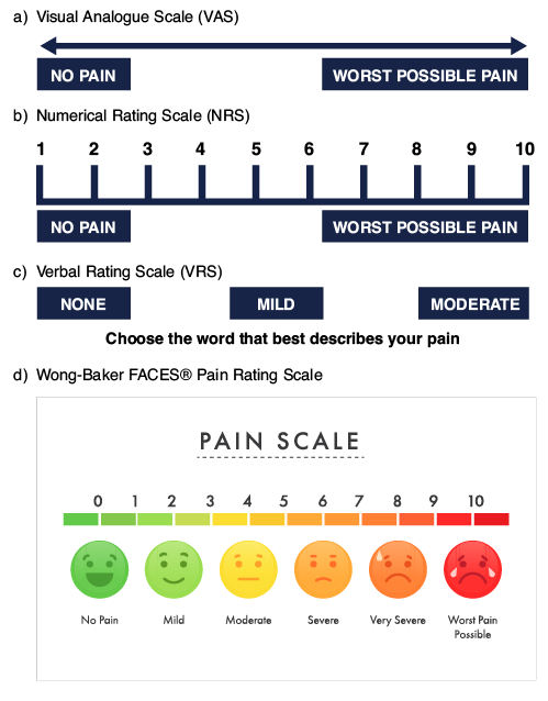
Commonly used pain scoring systems and scales are detailed in Table 2 and schematic representations of some of these are shown in Figure 1. These range from simple ratings, either numerical or visual—such as the Numeric Rating Scale (NRS) or Visual Analogue Scale (VAS)—to more complex characterization of pain such as the McGill Pain Questionnaire. Each has benefits depending on patient groups and setting, with the multiple assessments providing useful adjunctive guidance as research tools. Assessment of WRP is complex and multidimensional therefore it is recommended that an assessment tool that incorporates pain intensity and quality of life measures be used.
Managing Wound-Related Pain
There is a strong correlation between patient satisfaction with analgesia and adherence to wound treatment plans and healing outcomes. Inadequate analgesia may result in a patient becoming exhausted and nonadherent.36 Such patients may anticipate pain and discomfort at every subsequent intervention causing them to skip clinic appointments or directed dressing changes leading to disruptions in care. In addition, pain intensity due to injury is significantly correlated to poor sleep quality and duration37 and increased pain is associated with increased anxiety, distress, and worry during transport.38
Pharmacological. A variety of therapies and strategies are available to treat, manage, prevent, and lessen WRP. The first step in successfully treating WRP is appropriately addressing the underlying wound etiology. Controlling inflammation, limb ischemia, infection, venous hypertension, and pressure are important components of a wound management plan.
Commonly, providers prescribe pharmacological analgesics such as nonsteroidal anti-inflammatory drugs (NSAIDs) and opioid medications to manage WRP. The World Health Organization (WHO) Analgesic Dosing Ladder, originally developed for cancer patients, frequently applies for effective wound pain management.13,39 This ladder employs a 3-step system to recommend increasingly potent treatments based on increasing pain severity scoring.
Step 1: A non‐opioid analgesic (NSAID) with or without an analgesic adjuvant. Adjuvants may include tricyclic antidepressants, anticonvulsants, antihistamines, benzodiazepines, steroids, and phenothiazines. Adjuvants are given for their indirect benefits in pain management.
Step 2: If pain is not controlled, continue the initial medication and add an opioid, such as codeine or tramadol, and an adjuvant.
Step 3: When a patient does not respond to second‐step medications, the provider should discontinue the current regimen, and a more potent oral narcotic.
The utilization of the WHO dosing ladder to control non–cancer-related pain may contribute to opioid overuse. Serious side effects such as addiction, contraindications of comorbid conditions, and concurrent medications limit the use of oral analgesics and opioid drugs. Aside from the potentially serious complications, their effectiveness to control WRP has been questioned, especially in ischemic wounds.40 The use of opioids can also have a negative impact on tissue repair. Narcotic pain medications can reduce immune system activation, decrease endothelial proliferation, impact tissue oxygenation, alter fibroblast recruitment, and impact keratinocyte function, interfering with the wound repair process.41 Furthermore, the pharmacological treatment for leg ulcer pain is limited, and is sometimes considered refractory due to the type of pain involved.42,43
Since the introduction of the WHO analgesic ladder in 1986 notions about pain physiology and pain management have greatly changed. In 2020 Yang and colleagues updated the dosing ladder to incorporate multimodal and multidisciplinary approaches to appropriately address non–cancer related pain.44 (Figure 2) The investigators felt that the major deficiency in the WHO analgesic ladder is that it only emphasizes the pharmacological treatment for pain, instead of adequately addressing the importance of nonpharmacological therapy. Thus, both nonpharmacological and nonopioid therapies became first-line therapeutic modalities in this updated algorithm.44
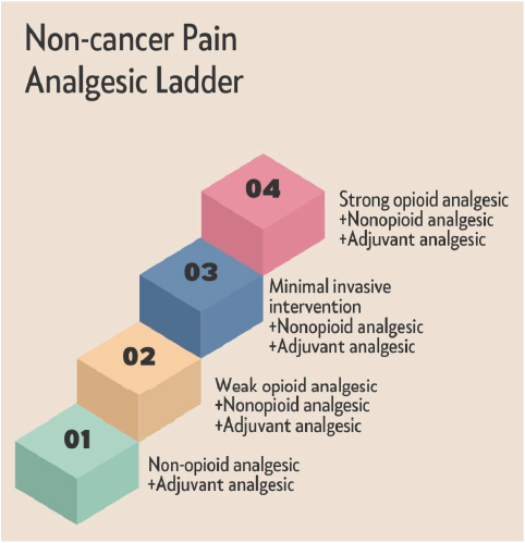
All steps include the use of adjunctive integrative medical therapies such as acupuncture, massage, yoga, relaxation, tai chi, and spinal manipulation for chronic pain management, supporting a holistic approach to pain management. Step 3 of Yang and colleagues’ dosing ladder44 includes minimally invasive interventional therapies, such as nerve block, radiofrequency, spinal cord stimulation, spinal (epidural and subarachnoid) administration of local anesthetics, surgical intervention, and disc decompression. Finally, in step 4, if the above modalities fail, strong opioid medication should be prescribed to the chronic non-cancer pain (CNCP) patient as “the last resort.”44
Topical analgesics. The use of topical analgesics and local anesthetics can reduce wound related pain. These products are particularly useful in mitigating procedural pain resulting from wound dressing changes and debridement. The use of an eutectic mixture of local anesthetics (EMLA) cream such as lidocaine and prilocaine, prior to wound dressing changes has been commonly used to reduce wound related procedural pain.45,46 Such products have utility in the space, although EMLA cream onset of analgesia is slow and may take up to 30 minutes between application and procedure.41
Application of topical sevoflurane on painful wounds produces an intense analgesic effect within a few minutes and lasts for several hours. It has been reported to be effective on both venous and ischemic wounds, both for pain at rest and pain caused by debridement. Sevoflurane acts on the nociceptive nerves inhibiting the transmission of a painful stimulus.47
Medicated dressings. Ibuprofen-impregnated foam dressings may help reduce WRP by providing localized anti-inflammatory effects. The foam dressing releases a continuous low dose of ibuprofen in the presence of wound exudate.48 Evidence suggests that the dressing provides pain relief for exudating acute and chronic wounds of various etiologies and therefore is a safer alternative to systemic pain treatment.49,50
Nonpharmacological. Jebril and colleagues51 published on continuous topical oxygen therapy (cTOT) to treat chronic pain associated with lower leg ulcers. This 20-patient retrospective study noted that adjunctive use of cTOT contributed to a 76% reduction in substantial pain in the study cohort. Additionally, investigators reported that 69% of patients stopped opioid use while 53% had complete pain resolution.51 While the exact mechanism of action is not yet understood, it is widely accepted that higher oxygen levels are required to reverse local hypoxia in wounded tissues and facilitate the host response to wound infection, aid antibiotic effectiveness and wound progression through the healing cascade.52,53 Thus, it stands to reason that the use of supplemental oxygen in wound management may reverse physiological features of chronic wounds affecting tissue oxygenation such as high metabolic activity in the tissues, edema, poor microcirculation, diffusion constraints, and oxygen consumption by bacteria. This initial trial illustrates the potential of topical oxygen therapy as a treatment option to support not only wound healing, but chronic wound pain management.
A systematic review by Peplow and colleagues reported on low-level laser therapy (LLLT) for pain relief and wound healing. LLLT uses red and near-infrared light to enhance the body’s natural healing process.54 The light penetrates the tissue and triggers cellular changes. Though the exact mechanism is unclear, it may involve increased ATP production and low levels of reactive oxygen species which activate transcription factors. LLLT has shown promise in promoting osteogenesis, wound healing, eliminating bacterial biofilms and could decrease pain.55 However, more research is required to establish its effectiveness in wound care and wound-related pain.
Many breakthroughs in nonpharmacological pain management have been in patients with burn wounds due to the immense pain and anxiety associated with this wound type and the multiple types and long duration of interventions required.56 The use of virtual reality (VR) or similar to distract patients during dressing changes has been reported in burns patients with clinically meaningful (33% or greater) reductions in pain during VR distraction.57 This was further confirmed in a meta-analysis of studies that highlighted equivocal evidence for the effect of VR in conjunction with pharmacologic analgesics on reducing anxiety in burn injury patients during wound dressing changes and physiotherapy,58 More recently, Norouzkhani and colleagues59 showed VR significantly decreased pain severity in the intervention group compared to the control group following meta-analysis of 1293 patients with burns across 30 studies. Other nonpharmacological approaches to reduce pain and anxiety include breathing exercise techniques (BET)60 and aromatherapy61 although further larger studies are needed to confirm current findings. Learnings from burn management should be assessed and potentially adapted for other painful wounds going forward.
Dressing changes. WRP is often exacerbated by wound management procedures such as debridement, wound cleansing, and dressing changes. Gardner and colleagues62 reported that wound care procedures such as dressing changes can cause moderate to severe pain in 74% of patients, with nearly half (36%) of those patients experiencing severe pain (rated as 8–10 on a 10-point numeric rating scale) during dressing change.62 A survey conducted by Price and colleagues63 focused on the patients’ perspective on pain during dressing changes. These investigators found that >30% of patients related dressing-related pain some or all of the time. Additionally, 60% of those surveyed reported that dressing-related pain took more than an hour to abate.63
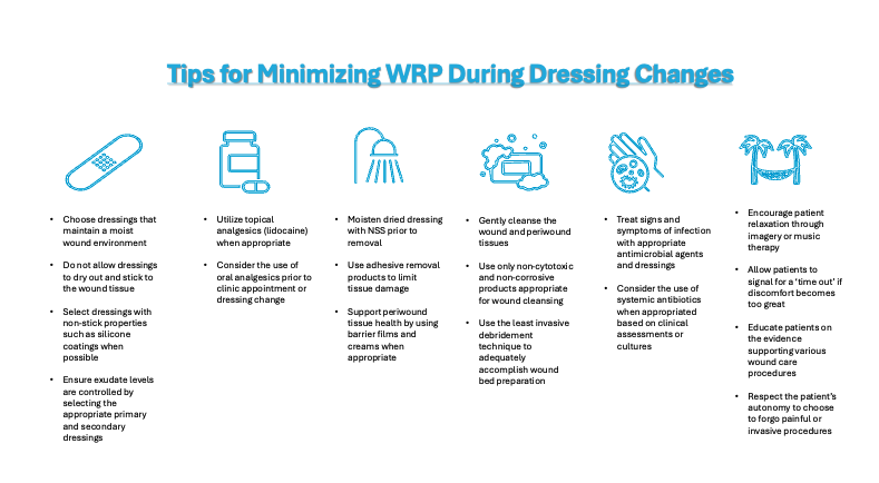
It has been the authors’ experience that variables such as wound size, amount of tissue loss, extent of debridement, wound etiology, cleansing agents, periwound tissue status, and the presence of inflammation or infection can compound patient pain during wound management procedures. A recent systematic review of pain management in resource-limited settings reminds us of the basics that can help to minimize pain, such as use of non-adhesive dressings, introduction of time-out moments, cleaning wounds gently with warm water, treating comorbidities, preserving a moist wound environment, avoiding unnecessary exposure, and protecting surrounding skin).64 Thus, the authors offer the following tips to minimize WRP during dressing changes based on their clinical experience (Figure 3).9
In Conclusion
Pain is a common symptom endured by many chronic wound patients. If not well managed, pain can result in a detrimental cycle of physical and emotional stress that will further derail the wound healing cascade and reduce the patient’s quality of life. Wound care practitioners must be diligent in addressing WRP early in the treatment pathway employing multimodal treatment strategies to maximise comfort and engagement with the patient.
Health care providers should first acknowledge the individual patient’s experience. Everyone experiences pain differently. By employing the most suitable assessment tool for the given situation, clinicians can help acknowledge and validate the patient’s level and severity of pain. Routinely assessing, tracking, and managing pain will assist the clinician in determining the most appropriate treatment pathway throughout the patient journey. Whether it be through pharmacological agents or non-pharmacological modalities, minimizing WRP supports wound healing and increases patients’ QoL in a variety of ways. As our WRP knowledge base grows, so should the available treatment options. Future investigations are necessary to validate new and emerging approaches to WRP.
Windy Cole, DPM, CWSP, is the Director of Wound Care Research at Kent State University College of Podiatric Medicine.
Nicoletta Frescos, PhD, MPH BAppSci(Pod) FWA, is Research Development Lead in Faculty of Pain Medicine at Australian and New Zealand College of Anesthetists (ANZCA).
Emma Woodmansey, PhD, is the Global Clinical Director of Natrox Wound Care in Cambridge, UK.
References
- International Association for the Study of Pain. Definition of Pain. (2020).
- Cohen M, Quintner J, Buchanan D. Is chronic pain a disease?. Pain Med. 2013;14(9):1284-1288. doi:10.1111/pme.12025
- Newbern S. Identifying pain and effects on quality of life from chronic wounds secondary to lower-extremity vascular disease: an integrative review. Adv Skin Wound Care. 2018;31(3):102-108. doi:10.1097/01.ASW.0000530069.82749.e5
- Giordano J. The neuroscience of pain, and a neuroethics of pain care. Neuroethics. 2010; 3:89–94.
- Frescos N. Assessment of persistent pain in chronic lower limb wounds. Doctoral dissertation. (La Trobe University, Victoria, 2019).
- Bechert K, Abraham SE. Pain management and wound care. J Am Col Certif Wound Spec. 2009;1(2):65-71. Published 2009 May 23. doi:10.1016/j.jcws.2008.12.001
- Kim TJ, Freml L, Park SS, Brennan TJ. Lactate concentrations in incisions indicate ischemic-like conditions may contribute to postoperative pain. J Pain. 2007;8(1):59-66. doi:10.1016/j.jpain.2006.06.003
- Leren L, Eide H, Johansen EA, Jelnes R, Ljoså TM. Background pain in persons with chronic leg ulcers: An exploratory study of symptom characteristics and management. Int Wound J. 2022;19(6):1357-1369. doi:10.1111/iwj.13730
- Sabah L, Burian EA, Kirketerp-Møller K, Thomsen SF, Moltke FB. Prevalence and characteristics of pain in patients with lower-extremity ulcers-A cross-sectional study. Wound Repair Regen. Published online January 23, 2024. doi:10.1111/wrr.13153
- Briggs M, Closs SJ. Patients’ perceptions of the impact of treatments and products on their experience of leg ulcer pain. J Wound Care. 2006;15(8):333-337. doi:10.12968/jowc.2006.15.8.26941
- Holtman D, Gahtan V. Peripheral arterial perfusion: is it adequate for wound healing?. Wounds. 2008;20(8):230-235.
- Barron GS, Jacob SE, Kirsner RS. Dermatologic complications of chronic venous disease: medical management and beyond. Ann Vasc Surg. 2007;21(5):652-662. doi:10.1016/j.avsg.2007.07.002
- Price P, Fogh K, Glynn C, Krasner DL, Osterbrink J, Sibbald RG. Managing painful chronic wounds: the Wound Pain Management Model. Int Wound J. 2007;4 Suppl 1(Suppl 1):4-15. doi:10.1111/j.1742-481X.2007.00311.x
- Moffatt CJ, Franks PJ, Hollinworth H. An international perspective on wound pain and trauma. Ostomy Wound Manage. 2003;49(4):12-14.
- Sternberg EM. Neural regulation of innate immunity: a coordinated nonspecific host response to pathogens. Nat Rev Immunol. 2006;6(4):318-328. doi:10.1038/nri1810
- Sussman C, Bates-Johnson B. Wound Care: A Collaborative Practice Manual for Health Professionals. ( Lippincott Willams and Wilkins, Baltimore, 2007).
- Broadbent E, Petrie KJ, Alley PG, Booth RJ. Psychological stress impairs early wound repair following surgery. Psychosom Med. 2003;65(5):865-869. doi:10.1097/01.psy.0000088589.92699.30
- Klein TM, Andrees V, Kirsten N, Protz K, Augustin M, Blome C. Social participation of people with chronic wounds: A systematic review. Int Wound J. 2021;18(3):287-311. doi:10.1111/iwj.13533
- Ribu L, Wahl A. Living with diabetic foot ulcers: a life of fear, restrictions, and pain. Ostomy Wound Manage. 2004;50(2):57-67.
- Proctor WR, Hirdes JP. Pain and cognitive status among nursing home residents in Canada. Pain Res Manag. 2001;6(3):119-125. doi:10.1155/2001/978130
- Caplan Z. U.S. Older Population Grew From 2010 to 2020 at Fastest Rate Since 1880 to 1890. (2023).
- Siersma V, Thorsen H, Holstein PE, et al. Health-related quality of life predicts major amputation and death, but not healing, in people with diabetes presenting with foot ulcers: the Eurodiale study. Diabetes Care. 2014;37(3):694-700. doi:10.2337/dc13-1212
- Karcioglu O, Topacoglu H, Dikme O, Dikme O. A systematic review of the pain scales in adults: Which to use? Am J Emerg Med. 2018;36(4):707-714. doi:10.1016/j.ajem.2018.01.008
- Dickinson AM, Frescos N, Firth JC, Hamblin PS. The characteristics of wound pain associated with diabetes-related foot ulcers: a pilot study. Wound Practice Research. 2016; 24(3):138–148.
- Bengtsson L, Jonsson M, Apelqvist J. Wound-related pain is underestimated in patients with diabetic foot ulcers. J Wound Care. 2008;17(10):433-435. doi:10.12968/jowc.2008.17.10.31306
- Downie WW, Leatham PA, Rhind VM, Wright V, Branco JA, Anderson JA. Studies with pain rating scales. Ann Rheum Dis. 1978;37(4):378-381. doi:10.1136/ard.37.4.378
- Huskisson EC. Measurement of pain. Lancet. 1974;2(7889):1127-1131. doi:10.1016/s0140-6736(74)90884-8
- Wong-Baker FACES pain rating scale.
- Keele KD. The pain chart. Lancet. 1948;2(6514):6-8. doi:10.1016/s0140-6736(48)91787-5
- Fink R. Pain assessment: the cornerstone to optimal pain management. Proc (Bayl Univ Med Cent). 2000;13(3):236-239. doi:10.1080/08998280.2000.11927681
- Melzack R. The McGill Pain Questionnaire: major properties and scoring methods. Pain. 1975;1(3):277-299. doi:10.1016/0304-3959(75)90044-5
- Poquet N, Lin C. The Brief Pain Inventory (BPI). J Physiother. 2016;62(1):52. doi:10.1016/j.jphys.2015.07.001
- Gomarverdi S, Sedighie L, Seifrabiei MA, Nikooseresht M. Comparison of two pain scales: behavioral pain scale and critical-care pain observation tool during invasive and noninvasive procedures in intensive care unit-admitted patients. Iran J Nurs Midwifery Res. 2019;24(2):151-155. doi:10.4103/ijnmr.IJNMR_47_18
- Abbey J, Piller N, De Bellis A, et al. The Abbey pain scale: a 1-minute numerical indicator for people with end-stage dementia. Int J Palliat Nurs. 2004;10(1):6-13. doi:10.12968/ijpn.2004.10.1.12013
- Warden V, Hurley AC, Volicer L. Development and psychometric evaluation of the Pain Assessment in Advanced Dementia (PAINAD) scale. J Am Med Dir Assoc. 2003;4(1):9-15. doi:10.1097/01.JAM.0000043422.31640.F7
- Emflorgo CA. The assessment and treatment of wound pain. J Wound Care. 1999;8(8):384-385. doi:10.12968/jowc.1999.8.8.25911
- Ritland BM, Judkins JL, Naylor JA, Kardouni JR, Pasiakos SM, Jayne JM. The relationship between sleep, pain,and musculoskeletal injuries in US Army Soldiers. BMJ Mil Health. Published online February 15, 2023. doi:10.1136/military-2022-002281
- Buckenmaier CC 3rd, Rupprecht C, McKnight G, et al. Pain following battlefield injury and evacuation: a survey of 110 casualties from the wars in Iraq and Afghanistan. Pain Med. 2009;10(8):1487-1496. doi:10.1111/j.1526-4637.2009.00731.x
- Anekar AA, Hendrix JM, Cascella M. WHO Analgesic Ladder. In: StatPearls. Treasure Island (FL): StatPearls Publishing; April 23, 2023.
- Rosenblum A, Marsch LA, Joseph H, Portenoy RK. Opioids and the treatment of chronic pain: controversies, current status, and future directions. Exp Clin Psychopharmacol. 2008;16(5):405-416. doi:10.1037/a0013628
- Shanmugam VK, Couch KS, McNish S, Amdur RL. Relationship between opioid treatment and rate of healing in chronic wounds. Wound Repair Regen. 2017;25(1):120-130. doi:10.1111/wrr.12496
- Kogure T, Sumitani M, Abe H, et al. Ischemic ulcer pain is both nociceptive and neuropathic pain based on a discriminant function analysis using the McGill Pain Questionnaire. J Pain Palliat Care Pharmacother. 2017;31(2):98-104. doi:10.1080/15360288.2017.1304495
- Frykberg R, Andersen C, Chadwick P, et al. Use of topical oxygen therapy in wound healing. J Wound Care. 2023;32(Sup8b):S1-S32. doi:10.12968/jowc.2023.32.Sup8b.S1
- Yang J, Bauer BA, Wahner-Roedler DL, Chon TY, Xiao L. The Modified WHO Analgesic Ladder: is it appropriate for chronic non-cancer pain? J Pain Res. 2020;13:411-417. Published 2020 Feb 17. doi:10.2147/JPR.S244173
- Briggs M, Nelson EA, Martyn-St James M. Topical agents or dressings for pain in venous leg ulcers. Cochrane Database Syst Rev. 2012;11(11):CD001177. Published 2012 Nov 14. doi:10.1002/14651858.CD001177.pub3
- Purcell A, Buckley T, King J, Moyle W, Marshall AP. Eutectic mixture of local anaesthetics (EMLA®) as a primary dressing on painful chronic leg ulcers: a pilot randomised controlled trial. Pilot Feasibility Stud. 2018;4:123. Published 2018 Jul 7. doi:10.1186/s40814-018-0312-6
- Martínez-Monsalve A, Selva-Sevilla C, Gerónimo-Pardo M. Analgesic effectiveness of topical sevoflurane to perform sharp debridement of painful wounds. J Vasc Surg. 2019;69(5):1532-1537. doi:10.1016/j.jvs.2018.08.175
- Palao i Domenech R, Romanelli M, Tsiftsis DD, et al. Effect of an ibuprofen-releasing foam dressing on wound pain: a real-life RCT. J Wound Care. 2008;17(8):342-348. doi:10.12968/jowc.2008.17.8.30797
- Arapoglou V, Katsenis K, Syrigos KN, et al. Analgesic efficacy of an ibuprofen-releasing foam dressing compared with local best practice for painful exuding wounds. J Wound Care. 2011;20(7):319-325. doi:10.12968/jowc.2011.20.7.319
- Romanelli M, Dini V, Polignano R, Bonadeo P, Maggio G. Ibuprofen slow-release foam dressing reduces wound pain in painful exuding wounds: preliminary findings from an international real-life study. J Dermatolog Treat. 2009;20(1):19-26. doi:10.1080/09546630802178232
- Jebril W, Nowak M, Palin L, Nordgren M, Bachar-Wikstrom E, Wikstrom JD. Topical oxygen treatment relieves pain from hard-to-heal leg ulcers and improves healing: a case series. J Wound Care. 2022;31(1):4-11. doi:10.12968/jowc.2022.31.1.4
- Castilla DM, Liu ZJ, Velazquez OC. Oxygen: implications for wound healing. Adv Wound Care (New Rochelle). 2012;1(6):225-230. doi:10.1089/wound.2011.0319
- Sen CK. Wound healing essentials: let there be oxygen. Wound Repair Regen. 2009;17(1):1-18. doi:10.1111/j.1524-475X.2008.00436.x
- Peplow PV, Chung TY, Baxter GD. Application of low level laser technologies for pain relief and wound healing: overview of scientific bases. Physical Therapy Reviews. 2010; 15:253–285.
- Boateng J, Catanzano O. Advanced therapeutic dressings for effective wound healing--a review. J Pharm Sci. 2015;104(11):3653-3680. doi:10.1002/jps.24610
- Sahin AT, Sahin SY. Influence of burn specific pain anxiety on pain experienced during wound care in adult outpatients with burns. Burns. 2023;49(6):1335-1343. doi:10.1016/j.burns.2022.12.009
- van Twillert B, Bremer M, Faber AW. Computer-generated virtual reality to control pain and anxiety in pediatric and adult burn patients during wound dressing changes. J Burn Care Res. 2007;28(5):694-702. doi:10.1097/BCR.0B013E318148C96F
- Morris LD, Louw QA, Grimmer-Somers K. The effectiveness of virtual reality on reducing pain and anxiety in burn injury patients: a systematic review. Clin J Pain. 2009;25(9):815-826. doi:10.1097/AJP.0b013e3181aaa909
- Norouzkhani N, Chaghian Arani R, Mehrabi H, et al. Effect of virtual reality-based interventions on pain during wound care in burn patients; a systematic review and meta-analysis. Arch Acad Emerg Med. 2022;10(1):e84. Published 2022 Oct 24. doi:10.22037/aaem.v10i1.1756
- Miri S, Hosseini SJ, Takasi P, et al. Effects of breathing exercise techniques on the pain and anxiety of burn patients: A systematic review and meta-analysis. Int Wound J. 2023;20(6):2360-2375. doi:10.1111/iwj.14057
- Farzan R, Firooz M, Ghorbani Vajargah P, et al. Effects of aromatherapy with Rosa damascene and lavender on pain and anxiety of burn patients: A systematic review and meta-analysis. Int Wound J. 2023;20(6):2459-2472. doi:10.1111/iwj.14093
- Gardner SE, Blodgett NP, Hillis SL, et al. HI-TENS reduces moderate-to-severe pain associated with most wound care procedures: a pilot study. Biol Res Nurs. 2014;16(3):310-319. doi:10.1177/1099800413498639
- Price PE, Fagervik-Morton H, Mudge EJ, et al. Dressing-related pain in patients with chronic wounds: an international patient perspective. Int Wound J. 2008;5(2):159-171. doi:10.1111/j.1742-481X.2008.00471.x
- Admassie BM, Ferede YA, Tegegne BA, Lema GF, Admass B A. Wound-related procedural pain management in a resource limited setting: Systematic review. International J Surgery Open. 2022; 47:100549.