Is MIS a Viable Alternative to the Lapidus for Larger Bunions?

Created in partnership with the American Society of Podiatric Surgeons (ASPS)
With advancing technology, new emerging bunion techniques have evolved, the newest concept being innovations in minimally invasive surgery (MIS). The question, however, is, “What procedure should surgeons do for a larger bunion deformity?”
Traditionally, surgeons used the Lapidus and proximal osteotomies of the first metatarsal to correct larger bunion deformities. In the last several years, it has become increasingly common to correct larger bunion deformities with distal MIS osteotomies. Here, the authors review the current literature on correcting larger bunion deformities with both Lapidus and MIS techniques.
 The Literature on the Lapidus
First described by Paul Lapidus in 1934, the Lapidus was originally described as a fusion of the first metatarsocuneiform joint and a fusion of the first and second metatarsal base.1 Modern day Lapidus procedures involve only the fusion of the first metatarsocuneiform joint, but they can also include hardware extending into the cuneiforms if instability is present.2 Advantages of this procedure include the ability to correct the intermetatarsal angle, plantarflex the first metatarsal, and stabilize the medial column. Fusing the first tarsometatarsal joint corrects the center of the bunion deformity, also known as the center of rotation angulation (CORA). Along with transverse and sagittal plane deformity, the Lapidus is a strong corrector of the frontal plane. Kim and colleagues found eighty seven percent of patients with a bunion deformity had a frontal plane deformity.3
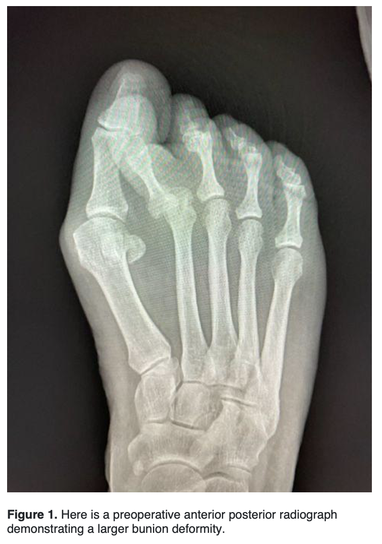
Opponents of the procedure argue that fusion of that joint could lead to early arthritic changes at other joints, higher wound complications due to a larger incision, and the nonunion rate being as high as 12%.4 Despite the complications and newer MIS techniques, the Lapidus continues to be the gold standard for high intermetatarsal angles with hypermobility.
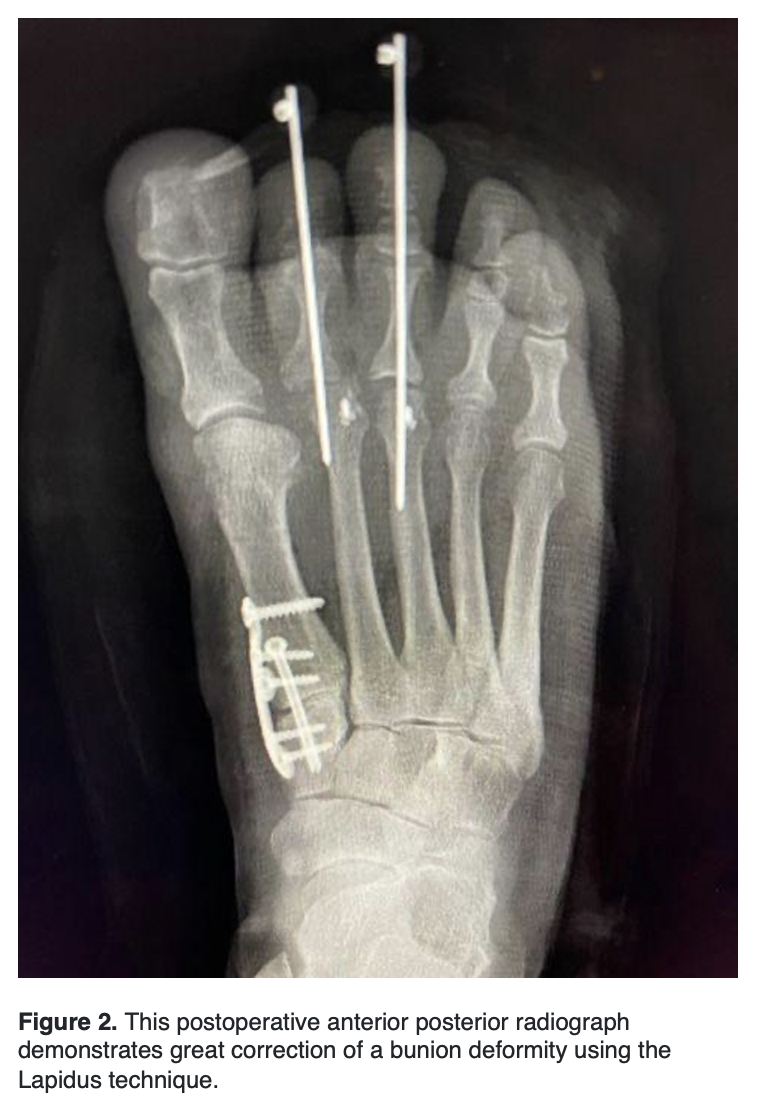 Reports of intermetatarsal angle correction have been as high as 10 degrees. Langdan and colleagues in Foot and Ankle International reported a 6.8 degrees improvement in the IM angle and a fusion rate of 96.7%.5 While comparing crossing screws to a locking plate and plantar compression screw, Saxena and colleagues found a mean decrease of 10.3 degrees in the intermetatarsal angle.6 These studies demonstrate a high degree of correction that surgeons can obtain for larger bunion deformities.
Although nonunion rates have been reported as high as 12%, promising results continue to be seen with the fusion rates. Colleagues in Ohio had 80 patients weight bear two weeks after a Lapidus and found a hundred percent fusion rate a little over 40 days after surgery.7 Patel and colleagues found a 5.3% nonunion rate of the 227 patients undergoing a Lapidus using screw fixation.4 Contrary to traditional fixation, staple fixation has reported success with union rates higher than 92% after undergoing a Lapidus.8 With the addition of Kim’s study demonstrating metatarsal pronation averaging more than 21.9 degrees in bunion feet, more literature has come out regarding correction of the third plane. Dayton and colleagues found an 18.7-degree change in the proximal articular set angle (PASA), a vital component of the frontal plane. Without breaking the bone, the Lapidus procedure can translate, rotate, and shift.9 These are all essential parts to correcting a bunion deformity at its underlying deformity.
Despite the potential complications of nonunion, the Lapidus is still the procedure of choice for larger bunion deformities.
 How Surgeons Have Used MIS in Recent Years
Minimally invasive bunion surgery is relatively new in the United States as the required burrs currently used were only approved by the FDA in 2017.10 Minimally invasive surgery involves little soft tissue dissection with bony correction while correcting the bunion deformity. Benefits of this technique include greater cosmesis results, minimal soft tissue dissection, shorter recovery period, and great patient satisfaction. It has been established that MIS technique is comparable to open technique in a randomized control trials comparing American Orthopedic Foot and Ankle Scores (AOFAS), visual analogue scale (VAS) and pain scores 5 years postoperatively.11 Opponents of this technique warn of complications such as nonunion, malunion, infection, and soft tissue irritation.12 Most of the soft tissue irritation occurs at the distal medial corner of the metatarsal shaft that is usually not shaved after performing the osteotomy, but the reported incidence is low.13
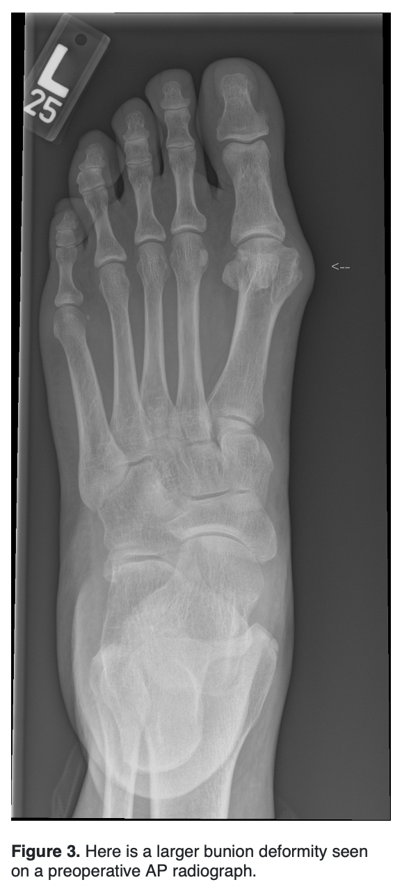
Severe hallux valgus is categorized as an intermetatarsal (IM) angle of greater than or equal to 20 degrees and a hallux valgus angle (HAV) of greater than 40 degrees.
In 2011, De Lavigne looked at 6 patients who underwent percutaneous double osteotomy for severe hallux valgus deformity.14 With a minimum 2-year follow-up, AOFAS scores improved from 34 to 84, postoperative radiological assessment showed significant improvement, no complications were encountered, and all patients were satisfied.
In 2016, Veronis and Redfern described correcting severe hallux valgus deformity with percutaneous procedures. The procedures described later became the currently used third generation of minimally invasive surgery for hallux valgus known as minimally invasive chevron-Akin (MICA). They focus heavily on evaluation of the width between the lateral first metatarsal head and medial second metatarsal head. They recommend 100% displacement as the maximum shift, which offers a reliable technique for all deformities. They report a large IM angle with a small metatarsal head as a limitation to this technique. For this pathology, they recommend a percutaneous basal closing wedge osteotomy. Veronis goes further suggesting that weight-bearing radiographs undervalue the instability of the tarsometatarsal (TMT) joint. He recommends a clinical squeeze test to assess the true potential IM angle. This allows the surgeon to determine the required lateral displacement of the metatarsal head to correct the deformity.15
In 2021, Lewis and colleagues looked at 53 feet with severe hallux who underwent percutaneous chevron and Akin (PECA) with a mean follow-up of 3 years.16 There was a statistically significant decrease in IM angle from 17.5 to 5.1 and HV angle from 44.1 to 11.5 degrees. All patients were satisfied with their outcome and 76.8% were highly satisfied. The recurrence rate was 7.5%. This recurrence rate is similar to open surgery for severe hallux valgus (2.7%-16%).17
 In 2022, Lewis and colleagues looked at 106 patients prospectively undergoing third generation MICA with severe hallux valgus deformity. They looked at IM angles and HVA before and 6 weeks after surgery, as well as complications and clinical passive ranges of motion (PROM) 2 years following surgery. They found the mean IMA improved from 18.2 to 6.3 and the mean HVA improved from 45.3 to 10.9. They had a complication rate of 18.8% and screw removal rate of 5.6%. Two-year PROMs scores significantly improved for pain, walking and standing, and social interaction from 39.2 to 7.5, 38.2 to 5.9 and 48.6 to 5.5 respectively. The largest limitation to this study was the lack of long-term radiographic recurrence assessment.18
There is limited literature specifically looking at severe hallux valgus deformities (IM > 20 degrees, HV > 40 degrees) with minimally invasive surgical techniques. However, the technique currently utilized was first described only 6 years ago. The title of the paper first describing the widely used technique was “Percutaneous Surgery for Severe Hallux Valgus” indicating the technique was first utilized for large deformity.15 More literature is needed looking specifically at this subset of patients with hallux abductovalgus.
Closing Thoughts
In conclusion, the Lapidus procedure continues to be at the forefront of larger bunion deformities as it significantly lowers the IM angle while correcting sagittal and frontal plane deformities. However, as MIS surgery continues to evolve with the technique, it too can correct in multiple planes in larger bunion deformities.
Dr. Hoffler is a Fellow at the Southeast Permanente Foot & Ankle Trauma & Reconstructive Fellowship Program at the Southeast Permanente Medical Group in Atlanta.
Dr. King is in private practice at Ankle and Foot Centers of America in Nashville.
Dr. Black is an attending in the Southeast Permanente Foot & Ankle Trauma & Reconstructive Fellowship Program at the Southeast Permanente Medical Group in Atlanta.
References
1. Lapidus PW. The operative correction of the metatarsus varus primus in hallux valgus. Surg Gynecol Obstet. 1934; 58:183-1-7
2. Fleming JJ, Kwaadu KY, Brinkley JC, Ozuzu Y. Intraoperative evaluation of medial intercuneiform instability after Lapidus arthrodesis: intercuneiform hook test. J Foot Ankle Surg. 2015; 54(3):464-472.
3. Kim Y, Kim SK, Young KW, et al. A new measure of tibial sesamoid position in hallux valgus in relation to coronal rotation of the first metatarsal in CT scans. Foot Ankle Int. 2015;36(8):944-952.
4. Patel S, Ford LA, Etcheverry J, Rush SM, Hamilton GA. Modified lapidus arthrodesis: rate of nonunion in 227 cases. J Foot Ankle Surg. 2004 Jan-Feb;43(1):37-42.
5. Langan TM, Greschner JM, Brandão RA, Goss DA Jr, Smith CN, Hyer CF. Maintenance of correction of the modified Lapidus procedure with a first metatarsal to intermediate cuneiform cross-screw technique. Foot Ankle Int. 2020 Apr;41(4):428-436.
6. Saxena A, Nguyen A, Nelsen E. Lapidus bunionectomy: Early evaluation of crossed lag screws versus locking plate with plantar lag screw. J Foot Ankle Surg. 2009 Mar-Apr;48(2):170-9.
7. Blitz NM, Lee T, Williams K, Barkan H, DiDimenico LA. Early weight bearing after modified lapidus arthrodesis: a multicenter review of 80 cases. J Foot Ankle Surg. 2010 Jul-Aug;49(4):357-62.
8. Mallette JP, Glenn CL, Glod DJ. The incidence of nonunion after Lapidus arthrodesis using staple fixation. J Foot Ankle Surg. 2014 May-Jun;53(3):303-6.
9. Dayton P, Feilmeier M, Kauwe M, Hirschi J. Relationship of frontal plane rotation of first metatarsal to proximal articular set angle and hallux alignment in patients undergoing tarsometatarsal arthrodesis for hallux abducto valgus: a case series and critical review of the literature. J Foot Ankle Surg. 2013 May-Jun;52(3):348-54.
10. Cody EA, Caolo K, Ellis SJ, Johnson AH. Early Radiographic outcomes of minimally invasive chevron bunionectomy compared to the modified Lapidus procedure. Foot Ankle Orthop. 2022 Jul 21;7(3):24730114221112103.
11. Kaufmann G, Mörtlbauer L, Hofer-Picout P, Dammerer D, Ban M, Liebensteiner M. Five-year follow-up of minimally invasive distal metatarsal chevron osteotomy in comparison with the open technique: A randomized controlled trial. J Bone Joint Surg Am. 2020 May 20;102(10):873-879.
12. Kadakia AR, Smerek JP, Myerson MS. Radiographic results after percutaneous distal metatarsal osteotomy for correction of hallux valgus deformity. Foot Ankle Int. 2007;28(3):355-60.
13. Bosch P, Wanke S, Legenstein R. Hallux valgus correction by the method of Bosch: a new technique with a seven-to-ten-year follow-up. Foot Ankle Clin. 2000;5(3):485-98
14. De Lavigne C, Rasmont Q, Hoang B. Percutaneous double metatarsal osteotomy for correction of severe hallux valgus deformity. Acta Orthop Belg. 2011 Aug;77(4):516-21.
15. Vernois J, Redfern DJ. Percutaneous surgery for severe hallux valgus. Foot Ankle Clin. 2016 Sep;21(3):479-93.
16. Lewis TL, Ray R, Robinson P, Dearden PMC, Goff TJ, Watt C, Lam P. Percutaneous chevron and Akin (PECA) osteotomies for severe hallux valgus deformity with mean 3-year follow-up. Foot Ankle Int. 2021 Oct;42(10):1231-1240.
17. Raikin SM, Miller AG, Daniel J. Recurrence of hallux valgus: A review. Foot Ankle Clin. 2014 Jun;19(2):259-74.
18. Lewis TL, Ray R, Gordon DJ. Minimally invasive surgery for severe hallux valgus in 106 feet. Foot Ankle Surg. 2022 Jun;28(4):503-509.