


Reconstructive Surgical Option for a Comminuted Fifth Metatarsal Fracture
Fractures of the fifth metatarsal are commonly encountered injuries sustained in the foot and have been abundantly documented in trauma literature and surgical textbooks.1–3 A spiral oblique fracture of the fifth metatarsal shaft, also known as a dancer’s fracture, is often observed with a butterfly fragment.4 Due to the configuration and inherent instability of this fracture pattern, surgical intervention is typically warranted.4 Multiple techniques have been reported and utilized for open reduction and internal fixation (ORIF) of fifth metatarsal fractures, particularly of the base.3
The literature is not as robust for revisional surgeries for refracture or hardware failure after prior ORIF of fifth metatarsal shaft fractures in the setting of hardware failure, osteopenia due to non-weight-bearing status, and advanced comminution. While some research suggests a conservative approach to diaphyseal fifth metatarsal fractures, several practitioners continue to opt for surgical intervention in these cases.
The authors present the intraoperative surgical techniques, materials, and pearls that may be utilized in the future for similar revisional surgical cases.
Case Report: A Displaced Fifth Metatarsal Fracture of the Shaft
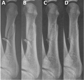
A 58-year-old female with past medical history of gastroesophageal reflux disease (GERD), hypothyroidism, and bronchitis presented to the podiatry clinic with chief complaint of pain to the lateral aspect of the right foot after suffering a fall. She was a former smoker with a one-pack-per-day, 18-pack-year history, having quit 20 years ago. On exam, the patient had palpable 2/4 pedal pulses and no neurological deficits. Ecchymosis and swelling were noted to the lateral aspect of the foot. X-rays were obtained, demonstrating a closed, displaced fracture of the fifth metatarsal shaft with a butterfly fragment and shortening (Figure 1).
The treatment plan consisted of surgical intervention due to the nature of the fracture. Appropriate ORIF was performed utilizing a lag screw and neutralization plate construct (Figure 2). The patient was instructed to remain non-weightbearing to the surgical foot.
The patient progressed well in the postsurgical phase with no significant initial events. About 3 weeks after the surgery, the patient presented to the office with acute pain and swelling after bearing weight on the surgical foot. X-rays were obtained, showing loosening and failure of the hardware with increased diastasis across the previous fracture fragments (Figure 3). The decision was made for revisional surgery and repeat ORIF of the fracture.
Intraoperatively, a longitudinal incision was placed utilizing the previous incision site. Dissection was carried through the deeper tissues until the hardware was encountered and removed. Significant comminution and displacement of the fracture fragments was appreciated as well as osteopenic changes of the metatarsal (Figure 4). Appropriate reduction and temporary fixation proved to be difficult due to these changes.
The decision was made to attempt reconstitution of the fifth metatarsal and large defects secondary to the repeat fracture. An injectable bone substitute (HydroSet®, Stryker Corporation) was used to fill the various defects between the fracture fragments and remodel the shaft and head of the fifth metatarsal as close to anatomical position as possible (Figure 5). The HydroSet was allowed to harden and convert to hydroxyapatite prior to fixation being attempted as per manufacturer recommendations. A 3-0 Vicryl suture was tied circumferentially around the distal shaft of the fifth metatarsal in order to further secure the bone fragments and HydroSet together. The bone substitute was allowed to set for approximately eight minutes per manufacturer and company representative suggestion.
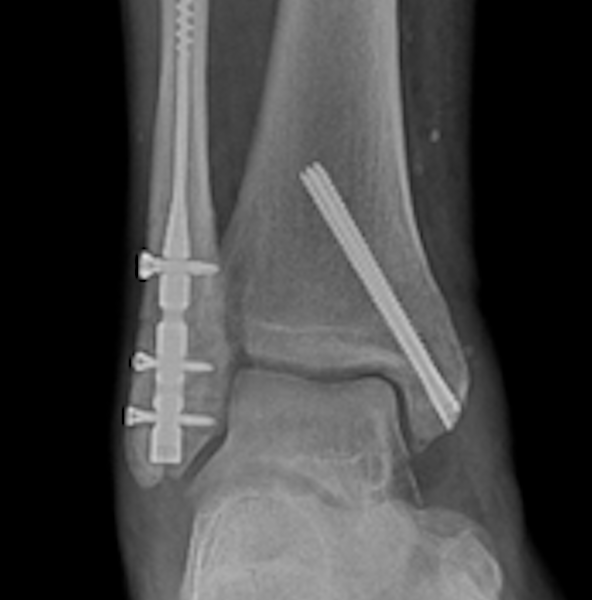
At this time, a 7-hole Y-plate was applied to the dorsal aspect of the remodeled fifth metatarsal and was fixated with locking screws. Following this, a 6-hole straight plate was applied to the lateral aspect of the fifth metatarsal with locking screws, avoiding the site of most comminution and reconstruction via bone substitute (Figure 6). The surgical site was appropriately irrigated and closed, and the extremity was splinted. Immediate postoperative films demonstrated the metatarsal cage construct (Figure 7). The patient was given instructions again for strict non-weight-bearing with the use of crutches and/or a knee roller.
The patient progressed appropriately during the postoperative course. She remained non-weight-bearing initially with frequent evaluation and short leg cast changes. Progressive advancement of weight-bearing status was tolerated well. The metatarsal cage remained intact due to patient compliance and construct rigidity, and the metatarsal continued to show bone callus formation on follow up radiographs and visits (Figure 8). The patient had a minor postoperative complication of distal wound dehiscence that was treated with local wound care until resolution. The patient was informed about potential discomfort from prominent hardware and returned several months for hardware removal without complications (Figure 9).
What You Should Know About Fifth Metatarsal Fractures
Fractures of the fifth metatarsal are common injuries sustained in the foot and have been abundantly documented in trauma literature and surgical textbooks.1–3 A spiral oblique fracture of the fifth metatarsal shaft, also known as a dancer’s fracture, is frequently observed with a butterfly fragment.4 The configuration and inherent instability of this fracture pattern often warrants surgical intervention.3,4
As reported, initial fixation was conducted with a lag screw and neutralization plate implementing standard AO principles. In the revision ORIF, significant comminution and advanced osteopenia was noted given the repeat fracture, previous sites of screw fixation, and non-weightbearing status of the patient’s extremity. Bone substitute and grafting has been described in the literature for use in comminuted fracture patterns to augment fracture healing in long bones and was implemented in this surgical scenario.5 Bone substitute and grafting has also been shown to improve outcomes in complex fractures.6
While typically used as supplementation, the HydroSet injectable bone matrix was utilized in the reported case to partially remodel the fifth metatarsal shaft and head to anatomic position and fill the large defects present within the damaged bone. The HydroSet tetra-calcium phosphate crystalline structure self-sets into hydroxyapatite and allows both osteoconductive and osteointegrative properties.7 In the authors’ experience, allowing the HydroSet to set for longer than the suggested eight minutes after implantation will let the crystalline structure fully develop, allowing for better drilling and screw fixation.
The authors recommend a stronger fixation construct in revision fifth metatarsal surgeries that exhibit comminution, osteopenia and large osseous defects: utilization of a dorsal locking plate enhanced with a lateral locking plate, resulting in a strong metatarsal cage construct. Double plate techniques have been described and implemented with good results for comminuted distal fibular fractures for additional rigidity and decreased risk of fixation failure.8,9 This construct has also been reported with good outcomes for fixation of a comminuted second metatarsal fracture in an overweight and diabetic patient.9 Plantar plating via a compression plate at the tension side of the tension side of proximal fifth metatarsal fractures has been described in elite athletes to address rotational forces and plantar lateral gapping—this technique may be incorporated into the dual plate construct.10 Additionally, cerclage wire or larger diameter suture may be utilized around the metatarsal for circumferential compression to further add stability to constructs.
Conclusion
Reconstitution and remodeling of fifth metatarsal fractures featuring large defects, advanced osteopenia, and significant comminution can be appropriately conducted with an injectable bone substitute. A dual locking plate metatarsal cage construct is a viable and replicable option for stable ORIF with the described pattern of injury. The authors recommend thorough evaluation of patients undergoing revisional surgery and determining the best possible intervention and techniques for each individual case, and additionally weigh the options of conservative versus surgical intervention.
Dr. Irfan is a Fellow at Foot and Ankle Specialists of Central Ohio.
Dr. Patel is affiliated with Henry Ford Health in Trenton, MI.
References
1. Buddecke DE, Polk MA, Barp EA. Metatarsal fractures. Clin Podiatr Med Surg. 2010 Oct;27(4):601–24. doi: 10.1016/j.cpm.2010.07.001. PMID: 20934107.
2. Beck M, Wichelhaus A, Rotter R, Gierer P, Mittlmeier T. Mittel- und Vorfußfrakturen [Metatarsal and toe fractures]. Unfallchirurg. 2019 Apr;122(4):309–322. German. doi: 10.1007/s00113-019-0620-1. PMID: 30847497.
3. Vance DD, Vosseller JT. Double plating of distal fibula fractures. Foot Ankle Spec. 2017 Dec;10(6):543–546. doi: 10.1177/1938640017692416. Epub 2017 Feb 7. PMID: 28173717.
4. Carter S. Distal Fractures of the Fifth Metatarsal. Podiatry Institute. 1996. http://www.podiatryinstitute.com/pdfs/Update_1996/1996_40.pdf
5. Ozer K, Chung K. The use of bone grafts and substitutes in the treatment of distal radial fractures. Hand Clinics. 2012; 28(2):217–223.
6. Ollivier M, Bulaïd Y, Jacquet C, Pesenti S, Argenson JN, Parratte S. Fixation augmentation using calcium-phosphate bone substitute improves outcomes of complex tibial plateau fractures. A matched, cohort study. Int Orthop. 2018 Dec;42(12):2915–2923. doi: 10.1007/s00264-018-3926-7. Epub 2018 Apr 7. PMID: 29627848.
7. Lee M, Ho L. Metatarsal fractures. In: McGlamrys Comprehensive Textbook of Foot and Ankle Surgery. Vol 1. 4th ed. Chapter 104. Lippincott, Williams & Wilkins; 2013:1658–1660
8. Singh SK, Wilson MG. A double plate technique for the management of difficult fibula fractures. Techniques Foot Ankle Surg. 2005; 4(4):235–239. doi: 10.1097/01.btf.0000176001.88953.fb
9. Chesser A, Saltrick K. Dual plating technique for comminuted second metatarsal fracture in the diabetic obese patient: A case report. Foot and Ankle Online Journal: 2017
10. Mitchell RJ, Duplantier NL, Delgado DA, Lambert BS, McCulloch PC, Harris JD, Varner KE. Plantar plating for the treatment of proximal fifth metatarsal fractures in elite athletes. Orthopedics. 2017 May 1;40(3):e563–e566. doi: 10.3928/01477447-20170327-04. Epub 2017 Mar 31. PMID: 28358977.


