


Tibio-Calcaneal Arthrodesis Using a Femoral Head Allograft and a Proximal Humerus Locking Plate For Talar Extrusion
© 2023 HMP Global. All Rights Reserved.
Any views and opinions expressed are those of the author(s) and/or participants and do not necessarily reflect the views, policy, or position of Podiatry Today or HMP Global, their employees, and affiliates.
Complete extrusion of the talus is an uncommon injury with controversy as to its optimal management. Definitive treatment ultimately depends on the extent of the extruded talus as well as recovery of the talus and retention of the talar head and neck.1 When re-implantation takes place, infection, talar subsidence, and post-traumatic arthritis are commonly encountered sequelae. However, reimplantation of the talus, when possible, helps maintain limb length for future reconstructive procedures which coincides with improved functional outcomes.2
When complete extrusion of the talus occurs, and re-implantation of the native talus is not possible, surgical reconstruction pathways vary widely. There are descriptions of tibio-calcaneal arthrodesis using an intramedullary nail,3 a number of plating systems,4–6 and even autogenous fibular onlay.7 Various techniques to maintain limb length also exist, some of which include custom titanium trusses filled with bone graft,8 femoral head allografts,9 and more recently total talus replacement.10
Proximal humerus locking plates for complex hindfoot fusions yield a fusion rate of 85-95%.11–13 However, these small case series did not use large structural allografts. In this case report we discuss the surgical technique and outcome for a tibio-calcaneal arthrodesis with a femoral head allograft and a proximal humerus locking plate.
When A Motor Vehicle Accident Results in Total Talar Extrusion
The primary surgeon was consulted on the case of a 55-year-old African American male who suffered an epileptic seizure and a resultant high speed motor vehicle accident. He was ejected from the vehicle and sustained a complete extrusion of the right talus in February 2020. The talus was not recoverable from the site of the accident. After a comprehensive evaluation of the injury the patient was taken for immediate surgical washout and exploration of his wound and an antibiotic cement spacer and external fixator were applied (Figure 1) with plans for more definitive fixation once his acute condition had stabilized.
During his admission the patient insisted the external fixator be removed. Once the external fixator was removed, he left the hospital against medical advice with his sutures and antibiotic spacer in place. He was lost to follow up for 2 years and admitted to walking on the limb with the assistance of a walker that he obtained from a friend.
The patient then presented March 2022 complaining of continued pain to the right ankle requesting surgical correction. The surgical reconstruction and postoperative period then occurred between March and October 2022. During preoperative reevaluation the patient reported a past medical history of epilepsy for which he was taking carbamazepine. He denied allergies and pertinent family or past surgical history other than the acute injury for which he had been treated 2 years prior. On physical exam he complained of significant pain on palpation and attempted range of motion at the right ankle and subtalar joint. Preoperative radiographs were obtained and significant post-traumatic arthrosis involving the articular surface of the calcaneus, tibia, and navicular was appreciated. The patient was considered a poor candidate for total talus replacement and the surgeon elected to remove the antibiotic cement spacer and perform a tibio-calcaneal arthrodesis with a femoral head allograft to maintain limb length.
Key Notes on the Surgical technique
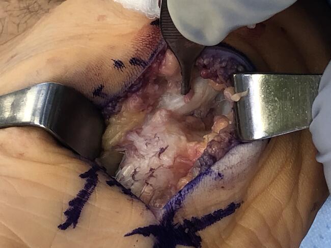
The team placed the patient on the operating room table in the supine position with a bump under the ipsilateral hip. The patient underwent general anesthesia and received a popliteal nerve block by the anesthesia team after which a pneumatic thigh tourniquet was applied to the thigh for hemostasis. Aseptic preparation and draping then took place. First, the surgeon harvested bone marrow aspirate from the tibial. Then the surgeon created a 17-cm full-thickness linear incision over the lateral aspect of the fibula coursing distally to the tip of the lateral malleolus taking care to identify and retract the superficial peroneal nerve anteriorly. An incision was then carried out down to bone over the fibula in line with the skin incision. A periosteal elevator was then used to help reflect the periosteum from the fibula and a sagittal saw was then utilized to create an osteotomy in the fibula approximately 8 cm proximal to the tibial plafond. The surgeon excised the fibula and passed to the back table to be morselized for autograft. He then sections and removed the antibiotic cement spacer from the ankle, taking care not to violate the medial malleolus, posterior tibial artery, or tibial nerve. The articular surfaces of the tibial plafond along with the posterior, middle, and anterior facets of the calcaneus then underwent preparation for arthrodesis by debriding any eburnated subchondral bone, and any remaining articular cartilage with curettes, osteotomes, and rongeurs. The surgeon fenestrated any remaining subchondral bone with a small drill and fish-scaled with an osteotome.
The surgeon then cut a thawed femoral head allograft to proper dimensions to fit the arthrodesis site, later fenestrating and soaking it in bone marrow aspirate (Figure 2). The surgeon then introduced the allograft into the arthrodesis site. The ankle joint and subtalar joint were then reduced with the ankle joint in neutral and the subtalar joint in 3 to 5 degrees of valgus. Two 4.5-mm, partially threaded, headless compression screws inserted across the medial malleolus into the femoral head allograft secured the position of the ankle joint and added compression along the medial gutter. All small areas of gapping between the bone allograft interface were then packed with bone marrow aspirate soaked morselized fibular autograft. A 12-hole reverse proximal humerus locking plate was then placed over the lateral tibia and calcaneus and filled with 4.0-mm locking screws (Figure 3).
Pertinent Aspects of the Postop Course
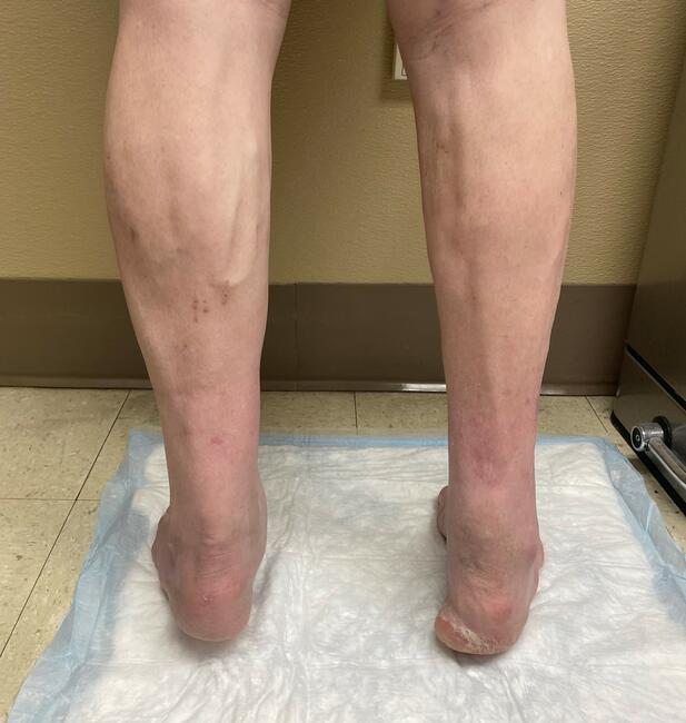
Immobilization in a posterior splint began immediately postoperatively and progressed to non-weight-bearing in a boot at the patient's 2-week post-op appointment. Non-weight-bearing continued for a total of 12 weeks, after which he began protected weight-bearing in a boot for an additional 6 weeks. Cortical bridging on plain film radiographs was appreciated at 12 weeks and a postoperative CT was obtained at week 20, which demonstrated 70% fusion of the tibia-allograft interface and 85% fusion along the calcaneus-allograft interface. At 6 months post-op the patient had no pain and was able to bear weight in normal shoe gear.
Points to Consider
Severe deformity of the rearfoot is difficult to address using more traditional fixation constructs, such as compression screws or traditional plating. This has led to the development of more rigid fixation techniques, such as intramedullary nailing, blade plate fixation, and the use of locking plates.12
Locking plates act as fixed angle devices in multiple planes and have shown benefits when compared with traditional plating techniques, in particular, in patients with highly comminuted, unstable, and osteoporotic fractures. The screws lock into the plate itself and may lead to less overall contact pressure and reduced periosteal devascularization.14
According to Buck and colleagues, valgus position of the ankle arthrodesis is more advantageous and helps provide a more normal gait, particularly on uneven ground. The ideal position for fusion of the ankle was found to be neutral in flexion and slight (0 to 5 degrees) in valgus angulation of the hindfoot.15 The surgeon has found that one of the difficulties in performing this surgery is maintaining the rectus to slight valgus alignment of the calcaneus when inserting a large structural allograft from the lateral aspect of the ankle. The proximal humerus locking plate is designed with a bend to accommodate the greater tuberosity of the humerus in humeral neck fractures. Kim and colleagues determine that the bend in the plate to accommodate for the greater tuberosity equates to 8 degrees.16 However, if you measure the angle between the bend and the most distal aspect of the plate where there is a small flare this translate to approximately 5 degrees of valgus when used for hindfoot fusions and helps prevent varus alignment of the rearfoot when the calcaneus can be reduced and locked to the plate.
O’Neil and colleagues compared the rigidity of locking plates to intramedullary nail fixation for the use of tibio-talo-calcaneal (TTC) arthrodesis.17 They found no difference in the stability of TTC fusions fixed with an augmented intramedulary nail or a locking plate and concluded that locking plates are an acceptable alternative to IM nails.
A major issue we find in these complex cases is the presence of bony gaps resulting from the severe bone loss, which results in the need for grafting. Locking plates might be advantageous in that the locking screws are less likely to loosen from the plate even in the presence of a bony gap or an unstable graft. This could decrease the incidence of problems resulting from hardware loosening, which can increase the risk of inflammation, infection, and nonunion.14 Finally, placement of the screws from different angles and directions provides multiplanar stability.
Final Thoughts
While the use of proximal humerus locking plates for complex hindfoot fusions has been adequately described,18 our review of current literature revealed no description of the use of these plates in combination with large structural allografts. We have concluded that the use of proximal humerus locking plates with combined femoral head allograft is a viable option for limb salvage and surgical reconstruction following talar extrusion.
Dr. Duffin is a third-year podiatric resident at Hunt Regional Medical Center in Greenville Texas.
Dr. Lampe is an Attending Physician at the Hunt Regional Medical Center residency program and is Chief of Podiatry at Dallas Methodist Hospital in Dallas Texas.
References
1. Schuberth JM, Jennings MM. Reconstruction of the extruded talus with large allograft interfaces; a report of 3 cases. J Foot Ankle Surg. 2008:47(5)476-82.
2. Smith CS, Nork SE, Sangeorzan BJ, The extruded talus: results of reimplantation. J Bone Joint. Surg. 2006; 88(11):2418-24.
3. Jaffe KA, Conlan TK, Sardis L, Meyer RD. Traumatic talectomy without fracture: four case reports and review of the literature. Foot Ankle Int. 1995;16(9): 583-587.
4. Alvarez R, Gaines D, Easley ME. Tibiocalcaneal arthrodesis using blade plate fixation. In: Operative Techniques in Orthopaedic Surgery. Lippincott Williams & Wilkins; 2011: 4179-4190.
5. DiDomenico LA, Wargo-Dorsey M. Tibiotalocalcaneal arthrodesis using a femoral locking plate. J Foot Ankle Surg. 2012;51(1):128–138.
6. Lui TH. Tibiotalocalcaneal arthrodesis with combined retrograde intramedullary nail and lateral L-plate. J Foot Ankle Surg. 2012;51(5):693–695.
7. Ley D, Hassan M, DiDomenico L. Can biological fibular plates provide viable fixation for tibiocalcaneal arthrodesis? Podiatry Today. Published April 2020. Accessed May 30, 2023.
8. Lachman J, Adams S. Tibiotalocalcaneal arthrodesis for severe talar avascular necrosis. Foot Ankle Clin. 2019;24(1):143-161.
9. Cuttica DJ, Hyer CF. Femoral head allograft for tibiotalocalcaneal fusion using a cup and cone reamer technique. J Foot Ankle Surg. 2010; 50(1): 126–129.
10. West TA, Rush SM. Total talus replacement: case series and literature review. J Foot Ankle Surg. 2021; 60(1):187–193.
11. Shearman AD, Kyriakos L, Patel A, Pradhan R, Rosenfeld P. Use of a proximal humeral locking plate for complex ankle and hindfoot fusion. J Foot Ankle Surg. 2016;55(3):612–618.
12. Ahmad J, Pour AE, Raikin SM. The modified use of a proximal humeral locking plate for tibiotalocalcaneal arthrodesis. Foot Ankle Int. 2007;28(9):977-983.
13. Fan J, Zhang X, Luo Y, You GW, Ng WK, Yang YF. Tibiotalocalcaneal (TTC) arthrodesis with reverse PHILOS plate and medial cannulated screws with lateral approach. BMC Musculoskelet Disord. Available at: https://bmcmusculoskeletdisord.biomedcentral.com/articles/10.1186/s12891-017-1666-2. Published 2017. Accessed Sept 29, 2022.
14. Greiwe RM, Archdeacon MT. Locking plate technology: current concepts. J Knee Surg. 2007;20:50–55.
15. Buck P, Morrey BF, Chao EY. The optimum position of arthrodesis of the ankle. A gait study of the knee and ankle. J Bone Joint Surg Am. 1987 Sep;69(7):1052-62.
16. Kim H, Chung YG, Jang J, Kim Y, Park S, Song HS. Why locking plates for the proximal humerus do not fit well. Arch Orthoped Trauma Surg. 2022;142. 1-8. 10.1007/s00402-020-03676-0.
17. O'Neill PJ, Logel KJ, Parks BG, Schon LC. Rigidity comparison of locking plate and intramedullary fixation for tibiotalocalcaneal arthrodesis. Foot Ankle Int. 2008; 29(6):581-586.


