The Top 10 Contact Allergens In Footwear
Allergic contact dermatitis (ACD) caused by exposure to footwear may occur from a variety of agents found in any part of the shoe. Also known as shoe dermatitis, the most common allergens are leather, rubber, and adhesives.1-5 One must also not overlook socks as a source of allergens, as reports exist of patients reacting to rubbers and dyes in socks.3 Shoe dermatitis can present as either an acute or chronic reaction. Clinicans can use pruritus and lesion severity as differentiating markers for mimickers, including tinea pedis and lichen planus.2,6 The impact of shoe dermatitis is not limited to particular races or cultures. It is essential to identify the offending agent or agents to help patients avoid further exposure, a critical initial step in treating ACD.1-5,7-11 If conservative treatments are not effective, available pharmacologic agents may ease the burden of this skin disease.2,8,12
The terms “dermatitis” and “eczema” are interchangeable with one another, and represent a blanket term that is not specific. A form of dermatitis, contact dermatitis (CD) is a broad term for the inflammatory skin reaction due to direct contact with a noxious agent in the environment.2,6 CD includes both non-immunologic irritant contact dermatitis (ICD) and immunologically mediated allergic contact dermatitis (ACD).2,6 This article will focus on ACD specifically related to footwear.
Various allergens cause ACD, and the causative allergens vary with the population considered. Among the various possible allergens, top contributors are often included in the shoe materials, such as rubber, dye, and fragrances.1-5 Studies show that throughout the history of shoe dermatitis, allergens with the highest number of positive patch test results are potassium dichromate, mercapto mix, thiuram mix, and fragrance mix.13,14 One study showed that among the British Contact Dermatitis Society Standard series, potassium dichromate was the most prevalent cause of contact dermatitis with the highest number of populations reacting to patch test, followed by neomycin, mercapto mix, and thiuram mix, in decreasing order.1
With new shoe products on the market, research identified a number of new allergens as causes of ACD.15-17 Dimethyl-thiocarbamylbenzothiazole sulfide (DMTBS) is a material used in the newer canvas Sperry® boat shoe model. After analyzing the shoe extracts, patch testing with varying degrees of dilution showed positive results with symptoms increasing in severity as the concentration of DMTBS increased.15
Shoe refreshers are another new allergen causing shoe dermatitis, with positive patch test results shown with diluted forms.16 In a patch test study using a sample of sports shin pad products, acetophenone azine showed positive results in both pediatric and adult populations.17 Other common contributory allergens of shoe dermatitis in India include leather and leather-related chemicals, cobalt chloride, paraphenylenediamine, epoxy resin, nickel sulfate, colophony, p-tert-butyle-formaldehyde resin, and formaldehyde.18
Clinical Presentation Points You Should Know
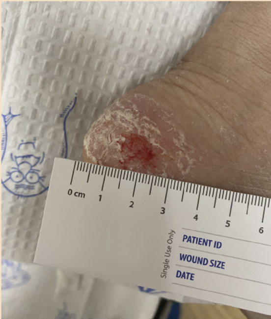
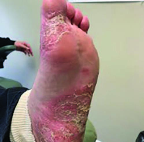
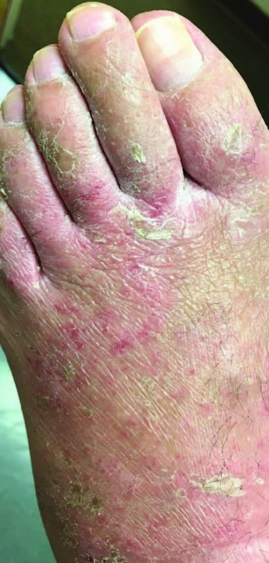
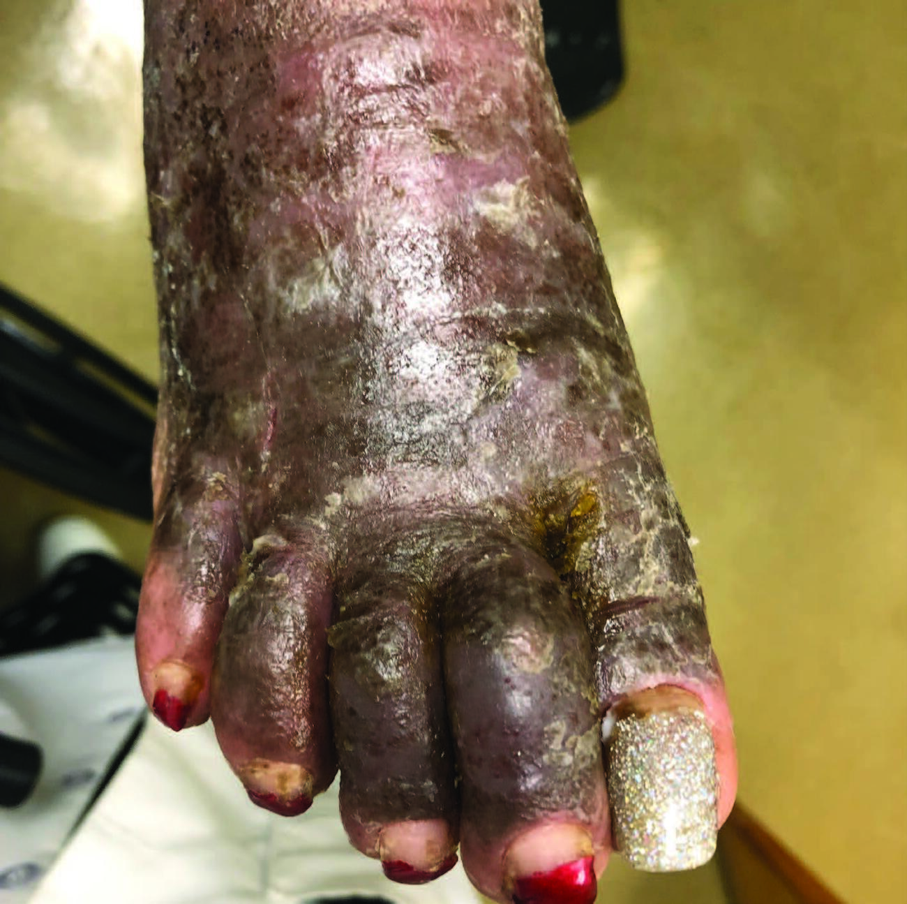
The common clinical presentation of a patient with dermatitis is a red and scaly rash. The patient interview best guides the physician to believe that it is an exposure-related skin reaction.19 In general, CD has acute, subacute, and chronic phases. An erythematous, scaling and oozing skin rash with or without vesicles characterizes the acute phase.20 The subacute form has a milder presentation of the same symptoms; a less erythematous, pruritic, scaling, and fissured skin rash. ACD involves previous exposure to an offending agent (allergen), causing a sensitization process. The sensitization process in ACD allows one or more exposures to the allergen before presenting as an eczematous skin reaction.20 Spread of the ACD reaction can occur by scratching the affected site and subsequently scratching another area of the body. ACD usually presents with bilateral, symmetric, eczematous lesions on the dorsum of the foot while sparing the interdigital space.2 The chronic form of ACD can cause the skin to become lichenified, fissured, or pigmented.2
Shoe ACD occurs in 1.5 to 24 percent of patch-tested patients.14,21 Shoe dermatitis has a predilection for men due to the wearing of tight or ill-fitting footwear for long periods, whereas women develop it due to variation in footwear.3,20 Erythema, papules, vesicles, oozing, scaling, and crusting at the site of contact characterize shoe ACD. Acral skin can facilitate the transfer of allergens to other parts of the body; therefore, these signs can be present anywhere on the body or foot, but are most common on the dorsum of the foot.2,3
What Is The Pathophysiology Of Allergic Contact Dermatitis?
Allergic contact dermatitis is a Type IV hypersensitivity, a T-cell-mediated delayed response. ACD develops more slowly than ICD, as antigen-specific effector T-cells require induced synthesis of effector molecules.22,23 Contact hypersensitivity antigens are typically highly reactive small molecules that can penetrate the skin, induced by itching and scratching. Antigens include small metal ions (nickel, chromate), and other haptens. Electrophilic chemicals, including compounds with polarized bonds, unsaturated compounds, or cations, including nickel and chromate, react to form haptens that lead to skin sensitization.22,23 Haptens react with self-proteins to form a protein-hapten complex that gets processed to hapten-peptide complexes by Langerhans cells, which bind to major histocompatibility complex (MHC) molecules to be presented to T-cells as foreign antigens. A local epidermal reaction then occurs, including erythema, cellular infiltrate, vesiculation, and intraepidermal abscess. This process divides into the sensitization phase and elicitation phase.2,6,22,24
The sensitization phase of the ACD hypersensitivity response process begins with the cutaneous Langerhans cells taking up and processing the antigen. The Langerhans cells then migrate to the regional lymph nodes where they activate T-cells, which then induce the production of memory T-cells.2,6,22,24 The T-cells then migrate to the site of effect in the dermis. Sensitization is proceeded by elicitation, in which further exposure to the sensitizing chemical leads to presentation to memory T-cells in the dermis, leading to activation and further recruitment of specific T-cells. Activation and recruitment occur with the release of T-cell cytokines such as IFN-y and IL- 17, stimulating keratinocytes of the epidermis to release cytokines. This leads to an increased inflammatory response with increased migration and maturation of monocytes into the lesion, resulting in attraction of more T-cells to the site.2,6,22,24
Understanding The Epidemiology Of ACD
Shoe dermatitis affects multiple ethnicities and geographical regions. In a study conducted by the North American Contact Dermatitis Group (NACDG), over 19,000 patients underwent testing with 45 allergens in 13 centers in North America, including significant allergens of shoe dermatitis (92.9 percent (17,803) Caucasian and 7.1 percent (1,360) African-American). The overall patch test positivity rates were similar for most allergens between the two groups. However, African-American patients reacted more frequently to shoe allergens. The United States African-American population had higher allergic reaction rates to bacitracin/neomycin, mercaptobenzothiazole (rubber), thiuram, and mercapto mix. However, the Caucasian population had a higher allergic reaction rate to dyes/fragrances and formaldehyde. The etiologies of these differences are unknown.25
According to a study conducted by the Contact and Occupational Dermatoses Forum of India (CODFI), among a total of 640 patch test patients, they found the proportion of shoe dermatitis was 24.22 percent, or 155 patients. There were more females, 61.93 percent or 96 patients, and male patients comprised only 38.06 percent, or 59 patients. Patient age distribution was from eight to 75 years old, showing that anyone who wears shoes is potentially at-risk for shoe dermatitis. The most prevalently involved age group was the fifth decade with 24.52 percent, or 38 patients. The most prevalently involved occupation group was housewives, comprising 42.58 percent, or 66 patients.18
Differential Diagnoses To Consider
The hallmark signs of dermatitis include calor, rubor, tumor, and pruritus. Pruritus can be a differentiating marker for other conditions that mimic dermatitis in visual presentation.2,6 A red and scaly rash, the appearance of foot contact dermatitis, may appear to be several diseases, including tinea pedis, psoriasis, or lichen planus.26 Other possibilities include mechanical irritant dermatitis, juvenile plantar dermatosis, and atopic eczema.27 However, patient history, physical, and clinical appearance may eliminate many of the possibilities. In cases where some differentials are eliminated, but one cannot discern the disease source, a patch test will distinguish allergic contact from irritant contact dermatitis.20,28
Atopic dermatitis, dyshidrotic eczema, and palmoplantar psoriasis are differential diagnoses of CD, but are diagnosable with a skin biopsy. Unlike CD, atopic dermatitis is more widespread and distributes along flexor surfaces. Dyshidrotic eczema also presents with deep-seated, tapioca-like vesicles and occurs on the hands more frequently than the feet. Palmoplantar psoriasis tends to present as localized plaques and pustules rather than a generalized rash.27
Furthermore, latex allergy, scabies, and tinea pedis may present like CD, but are diagnosable with allergy testing, skin scraping, and potassium hydroxide testing, respectively. Latex allergy may also present with a systemic reaction. Scabies presents with burrows and typically have distribution in the groin, axilla, waist, and hands in addition to the feet. Moreover, tinea pedis usually occurs between the toes rather than on the dorsum of the foot.27
Top 10 Allergens In Footwear And Their Relation To Shoe Construction
Rubber, both synthetic and natural, is in all types of footwear, ranging from sneakers to flip-flops, in the insoles, shoe elastics, and the soles. Allergens related to rubber can affect the dorsum of the foot and the plantar skin. It may also serve as cement in joining shoe uppers and linings.29 These chemicals are related to the vulcanization process. The rubber mix of allergens includes carba mix, thiuram mix, mercaptobenzothiazole, mercapto mix, mixed dialkyl ureas, and black rubber mix. Metal ornaments such as zippers, buckles, rivets, and snaps can cause dermatitis on the dorsum of feet and toes. Below are, in my experience, the top 10 contact allergens in footwear.
1. Carba mix has three allergens: diphenylguanidine; zincdibutyldithiocarbamate; and zincdiethyldithiocarbamate, which are pesticides and fungicides, but also used in rubber product manufacturing. With leather-based shoes, allergens can form to the products used in leather manufacturing as well as the gluing of the leather pieces.
2. Thiuram mix has four allergens: tetramethylthiuram monosulfide; disulfiram; tetramethylthiuram disulfide; and dipentamethylenethiuram disulfide. Similar to the carba mix, it is used for manufacturing rubber products.
3. Mercapto mix has the following allergens: N-cyclohexylbenzothiazyl-sulfenamide; dibenzothiazyl disulfide; and morpholinylmercaptobenzothiazole. This mix is found in the rubber components of work and athletic shoes.29
4 and 5. Cobalt (cobalt chloride hexahydrate) and nickel (nickel sulphate hexahydrate) are used to make metal alloys and are found in many of the same materials, so an allergy to both is typical. The most common cause of nickel allergy is direct contact with a piece of jewelry containing it, and perspiration exacerbates it. A patient who has had their ears pierced and becomes sensitized to nickel could become allergic to a component in their shoe. A patient who has a nickel allergy and wants to test a shoe for nickel content may use the Reveal & Conceal™ Nickel Spot Test (SmartPractice) and can follow this up by utilizing Nickel Clear Coat (Reveal & Conceal) to minimize direct contact with the nickel in the shoe item.
6. Chromium sulphate is used in leather tanning. Patients allergic to this can switch to chromium-free shoes, which can include vegetable-tanned shoes, or if not possible to wear non-leather shoes, the patient can try wearing two pairs of socks to minimize contact with the leather.29
7 and 8. Glutaraldehyde and formaldehyde are components of resins used to glue the leather and rubber components of the shoe. Like nickel, sweating is a factor in causing an allergy to formaldehyde.30 The shoe creates the perfect catalyst for this to occur.
9. Colophony or rosin is from the sap of pine trees. It is not only found in products to glue the layers underneath the insole but also for de-slicking dancers’ pointe shoes.29
10. Aceptophenone azine is the 2021 American Contact Dermatitis Society allergen of the year.31 This is an upcoming component found in EVA foam, shoe soles, shin guards in hockey and soccer players, and flip-flops. It begins as a local reaction but can become a generalized dermatitis over the body, which is possible with any type of ACD.
Treatment Pathways For Shoe Dermatitis
Elimination Of Allergens And Creating Physical Barriers. In treating ACD, one must first identify the causative agent, followed by avoiding the offending allergen(s) in the patient’s environment. In addition to obtaining a thorough patient history, the literature recommends patch testing to determine the offending agent or agents and better direct treatment and patient education.1-11 Suitable replacements for shoes and socks lacking the offending compounds may be readily available or may require custom manufacturing. Freeman and team reports that by replacing leather inner soles with cork inner soles and wearing moccasin-style footwear free of rubber adhesives, patients reported improvements in shoe dermatitis symptoms.1 When chromium salts, compounds commonly used in leather processing tanning and dyeing, cause ACD, it is best to direct patients to purchase shoes or boots made from chromium-free leather or alternative materials.3,5 In cases where one cannot implement an alternative shoe style for practical or safety reasons, patients may apply a double layer of socks to increase the barrier between dorsal and plantar foot skin and offending materials and adhesives contained in shoes. It is wise to advise patients that double socking may require shoes that are a half-size larger.1,3 High-quality leather shoes containing chromium compounds may be usable but may need to be discarded and replaced after a few months of use. Hyperhidrosis may shorten the usable lifespan of such shoes, as increased sweating may precipitate chromate lixiviating from the leather.3,5
Special fabrics are available to palliate the symptoms of ACD. Ricci and colleagues compared topical moisturizers and woven silk fabric (Microair DermaSilk® by Alpretec) and topical moisturizer with cotton fabric as a control in pediatric patients.32 Statistically, they noted significant improvement in patients using woven silk garments.32 A recent study by Corazza and coworkers examined another Microair multi-layer material used in socks and undergarments engineered by Alpretec to increase perspirability and block allergens and irritants.9 This material is a microporous membrane core interposed between polyester microfiber piqué. The majority of participants reported a reduction in itch (66 percent), pain reduction (77 percent), and improvement in their ability to walk without pain (100 percent) after eight weeks of wearing exclusively Microair barrier socks. It should be noted that the sample size of this study was relatively small and did not include a control group. Furthermore, 66 percent of the participants noted difficulties wearing Microair socks daily due to discomfort and aesthetic reasons.3,9
Pharmacologic Treatment. Emollients and humectants such as urea and glycerol are some of the most common therapeutics prescribed for ACD to maintain skin hydration by trapping water in the epidermis. Maintaining skin hydration is crucial in protecting and restoring skin integrity, thus maintaining the patient’s natural protective barrier. Anti-inflammatory, anti-pruritic, and epithelium growth-promoting agents may also be part of emollient or humectant preparations.2,33,34 Emollient preparations may contain vehicles such as propylene glycol that can trigger or exacerbate ICD, requiring close patient monitoring.2,3
Barrier creams are often included in ACD prophylaxis. The proposed mechanism is to establish a physical barrier between the epidermis and the outside world. Lipophilic preparations should offer protection against lipophobic irritants and allergens, and lipophobic preparations should offer protection against lipophilic irritants and allergens.12,34,35 Some preparations contain chelating agents. such as clioquinol, to decrease transdermal absorption of metals such as zinc and nickel. There is no strong consensus on the efficacy of barrier creams over a years-long debate, in part due to the large number of active compounds and vehicle combinations.2,3,7,12,35 Interestingly, Brendt and colleagues suggest that the active ingredients in protective creams are no more effective as skin protectants than preparation vehicle alone (no statistically significant difference found). They attribute this to glycerin (vehicle) being a good moisturizer, allowing for skin rehydration and regeneration.36
Topical corticosteroids are an important mainstay of ACD management. Corticosteroid-mediated inhibition of a localized immune response via inhibition of T-cell activation and leukocyte migration is a crucial process in arresting the hypersensitivity reaction causing ACD. A short-term course of a topical corticosteroid may be sufficient in the treatment of a patient with ACD.2,3,8,37 It is important to consider a paradoxical side effect of topical corticosteroid use. A short-term application of a potent corticosteroid such as clobetasol or betamethasone may result in epidermal thinning and compromise of the skin barrier.2,8
Final Thoughts
While there are numerous allergens causing shoe dermatitis, proper diagnosis and treatment of ACD are possible. One cannot underestimate the importance of a thorough patient history and physical. Furthermore, a patch test can distinguish ACD from ICD, which directs the source of the reaction and patient treatment course. Both conservative and pharmacological treatments, such as shoe gear changes, barrier creams, and topical corticosteroids, may lead to CD relief for our patients.
Dr. Vlahovic is a Clinical Professor in the Department of Podiatric Medicine at the Temple University School of Podiatric Medicine in Philadelphia.
1. Freeman S. Shoe dermatitis. Contact Dermatitis. 1997;36(5):247-251.
2. Bangash HK, Petronic-Rosic V. Acral manifestations of contact dermatitis. Clin Dermatol. 2017;35(1):9-18.
3. Matthys E, Zahir A, Ehrlich A. Shoe allergic contact dermatitis. Dermatitis. 2014;25(4):163- 171.
4. Lecamwasam K, Latheef F, Walker B, Wilkinson M. Contact allergy to reactive dyes in footwear. Contact Dermatitis. 2017;76(6):370- 371.
5. Landeck L, Uter W, John SW. Patch test characteristics of patients referred for suspected contact allergy of the feet – retrospective 10- year cross-sectional study of the IVDK data. Contact Dermatitis. 2012;66:271.
6. Vlahovic TC, Schleicher SM. Skin Disease of the Lower Extremities: A Photographic Guide. Malvern, PA:HMP Communications;2012.
7. Al-Otaibi ST, Alqahtani HAM. Management of contact dermatitis. J Dermatol Surg. 2015;19:86.
8. Cohen DE, Heidary N. Treatment of irritant and allergic contact dermatitis. Dermatol Ther. 2004;17:334.
9. Corazza M, Baldo F, Ricci M, Sarno O, Virgilii A. Efficacy of new barrier socks in the treatment of foot allergic contact dermatitis. Acta Derm Venereol. 2011;91(1):68-69.
10. Ibler KS, Jemec GBE, Diepgen TL, et al. Skin care education and individual counseling versus treatment as usual in healthcare workers with hand eczema: randomized clinical trial. BMJ. 2012;345:e7822.
11. Jacob SE, Burk CJ, Connelly EA. Patch testing: another steroid-sparing agent to consider in children. Ped Dermatol. 2008;25:81.
12. Alvarez MS, Brown LH, Brancaccio RR. Are barrier creams actually effective? Curr Allergy Asthma Rep. 2001;1:337.
13. Cockayne S, Shah M, Messenger A, et al. Foot dermatitis in children: causative allergens and follow-up. Contact Dermatitis. 1998;38:203- 206.
14. Holden CR, Gawkrodger DJ. 10 years’ experience of patch testing with a shoe series in 230 patients: which allergens are important? Contact Dermatitis. 2005;53:37-39.
15. Hulstaert E, Bergendorff O, Persson C, et al. Contact dermatitis caused by a new rubber compound detected in canvas shoes. Contact Dermatitis. 2018;78:12-17.
16. Mowitz M, Ponten A. Foot dermatitis caused by didecyldimethylammonium chloride in an shoe refresher spray. Contact Dermatitis. 2015;73:364-380.
17. Charlotte D, Bergendorff O, Raison-Peyron N, et al. Acetophenone azine: a new shoe allergen causing severe foot dermatitis. Contact Dermatitis. 2017;77:406-429.
18. Chowdhuri S, Ghosh S. Epidemio-allergological study in 155 cases of footwear dermatitis. Indian J Dermatol Venereol Leprol. 2007;73:319- 322.
19. Wilkinson M, Orton D. Allergic contact dermatitis. In: Griffiths C, Barjer J, Bleiker T, et al, eds). Rook’s Textbook of Dermatology, 9th ed. Indianapolis:John Wiley & Sons;2016;128.
20. Saha M, Srinivas CR, Shenoy SD, et al. Footwear dermatitis. Contact Dermatitis. 1993;28(5):260-264.
21. Rani Z, Hussain I, Haroon TS. Common allergens in shoe dermatitis: our experience in Lahore, Pakistan. Int J Dermatol. 2003;42(8):605- 607.
22. Divkovic M, Pease CK, Gerberick GF, Basketter DA. Hapten-protein binding: from theory to practical application in the in vitro prediction of skin sensitization. Contact Dermatitis. 2005;53(4):189–200.
23. Tan, C-H, Rasool S, Johnston GA. Contact dermatitis: allergic and irritant. Clin Dermatol. 2014;32(1):116–124.
24. Janeway C. Delayed-type hypersensitivity reactions are mediated by TH1 cells and CD8 cytotoxic T cells. In: Immunobiology: The Immune System in Health and Disease. London:Harcourt Brace;1999:12-17.
25. Deleo VA, Alexis A, Warshaw EM, et al. The association of race/ethnicity and patch test results: North American Contact Dermatitis Group, 1998-2006. Dermatitis. 2016;27(5):288- 292.
26. Onder M, Atahan AC, Bassoy B. Foot dermatitis from the shoes. Int J Dermatol. 2004;43:565– 567.
27. Usatine RP, Riojas M. Diagnosis and management of contact dermatitis. Am Fam Physician. 2010;82:249–255.
28. Lee HY, Stieger M, Yawalkar N, Kakeda M. Cytokines and chemokines in irritant contact dermatitis. Mediators Inflamm. 2013;2013:916497.
29. Footwear dermatitis. Contact Dermatitis Institute website. Available at: https://www.contactdermatitisinstitute.com/foot-dermatitis. php . Accessed November 8, 2021.
30. Ngan V. Formaldehyde allergy. DermNet NZ. Available at: https://dermnetnz.org/topics/ formaldehyde-allergy . Published 2002. Accessed November 8, 2021.
31. Reeder M. Acetophenone azine: The 2021 American Contact Dermatitis Society Allergen of the Year. Cutis. 2021;107:238-240.
32. Ricci G, Patrizi A, Bendandi B, et al. Clinical effectiveness of a silk fabric in the treatment of atopic dermatitis. Br J Dermatol. 2004;150:127.
33. Moncrieff G, Cork M, Lawton S, et al. Use of emollients in dry-skin conditions: consensus statement. Clin Exp Dermatol. 2013;38:231.
34. Zhai H, Maibach HI. Barrier creams and emollients. In: Chew AL, Maibach HI, eds. Irritant Dermatitis. Berlin:Springer;2006;179.
35. Schliemann S, Petri M, Elsner P. Preventing irritant contact dermatitis with protective creams: influence of the application dose. Contact Dermatitis. 2013;70:19.
36. Berndt U, Wigger-Alberti W, Gabard B, et al. Efficacy of a barrier cream and its vehicle as protective measures against occupational irritant contact dermatitis. Contact Dermatitis. 2000;42:77.
37. Hachem JP, De Paepe K, Vanpée E, et al. Efficacy of topical corticosteroids in nickel-induced contact allergy. Clin Exp Dermatol. 2002;27:47.