


Updates in PAD Diagnosis and Treatment
As our population grows and the proportion of people living past age 65 increases, so does the incidence of one of the most unappreciated chronic diseases, peripheral artery disease (PAD). Adding in the unhealthy lifestyle of many modern Americans, obesity, and diabetes, we are observing the consequences of those diseases that progress with time. This is especially true when it comes to PAD; the longer we live, the greater the progressive pathologic changes to our arterial system. As podiatrists, we are direct observers of these vascular changes in our patients, but we are also able to recognize and intervene in the development of this disease. This article will address some important concepts surrounding arterial insufficiency with the goal that our patients might benefit from an expanded perspective.
The prevalence of PAD in modern society is rising; for decades the most frequently quoted estimate has been 8 to 12 million in the United States for citizens aged 45 and over. This number was gleaned from 1985 and derived from the Criqui/PARTNERS studies and US population estimates circa 1995.1,2 Those estimates depended on population and diabetes data, both of which have grown in the past 28 years. More recently Yost has used the “Diabetes Method” calculation and determined that in 2020 the numbers increased to 21 million, with even more recent Medicare data now pointing to up to 26 million Americans having PAD.3 These newer calculations appear to reflect the clinical experience vascular specialists share within their patient base. But PAD is still an underdiagnosed disease and podiatrists are well suited to improve on this deficiency.
Atherosclerosis is a generalized disease affecting the arterial system, most commonly the coronary (CAD), cerebrovascular (CVD), and peripheral vascular systems, with coronary being most prevalent. There is overlap, where multiple anatomic arterial beds will suffer atherosclerosis (termed polyvascular disease), which affects about 20% of these patients.4 According to the 1996 CAPRIE STUDY, less than 25% of CAD patients have PAD, and 43–90% of PAD patients have CAD.5 Poredoš later found that 42% of patients with CAD also have PAD, with about 36% of patients with carotid disease having this peripheral involvement.6 Compared to coronary or carotid disease, PAD has the highest risk of progressing to polyvascular disease.7 In patients with PAD, the risk of suffering a major cardiovascular event, or “MACE” (defined as a myocardial infarction, stroke, or vascular death) will increase nearly 4 times within 4 years of diagnosis.8
Establishing and Assessing Risk
Well-established risk factors for PAD include age (increasing with each decade), hypertension, diabetes, dyslipidemia, obesity, tobacco use, renal insufficiency, and family history.9,10 This is especially true with our diabetic population where there is a 2- to 4-fold increased risk of developing PAD,11,12 and 1 out of 3 individuals with diabetes who are older than 50 years of age likely have this vascular disease.13-15 There are both macro- and microvascular components to PAD, with most present treatments aimed at the macro-level (surgical or endovascular interventions). Until recently, microvascular disease has basically been ignored though the manifestations are well-known: neuropathy, retinopathy, and nephropathy, conditions commonly seen in those with diabetes.16 Patients with microvascular disease alone have a 7 times greater risk of suffering an amputation, while those individuals with macrovascular disease alone have a 20-times greater risk of limb loss. But in those with both macro- and microvascular disease, the amputation risk skyrockets to 57 times higher compared to those without PAD.17
When interviewing our patients, the medical and family history will provide clues to PAD risk. In my practice, it is not uncommon for a patient to be on medications for hypertension, diabetes, or dyslipidemia, have had a coronary angioplasty, stroke or TIA, and have a tobacco history. As their age advances, so does their risk. Do they have a family history of atherosclerotic disease? All of these are risk factors for PAD, and I feel it is important to consider this patient “at-risk of PAD” until otherwise ruled out by vascular testing.
Addressing Deficits in Awareness
One of the most important challenges we face regarding PAD is simply “awareness” in both the public and medical professional arenas. A significant population is unaware of the dangers of PAD. Comparing the 5-year mortality rate and annual incident rate of critical limb ischemia (the most advanced stage of PAD) with 22 common cancers, only lung cancer had a more dangerous profile (Figure 1).18 Studies have also shown that over half of people who have arterial insufficiency may be unaware of their condition, and the same can be said for up to 30% of their physicians.19 Most persons with PAD do not present with “classic” ischemia symptoms such as claudication, wounds, or gangrene,20 and estimates state that there are 3 times as many asymptomatic PAD patients as there are symptomatic ones.21 Medical professionals are adept at recognizing PAD when ischemic symptoms (such as classic claudication or nonhealing wounds) are present, but up to 90% of people do not present with them.1
Since this disease is often asymptomatic or may present with atypical symptoms often confused with other conditions (arthritis, neuralgias, “just getting older”), it is of paramount importance to actually “look for” PAD. The podiatrist needs to be aware of signs and symptoms of asymptomatic or atypical PAD to best care for this at-risk population. The first step is the history and physical, evaluating past health episodes, medications, procedures, and family history with a focus on the known risk factors for PAD. Even if patients do not have significant PAD at the present time, due to its progressive nature it may become evident later in life. PAD does not just “happen,” but begins early in life and gradually, but silently, develops. Waiting until ischemic symptoms occur is not productive, but can be common. I also recommend sharing your concerns regarding PAD with the patient, their family if appropriate, and their primary care team.
A Progressive Process
Since PAD is chronic, progressive, begins early, and continues throughout life, clinicians tend to become aware of it only when symptoms develop. As shown by Hirsch and colleagues, after 50 years of age the disease worsens, leading to greater morbidity and mortality (Figure 2).22 Initially, 20–50% are asymptomatic, 30–40% have atypical leg pain, and 10–35% have claudication symptoms. Five years after diagnosis, 10–15% of this group will have died, the majority from cardiovascular causes, and 20% will have suffered a myocardial infarction (MI) or stroke. Of those that survive, 70–80% will develop stable claudication, 10–20% have worsening claudication, and 5–10% develop the most advanced stage of chronic PAD called chronic limb-threatening ischemia (CLTI).22
CLTI is the present preferred designation for the former term, critical limb ischemia (CLI). While both terms refer to the same subset of PAD, CLTI is when these persons (1–3% of patients) reach a level of ischemia demanding more intense treatment such as surgical revascularization, endovascular intervention, or amputation. One year after a CLTI diagnosis, 25% have died and 30% have undergone an amputation.23
Not all people will follow a linear progression through these stages of PAD. Some asymptomatic cases never become symptomatic while others are diagnosed only when ischemic symptoms develop. But one concept is known: once the process of PAD begins, it will continue until a person’s last breath. Peripheral arterial disease is never “cured.” One of the greatest gifts we can give these patients is to be vigilant in monitoring their legs for the rest of their lives.
The “Heart Attack of the Leg” and a Lifetime of Surveillance
There is a third level of PAD—an acute attack of ischemia so severe that intervention is urgently required. This presentation is termed acute limb ischemia (ALI) and is often called the “heart attack of the leg.”23 Symptoms will develop within 2 weeks of the episode, characterized by the “six P’s” of the extremity: pain at rest, paresthesias, pulselessness, paralysis, pale or mottled skin, and a cold extremity (polar).24 The ischemia is so severe that 15% of people die within 30 days, and 20% will require an amputation.25 The term “major adverse limb event” (MALE) refers to a severe limb ischemia where an intervention is required such as a surgical bypass, angioplasty, thrombolysis, or amputation proximal to the forefoot. For those who survive ALI but suffer a MALE, for the rest of their life they have a 7 times greater risk of hospitalization for a vascular issue, a 200 times increased risk of amputation, and are 3 times more likely to die.26
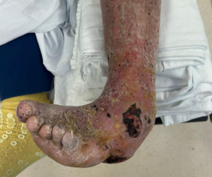
CLTI is a more severe form of PAD. It is defined as ischemic pain in the foot while a person is at rest with pain lasting two or more weeks, non-healing wounds, or gangrene that is attributable to objectively proven arterial occlusive disease.27
MALE is defined as either a major amputation (above the ankle) of the revascularized limb, and/or major reintervention of the revascularized segment (new bypass graft, jump/interposition graft revision, or thrombectomy/thrombolysis).28
The multiple techniques presently available to treat these severe cases of PAD surgically and endovascularly are remarkably successful in improving circulation and preserving the limb. Unfortunately, limb circulation is never “fixed.” In fact, after a successful intervention and limb preservation, there is a 4 times greater risk of an ALI event as soon as 1-year post-revascularization compared to those who have not undergone an intervention, and this risk continues for the rest of their lives.27–30 A review of over 393,000 patients after peripheral artery revascularization, revealed long-term risks of ischemic events, including MI, major amputation, or peripheral revascularization over the next 3 years, with the highest risk for limb events within 1 year.”31 After an arterial intervention, we need to remain diligent regarding the potential for a recurrent circulation reduction. We also need to educate our patients, their families, and the rest of the health care team regarding these findings and statistics.
How Pathogenesis Informs Treatment Development
There are several causal factors in the development of atherosclerosis, but the most powerful drivers are endothelial dysfunction, inflammation, dyslipidemia, and thrombogenesis. Each impacts the development of the atherosclerotic plaque, leading to obstruction of blood flow, rupture, or erosion, with the clinical outcome of ischemia.32 Understanding this pathophysiology has led to the development of treatments that intervene in the development of PAD.
Endothelial dysfunction occurs when the protective single layer of cells lining the internal aspect of the artery is damaged. This damage impacts vessel tone, growth, hemostasis, and generation of nitric oxide, among other cellular processes. The associated inflammatory responses can lead to the formation of atheroma, fibroatheroma, calcification, occlusion, thrombus, and atherothrombotic events. Understanding this process has led to therapeutic modalities that block these responses at different stages, whether removing blockages, stenting to counteract plaque, addressing nitric oxide, diabetes, smoking, lipids, or thrombogenesis. We now have viable medical or surgical options that affect the PAD process before, during, and after arterial insufficiency is present.
All PAD is Not the Same
Earlier in my career, I believed that atherosclerosis affected all arteries in a similar fashion. An artery was an artery, no matter where it may be in the body. That seemed a logical statement, since on histological examination all arteries had an intima (including endothelium), a media (with smooth muscle), and an externa (including adventitia and vasa vasorum) layer. I believed the difference between an artery in the heart and one in the leg was determined by their length. Today I have quite a different opinion. The REACH (Reduction of Atherothrombosis for Continued Health) Registry followed patients from 3647 centers in 29 countries for four years, evaluating complications and comorbities of the spectrum of atherothrombotic disease including coronary, cerebrovascular, and peripheral artery disease.33 One of their conclusions was “not all atherothrombosis is equal.” That study was the basis for further research that continues to this day.
Until recently, treatment of PAD in the legs was based on research focused on coronary artery disease. Antiplatelet therapy was found to significantly reduce the incidence of atherosclerosis in patients with coronary artery disease. This treatment is a mainstay for all patients with atherosclerosis in all vascular beds, but it seems to be more effective in the heart than in the legs. Antiplatelet therapy appears to be more effective after coronary interventions than after lower extremity procedures.34,35 Over the past 30 years, investigations focused on the legs (and not just corollaries of cardiac disease) revealed surprising data on why these arteries are different and how other therapies may provide more effective results.
Atherosclerotic vs. Atherothrombotic Disease
One of the most important recent findings is that atherosclerosis may develop differently depending on the anatomic level of the leg. In studies of CLTI patients who underwent amputation, histologic evaluation of arteries from above-knee amputations (AKA) compared to below-knee amputations (BKA) show marked differences.35 AKA arteries have a similar appearance to those of diseased coronary arteries and show greater medial calcification and acute thrombi, which are typical atherosclerotic changes within coronary plaque, while BKA arteries had a greater number of chronic thrombi (an atherothrombotic process) and less typical plaque signs.33 Although thrombin is a major factor in the development of PAD, antiplatelet therapy, the most common pharmacologic therapy utilized for PAD, does not have a significant effect on thrombin. The most effective inhibitor of thrombin are anticoagulant agents, but bleeding risk is of concern. Recently the Factor 10a inhibitor drug rivaroxaban, in a low “vascular dose” (2.5 mg) given twice daily along with low-dose (81 mg) aspirin given once daily has shown to be superior to aspirin alone in the treatment of PAD.36,37 This “dual pathway” combination has been approved by the US Food and Drug Administration (FDA) for the treatment of stable PAD, and following recent lower-extremity revascularization (LER) due to symptomatic PAD.38,39
Exercise: The Quiet Treatment
One therapeutic option that has shown benefit to stable symptomatic PAD but is not often utilized is supervised exercise therapy (SET), which can improve functional status, walking performance, and overall quality of life (QOL). The actual mechanism of this improvement is multifactorial and not well understood, but yields promise to augment traditional therapies.40 The SET typically uses a treadmill, though other protocols have been studied,41–43 and was approved for reimbursement by the Centers for Medicare & Medicaid Services (CMS) in 2017, with other insurances following suit. A Cochrane review comparing endovascular treatment with SET in 1,087 patients revealed both groups had similar benefit in QOL and functional improvement.44 Multiple studies utilizing SET after surgical or interventional revascularization have shown benefits with combination therapy compared with revascularization alone.45,46
In 2016 the ACC/AHA Guideline on the Management of Patients With Lower Extremity Peripheral Artery Disease (PAD) provided a class I (highest level) recommendation for the use of SET in those with symptomatic PAD.10 Unfortunately, unsupervised walking recommendations such as “just go out and walk” have not shown the same value.42 As podiatrists, we should become familiar with this conservative therapy and the associated insurance regulations. The recommendation of SET as an initial treatment of claudication, and before and after an intervention, is a well-established and valuable protocol, yet not commonly utilized. Discussing this option with primary care and the vascular team is an appropriate step to consider.
Looking to the Future
The understanding of PAD and the atherosclerotic process continues to grow as does potential intervention and treatment. The importance of thrombin, apolipoprotein B and other atherogenic lipoproteins, metabolic syndrome, uric acid and diabetes, renal insufficiency, inflammation, and calcium are all areas of research to note.47 Understanding microvascular disease has led to potential treatments directed at the cellular level, targeting angiogenesis within the plaque itself by inhibiting certain growth factors, VEGF and Ang2, and the antiatherogenic drug endostatin, all of which act by interrupting its vascular supply.48 Therapies to improve nitric oxide and endothelial health are in development, which may affect the progression of neuropathy, nephropathy, and retinopathy, as well.
With advances in research, it is important that our profession expand our knowledge of PAD and vascular medicine since so many of our patients suffer from this disease. Identifying PAD as early as possible provides the greatest opportunity to avoid the complications described as a “stairway to amputation” as referred to in the “Toe and Flow” model of limb preservation put forth by Rogers and Armstrong.49 Staying abreast of current literature in these areas can be difficult, but as podiatry is the “guardian of the foot” and limb, our patients and the medical community look to us for that purpose. We are a recognized vital partner in the PAD/CLTI collaborative team, and part of modern vascular and diabetes treatment guidelines. This achievement is well-deserved but also carries certain responsibilities, namely, expanding our knowledge and understanding of peripheral vascular disease and its prevention and treatment.
Dr. Evans is board certified by the American Board of Foot and Ankle Surgery and is Chief of Podiatry at Beaumont Hospital and Medical Center in Dearborn, MI. He is Chair-Emeritus of the American Podiatric Medical Association (APMA) Health Policy and Practice Committee and has served as a Medicare Carrier Advisory Committee (CAC) representative for over 20 years. He is active with the American Board of Foot and Ankle Surgery, including serving on the Board of Directors, and is the immediate Past-Chair of the Communications Committee. He is the 2018 recipient of the APMA Award of Excellence. Dr. Evans is the Podiatric representative to the PAD Guidelines Writing Committee of the American Heart Association and American College of Cardiology, and is a nationally recognized speaker on a variety of topics including Peripheral Arterial Disease, the Diabetic Foot, Limb Salvage, Physician Burnout, Dealing with the Difficult Patient, and Mindfulness in Medicine. Dr. Evans is in private practice in southeast Michigan.
References
1. Hirsch AT, Criqui MH, Treat-Jacobson D, et al. Peripheral arterial disease detection, awareness, and treatment in primary care. JAMA. 2001;286(11):1317-1324.doi:10.1001/jama.286.11.1317
2. Criqui MH, Fronek A, Barrett-Connor E, Klauber MR, Gabriel S, Goodman D. The prevalence of peripheral arterial disease in a defined population. Circulation. 1985;71(3):510-515. doi:10.1161/01.cir.71.3.510
3. Yost, M, The current U.S. prevalence of peripheral arterial disease. Vasc Dis Manage. 2023;20(4):E67-E73
4. Eagle KA, Hirsch AT, Califf RM, et al. Cardiovascular ischemic event rates in outpatients with symptomatic atherothrombosis or risk factors in the United States: Insights from the REACH registry. Crit Pathw Cardiol. 2009;8(2):91-97
5. CAPRIE Steering Committee. A randomised, blinded, trial of clopidogrel versus aspirin in patients at risk of ischaemic events (CAPRIE). Lancet.1996; 348(9038):1329-1339.
6. Poredoš P, Jug B. The prevalence of peripheral arterial disease in high risk subjects and coronary or cerebrovascular patients. Angiology. 2007;58(3):309-315.
7. Alberts MJ, Bhatt DL, Mas JL, et al. Three-year follow-up and event rates in the international REduction of Atherothrombosis for Continued Health Registry. Eur Heart J. 2009 Oct;30(19):2318-26. doi: 10.1093/eurheartj/ehp355.
8. Abtan J, Bhatt DL, Elbez Y, et al. Geographic variation and risk factors for systemic and limb ischemic events in patients with symptomatic peripheral artery disease: Insights from the REACH Registry. Clin Cardiol. 2017 Sep;40(9):710-718. doi: 10.1002/clc.22721.
9. Dormandy J, Mahir M, Ascady G, et al. Fate of the patient with chronic leg ischaemia. A review article. J Cardiovasc Surg (Torino). 1989;30(1):50-57.
10. Gerhard-Herman MD, Gornick HL, Barrett C, et al. 2016 AHA/ACC Guideline on the Management of Patients With Lower Extremity Peripheral Artery Disease: A Report of the American College of Cardiology/American Heart Association Task Force on Clinical Practice Guidelines. Circulation. 2017;135(12):e726.
11. Wattanakit K, Folsom AR, Selvin E, Weatherley BD, Pankow JS, Brancati FL, Hirsch AT. Risk factors for peripheral arterial disease incidence in persons with diabetes: the Atherosclerosis Risk in Communities (ARIC) Study. Atherosclerosis. 2005;180:389–397. doi: 10.1016/j. atherosclerosis.2004.11.024.
12. Hooi JD, Kester AD, Stoffers HE, Overdijk MM, van Ree JW, Knottnerus JA. Incidence of and risk factors for asymptomatic peripheral arterial occlusive disease: a longitudinal study. Am J Epidemiol. 2001;153:666–672.
13. Jude EB, Oyibo SO, Chalmers N, Boulton AJ. Peripheral arterial disease in diabetic and nondiabetic patients: a comparison of severity and outcome. Diabetes Care. 2001 Aug;24(8):1433-1437.
14. Hirsch AT, Criqui MH, Treat-Jacobson D, et al. Peripheral arterial disease detection, awareness, and treatment in primary care. JAMA. 2001;286(11):1317-1324.
15. Pande RL, Perlstein TS, Beckman JA, Creager MA. Secondary Prevention and Mortality in Peripheral Artery Disease: National Health and Nutrition Examination Study, 1999 to 2004. Circulation. 2011; 124(1):17-23.
16. Beckman JA, Creager MA. Vascular Complications of Diabetes. Circ Res. 2016; 118:1771.
17. Beckman JA, Duncan MS, Damrauer SM, et al. Microvascular disease, peripheral artery disease, and amputation. Circulation. 2019;140(6):449-458.
18. Mustapha JA, Katzen BT, Neville RF, Critical limb ischemia: a threat to life and limb. Endovasc Today. 2019;18(5):80-82.
19. Novo S. Classification, epidemiology, risk factors, and natural history of peripheral arterial disease. Diabetes Obes Metab. 2002;4 Suppl 2:S1-6.
20. Meijer WT, Hoes AW, Rutgers D. Peripheral arterial disease in the elderly: The Rotterdam Study. Arterioscler Thromb Vasc Biol. 1998;18:185-192.
21. Society for Vascular Surgery Lower Extremity Guidelines Writing Group, Conte MS, Pomposelli FB, et al. Society for Vascular Surgery practice guidelines for atherosclerotic occlusive disease of the lower extremities: management of asymptomatic disease and claudication. J Vasc Surg. 2015;61:2S.
22. Hirsch AT, Haskal ZJ, Hertzer NR, et al. ACC/AHA Guidelines for the Management of Patients with Peripheral Arterial Disease (lower extremity, renal, mesenteric, and abdominal aortic): a collaborative report from the American Associations for Vascular Surgery/Society for Vascular Surgery, Society for Cardiovascular Angiography and Interventions, Society for Vascular Medicine and Biology, Society of Interventional Radiology, and the ACC/AHA Task Force on Practice Guidelines (writing committee to develop guidelines for the management of patients with peripheral arterial disease)--summary of recommendations. J Vasc Interv Radiol. 2006 Sep;17(9):1383-97; quiz 1398.
23. Acar RD, Sahin M, Kirma C. One of the most urgent vascular circumstances: acute limb ischemia. SAGE Open Med. 2013;1:10.1177/2050312113516110.
24. Callum K, Bradbury A. ABC of arterial and venous disease: acute limb ischaemia. BMJ. 2000; 320(7237): 764–767.
25. Rajan DK, Patel NH, Valji K, et al.; CIRSE and SIR Standards of Practice Committees. Quality improvement guidelines for percutaneous management of acute limb ischemia. J Vasc Interv Radiol. 2005; 16(5): 585–595.
26. Anand SS, Bosch J, Eikelboom JW, et al. Rivaroxaban with or without aspirin in patients with stable peripheral or carotid artery disease: an international, randomised, double-blind, placebo-controlled trial. Lancet. 2018;391(10117):219-229.
27 Conte MS, Bradbury AW, Kolh P, et al. Global Vascular Guidelines on the Management of Chronic Limb-Threatening Ischemia. Eur J Vasc Endovasc Surg. 2019 Jul;58(1S):S1-S109.e33. doi: 10.1016/j.ejvs.2019.05.006. Epub 2019 Jun 8. Erratum in: Eur J Vasc Endovasc Surg. 2020 Mar;59(3):492-493. Erratum in: Eur J Vasc Endovasc Surg. 2020 Jul;60(1):158-159. PMID: 31182334; PMCID: PMC8369495.
28. Conte MS, Geraghty PJ, Bradbury AW, et al. Suggested objective performance goals and clinical trial design for evaluating catheter-based treatment of critical limb ischemia. J Vasc Surg. 2009 Dec;50(6):1462-73.e1-3. doi: 10.1016/j.jvs.2009.09.044. Epub 2009 Nov 7. PMID: 19897335.
29. Bonaca MP, Gutierrez JA, Creager MA, et al. Acute limb ischemia and outcomes with results from the Trial to Assess the Effects of Vorapaxar in Preventing Heart Attack and Stroke in patients with atherosclerosis-thrombolysis in myocardial infarction 50 (TRA2 degrees P-TIMI 50). Circulation. 2016;133:997-1005.
30. Jones WS, Baumgartner I, Hiatt WR, et al. Ticagrelor compared with clopidogrel in patients with prior lower extremity revascularization for peripheral artery disease. Circulation. 2017;135(3):241-250.
31. Hess C. Long-term outcomes and associations with major adverse limb events after peripheral artery revascularization. J Am Coll Cardiol. 2020;75(5):498–508
32. Libby P, Ridker PM. Inflammation and atherothrombosis: from population biology and bench research to clinical practice. J Am Coll Cardiol. 2006;48(9S):A33-A46.
33. Bhatt DL, Eagle KA, Ohman EM, et al. Comparative determinants of 4-year cardiovascular event rates in stable outpatients at risk of or with atherothrombosis. JAMA. 2010 Sep 22;304(12):1350-7. doi: 10.1001/jama.2010.1322. Epub 2010 Aug 30. PMID: 20805624.
34. Hess CN, Norgren L, Ansel GM, et al. A structured review of antithrombotic therapy in peripheral artery disease with a focus on revascularization: A TASC (InterSociety Consensus for the Management of Peripheral Artery Disease) Initiative. Circulation. 2017 Jun 20;135(25):2534-2555. doi: 10.1161/CIRCULATIONAHA.117.024469. Erratum in: Circulation. 2017 Nov 7;136(19):e347. PMID: 28630267.
35. Narula N, Dannenberg AJ, Olin JW, et al. Pathology of peripheral artery disease in patients with critical limb ischemia. J Am Coll Cardiol. 2018;72(18):2152-2163.
36. Liang Y, Zhu J, Liu L, et al. Efficacy and safety of rivaroxaban plus aspirin in women and men with chronic coronary or peripheral artery disease. Cardiovasc Res. 2021 Feb 22;117(3):942-949. doi: 10.1093/cvr/cvaa100. Erratum in: Cardiovasc Res. 2021 May 25;117(6):1577. PMID: 32289159.
37. Bonaca MP, Bauersachs RM, Anand SS, et al. Rivaroxaban in peripheral artery disease after revascularization. N Engl J Med. 2020;382(21):1994-2004. doi: 10.1056/NEJMoa2000052.
38. Angiolillo DJ, Goodman SG, Bhatt DL, et al. Antithrombotic therapy in patients with atrial fibrillation undergoing percutaneous coronary intervention: a North American perspective-2016 update. Circ Cardiovasc Interv. 2016;9(11):e004395.
39. XARELTO (rivaroxaban). Available at: https://www.accessdata.fda.gov/drugsatfda_docs/label/2021/215859s000lbl.pdf . Accessed July 18, 2023.
40. Harwood AE, Cayton T, Sarvanandan R, et al. A review of the potential local mechanisms by which exercise improves functional outcomes in intermittent claudication. Ann Vasc Surg. 2016;30:312-320.
41. Bronas UG, Regensteiner JG. Connecting the past to the present: A historical review of exercise training for peripheral artery disease. Vasc Med. 2022;27:174-185.
42. Hageman D, Fokkenrood HJ, Gommans LN, et al. Supervised exercise therapy versus home-based exercise therapy versus walking advice for intermittent claudication. Cochrane Database Syst Rev. 2018;4:CD005263.
43. Golledge J, Singh TP, Alahakoon C, et al. Meta-analysis of clinical trials examining the benefit of structured home exercise in patients with peripheral artery disease. Br J Surg. 2019;106:319-331.
44. Fakhry F, Fokkenrood HJ, Spronk S, et al. Endovascular revascularisation versus conservative management for intermittent claudication. Cochrane Database Syst Rev. 2018;(3):CD010512.
45. Fakhry F, Spronk S, van der Laan L, et al. Endovascular revascularization and supervised exercise for peripheral artery disease and intermittent claudication: a randomized clinical trial. JAMA. 2015;314:1936-1944.
46. Mazari FA, Gulati S, Rahman MN, et al. Early outcomes from a randomized, controlled trial of supervised exercise, angioplasty, and combined therapy in intermittent claudication. Ann Vasc Surg. 2010;24:69-79.
47. Marston NA, Giugliano RP, Melloni GEM, et al. Association of apolipoprotein b-containing lipoproteins and risk of myocardial infarction in individuals with and without atherosclerosis: distinguishing between particle concentration, type, and content. JAMA Cardiol. 2022;7(3):250-256.
48. Parma L, Baganha F, Quax PHA, de Vries MR. Plaque angiogenesis and intraplaque hemorrhage in atherosclerosis. Eur J Pharmacol. 2017;816:107-115.
49. Rogers LC, Andros G, Caporusso J, Harkless LB, Mills JL Sr, Armstrong DG. Toe and flow: essential components and structure of the amputation prevention team. J Vasc Surg. 2010;52(3 Suppl):23S-27S.


