Diagnosing and Treating Painful, Recurrent Blisters
A 56-year-old male presented to the clinic for a routine diabetic foot exam with a concern of painful blisters on his right arch. The blisters had formed in the last few weeks, but the patient related that this had happened a few times before when he had worn his work boots for extended periods of time. In those previous instances, the blisters had resolved on their own. Although the lesions were painful, he would work through the pain until they disappeared. He would use an unidentified over-the-counter cream, which would help. He expressed only mild concern about the blisters, but was curious to know the origin.
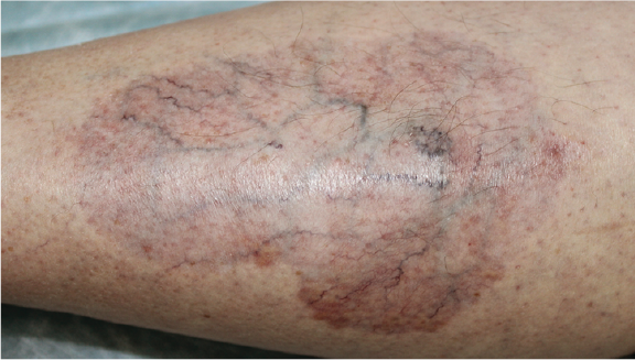
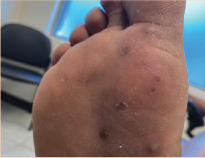
On physical exam, the patient had a mild rash at the right plantar foot, including the arch and first ray, with multiple vesicles, some ruptured and some unruptured. There was no drainage and minimal fluid in the blisters, but they were quite tender to palpation. Pedal pulses and epicritic sensation were both intact. His past medical history was significant for type 2 diabetes, with good glycemic control, reporting that his last HbA1c was 6.7%. The patient appeared very hygienic and in no acute distress.
Cultures and skin scrapings were performed and the patient was prescribed Loprox gel for application twice daily. Bacterial cultures came back negative, but KOH skin scrapings revealed dermatophytosis. Four weeks later, the patient achieved complete resolution of his rash, blistering and pain. The patient was instructed to use the Loprox gel twice daily for 4 weeks for future occurrences, avoid long periods in his boots, and to change them out during long work shifts to prevent recurrence.
Key Questions To Consider
- What is the diagnosis?
- What are the appropriate diagnostic methods to use for this condition?
- What are the most effective treatment options for this condition?
- What is the prognosis in these cases?
Answering the Key Diagnostic Questions
- Acute vesicular tinea pedis
- Skin scrapings and polymerase chain reaction
- Good hygiene practices and topical or oral antifungal agents
- Resolution is common, but so are recurrences
What You Need to Know About Acute Vesicular Tinea Pedis
Tinea pedis, or “athlete’s foot,” originated from southeast Asia, western Africa, and parts of Australia. It spread to the US early in the 19th century; the first case was documented in a World War I veteran in Birmingham, AL.1 Since then, the incidence of tinea pedis has increased significantly in both immunosuppressed and immunocompetent patients, and it currently affects 25% of the US population.1 Tinea pedis mostly occurs in men, but can impact any sex or age. Risk factors include male sex, immune suppression, diabetes, peripheral vascular disease, history of psoriasis or atopic dermatitis, occlusive footwear, activity in public sport facilities and pools without protective footwear, and living in a warm, humid climate.
Fungal infections can occur anywhere on the body, and they are caused most frequently by dermatophytes, although infection by yeasts (ie, Candida) and nondermatophyte molds is also possible. The dermatophytes are a group of fungi that survive on dead keratin, ie, the top layer of the skin, hair, and nails. The most common dermatophyte is Trichophyton rubrum, followed by T. mentagrophytes, Epidermophyton floccosum and T. tonsurans.1 While dermatophytes can go deep in immunocompromised patients, they have a hard time surviving on mucosal surfaces where the keratin layer doesn’t form. On the stratum corneum, the fungi have special enzymes that digest the keratin. As much as they don’t like mucosal surfaces, they thrive on acral surfaces, particularly the soles of the feet due to the lack of sebaceous glands with their antimicrobial properties.1
There are 4 types of pedal fungal infections: interdigital, moccasin, vesicular, and ulcerative.2 While each has its own specific presentation, they share many characteristics: for example, occlusive footwear is a major predisposing factor in all types of infections, as the shoes promote warmth and sweating, which facilitates fungal growth. Additionally, going barefoot in public places—eg, gyms, pools, and shared bathing facilities—increases the risk of infection. In the same way, spouses and family members are more susceptible to infection via fomites, ie, infected shower floors. These infections can produce “id reactions” or dermatophytid reactions, which cause a secondary reaction of itchy vesicles at a site remote to the primary infection, including the arms, chest, and sides of the fingers. Symptoms usually consist of annular erythematous patches (hence the term “ringworm”) with or without drainage, fissuring, or maceration. Patients may be asymptomatic, but will frequently complain of pruritus or odor, and possibly even pain.
Such is the case with acute vesicular tinea pedis, also known as vesiculobullous tinea pedis. Patients, in my experience, will more frequently complain of pain rather than pruritus or odor. While it does involve the characteristic annular rash, I find that vesicular tinea pedis usually arises in the instep and may radiate up to the dorsum of the foot. It is a more highly inflammatory fungal infection thanks to its high rate of involvement of T. mentagrophytes, and that inflammation causes not just the annular rash, but painful vesicles and blisters. Despite its medial arch location, I often note that it usually stems from a chronic web infection.
Tools to Diagnose Tinea Pedis
While most physicians diagnose tinea pedis clinically, some studies show low rates of accuracy in doing so.3 Therefore, it is prudent to confirm the diagnosis via various methods, such as a KOH wet-mount examination of skin scrapings from the active border of a lesion, or roof of a vesicle. While this method doesn’t distinguish the type of fungal infection, it is easy to perform and inexpensive, with results ready in hours. Still, fungal cultures are the gold standard, differentiating the infecting species; however, they are more expensive and take weeks to complete.
New methods include DNA-chip assay via multiplex polymerase chain reaction (PCR) amplification of fungal DNA. This method is also fast, can identify the infecting species, and is becoming more and more available via multiple labs, although it is quite expensive. Other methods include reflectance confocal microscopy and even old-fashioned punch biopsy, which gives a definitive answer, is technically easy to perform, and presents minimal risk.3,4
A Closer Look at the Treatment Options and Prognosis
Good hygiene practices are both an important treatment tool and the first line of defense against fungal infections. As fungus thrives in dark, warm, moist places, physicians should encourage patients to control perspiration and foot moisture as much as possible. This means airing out the feet when possible and changing shoes and socks at least once a day, possibly more depending on the amount of moisture, patient work requirements, etc. Patients should also keep their feet and shoes as clean as possible, making sure to clean their feet and in between their toes during bathing and drying them completely afterwards. Ultraviolet shoe sanitizers can provide not only antifungal protection, but also discourage bacterial and viral growth.4 Finally, physicians should encourage patients to wear water shoes/socks in communal areas such as public pools, bathing areas, etc. As was stated before, doing all of these things can both contribute to eradication of the fungal infection and prevent its recurrence.
Of course, treatment of acute vesicular tinea pedis should also include antifungal medication, whether oral or topical. Topical agents are usually first-line therapy, while oral medications address resistant or more severe cases. There are many topical agents available for the treatment of athlete’s foot, whether over-the-counter or prescription strength. An “oldie but goodie” treatment is wet Burow’s solution applied a few times a day for 30 minutes or so. Such a solution provides relief to inflamed skin, but also discourages bacterial and fungal growth.2 Foot powders, whether medicated or not, are also an accessible over-the-counter alternative.
Medicated creams include the allylamines (eg, terbinafine, butenafine) and the azoles (clotrimazole, miconazole, ketoconazole, etc.). These are usually applied to the affected areas once or twice daily for 2–6 weeks. Some are available over-the-counter and others only via prescription, but most are available in 1% concentrations. Studies have both confirmed and contested the similar efficacy of all these agents.5 Keratolytics such as ammonium lactate and urea cream can help increase their penetration and effectiveness, but usually aren’t necessary.4 Of course, the practitioner must decide whether a cream, gel or spray is best: classically, the saying goes, “If it’s wet, dry it. If it’s dry, wet it.” Creams are more moisturizing while gels and sprays have a drying effect.
For resistant cases of tinea pedis, or more severe or extensive cases, commonly utilized oral antifungal medications include terbinafine and itraconazole, usually given as a once-daily dose for 2–4 weeks. Studies have shown terbinafine to be more efficacious than itraconazole.5 Concurrent id reactions may require oral or topical steroids, but usually these reactions clear with resolution of the primary fungal infection. Secondary bacterial infections may require topical or oral antibiotics. As these infections have a high rate of relapse and can be passed to family members, patients should be well-educated on both good hygiene practices and the proper use of these medications for future occurrences.
In Conclusion
Acute vesicular tinea pedis involves a vesiculobullous annular rash, usually located in the instep, that causes itching and pain to affected patients. Diagnosis is usually by skin scrapings, but newer methods are becoming more available that both confirm the infection and accurately identify the infecting organism. Good hygiene practices, along with oral or topical antifungals, are the best way to both treat and prevent future infections. Patients should be well-educated on prevention and treatment as the recurrence rate is high and transmission to family members is easy.
Dr. Vella is a fellowship-trained podiatrist who practices in Gilbert and Sun City, AZ.
References
- Canavan TN, Elewski BE. Identifying signs of tinea pedis: a key to understanding clinical variables. J Drugs Dermatol. 2015;14(suppl 10): s42-s47.
- Habif TP. Clinical Dermatology: A Color Guide to Diagnosis and Therapy, 5th ed. 2010 Mosby Elsevier.
- Leung AKC, Barankin B, Lam JM, et al. Tinea pedis: an updated review. Drugs Context. 2023;12:2023-5-1.
- Vlahovic TC, Schleicher SM. Skin Disease of the Lower Extremities: A Photographic Guide. HMP Communications, Malvern PA, 2012.
- Ward H, Parkes N, Smith C, Kluzek S, Pearson R. Consensus for the treatment of tinea pedis: a systematic review of randomised controlled trials. J Fungi (Basel). 2022 Mar 29;8(4):351. doi: 10.3390/jof8040351. PMID: 35448582; PMCID: PMC9027577.