Current Concepts In Managing Diabetic Ankle Fractures

The prevalence of diabetes continues to rise in the U.S. with 2018 statistics demonstrating the incidence of diabetes to be approximately 34.2 million people, or 10.5 percent of the population.1 The total cost of diabetes management in 2017 was approximately 327 billion dollars, with the medical cost of patients with diabetes being approximately 2.3 times higher than those without.1 A patient with complicated diabetes (i.e. one with peripheral neuropathy, peripheral vascular disease, CAD, obesity or nephropathy) is at increased risk for developing complications following ankle fracture. A 2011 comparative study demonstrated that a patient with complicated diabetes had a 3.8 times higher likelihood of overall complications, 3.8 times higher likelihood of a non-infectious complication (malunion, nonunion, Charcot arthropathy), and a five times higher likelihood of requiring revisional surgery after ankle fracture.2
These complications usually lead to longer hospital stays and postop rehabilitation courses, thus placing even more strain on health care resources. Specifically, patients with complicated diabetes have been shown to stay 2.4 days longer and cost the health care system $6,895 more than those with uncomplicated diabetes after open reduction and internal fixation (ORIF) of ankle fractures.3 As a result of such complications of diabetic ankle fractures, the overall interest in appropriate surgical management and decision making for diabetic ankle fractures has increased.
Regardless of medical history, surgical versus non-surgical management of ankle fractures depends on fracture pattern, stability and displacement. However, it is thought that these factors carry a different weight in patients with complicated diabetes.4 An important factor in determining surgical necessity for ankle fractures is the computed tomography (CT) scan. Liberal use of CT is recommended for evaluation stability and displacement of ankle fractures, especially in those with diabetes.4 In fact, Kumar and team found that ankle fracture management changed in more than 23 percent of cases after CT examination of the injury. This study did not focus on diabetic ankle fractures, however use of CT becomes even more important in compromised patients.5
Newer literature does suggest operative management for diabetic ankle fractures primarily due to unacceptably high levels of complications with non-operative cast immobilization. Lovy and coworkers noted a complication rate for non-operative treatment of 75 percent, compared to 12.5 percent with operative treatment in displaced diabetic ankle fractures.6 Complications included nonunion, malunion, cast ulcerations, new onset Charcot arthropathy, unplanned operations (e.g. ORIF of fracture at least six weeks after initial injury) and deep infections.6 However, patients with stable isolated malleolar fractures can be managed conservatively via immobilization.4 Such cases however, warrant monitoring at 14-day intervals with serial imaging and thorough examinations to ensure appropriate recovery without early displacement, Charcot development or other skin complications.4
Achieving Optimal Fixation Constructs
There are several studied and suggested surgical fixation options for treating complicated diabetic ankle fractures. As mentioned earlier, inappropriate treatment of these injuries can often lead to complications including the worst case scenario of Charcot arthropathy, and potential resultant limb loss. Hence, the common fundamental goals currently used for Charcot reconstruction also apply in ankle fracture fixation for patients with complicated diabetes.7 Specifically, these include obtaining a fixation construct with maximum rigidity and absolute stability, extending the fixation beyond the immediate zone of injury and planning incisions to minimize soft tissue trauma and optimize healing.4 Some fixation constructs suggested in recent literature include: circular external frame fixation; internal fixation using screws and plates; tibiotalocalcaneal (TTC) nail construct; and even locked fibular intramedullary nail fixation.4,8-10
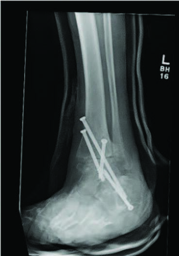
Facaros and colleagues presented a case report of a patient with diabetes and neuropathy who sustained a displaced fibular fracture with significant joint subluxation and syndesmotic injury.11 Subsequent surgical intervention consisted of ORIF with plate fixation, multiple pro-syndesmotic screws and application of a multiplanar circular external fixator. This allowed a maximally stable and rigid construct and also allowed the patient to fully weight bear postoperatively on the external fixator. The patient underwent removal of the external fixator after 12 weeks without any long-term sequelae.11The use of multiple syndesmotic screws regardless of a syndesmotic injury is repeatedly suggested in the literature, as it allows for a more mechanically stable construct.4
Alternately, the TTC nail allows a minimally invasive approach to a robust and rigid construct. A retrospective case series by Ebaugh and colleagues looked at 27 patients who underwent TTC nailing as a primary fixation method without joint preparation and followed them for an average of 2.5 years.8 Overall, their limb salvage rate was 96 percent, the mean time to weight bearing was 6.7 weeks and their fracture union rate was 88 percent. Their surgical complication rate was 18.5 percent, but none of the complications included malunions, nonunions or development of Charcot arthropathy.8
Similar to a TTC nail, the use of a locked fibular intramedullary nail also allows appropriate rigidity without the need for extensive surgical dissection and with earlier return to weight bearing.9 This technique can provide appropriate reduction without any postoperative complications, as seen in the study.9 Asloum and colleagues actually compared one year outcomes of ORIF with plate fixation versus fibular IM nailing for ankle fracture and found no significant difference in the rate of union between the two methods.10 The fibular IM nailing construct had far fewer complications (seven versus 56 percent) and better functional scores.10
Key Considerations And Challenges In Perioperative Management
In addition to proper and adequate surgical management of diabetic ankle fractures, medical optimization is also extremely vital. Glycemic control is essential to any foot and ankle surgery for a patient with diabetes. Patients with a hemoglobin A1c greater than 7% who underwent arthrodesis, osteotomy or fracture reduction are at a significantly higher risk of sustaining bone healing complications.12 Modifying A1c levels prior to surgical fixation of an ankle fracture is obviously not possible. We did not find any literature exploring postoperative complication rates in patients who keep or bring their A1c under control after surgery versus patients who did not. However, there has to be some benefit to encouraging tight glycemic control postoperatively.
Similarly, malnutrition and hypovitaminosis D both demonstrate impaired postoperative skin and bone healing. Similarly, due to the acute and emergent nature of these fractures, altering such factors preoperatively is not possible. More research is necessary to determine if postoperative correction in nutrition and vitamin D can actually lead to better functional outcomes. There are plenty of animal studies demonstrating reduction in re-fracture rates and increased mechanical strength of bone in animals who received vitamin D supplementation post-trauma as compared to animals who didn’t.13Unfortunately, none of these translated into human studies yet. Regardless, with diabetic ankle fractures it is imperative to perform due diligence and carry out any supplemental treatment that could potentially allow for improved outcomes, as long as it does not harm the patient.
Concluding Thoughts
The overall treatment of ankle fractures in patients with diabetes has gained more interest recently primarily due to the associated increase in health care costs. Management has evolved significantly with current guidelines now primarily suggesting some type of surgical intervention which can provide maximum rigidity across the zone of injury. The stronger construct does seem to lead an overall decrease in complications ranging from delayed wound healing to development of Charcot arthropathy. In addition to aggressive surgical intervention, postoperative medical management is also vital in ensuring optimum outcomes. This includes management and patient education regarding tight glycemic control, proper nutrition intake and correction of Vitamin D levels.
Dr. Schneider is an Assistant Professor of Surgery at Harvard Medical School and the Director of the Podiatric Residency Program at Cambridge Health Alliance in Cambridge, Mass. He is a Fellow of the American College of Foot and Ankle Surgeons.
Dr. Malani is a second-year podiatric resident at Cambridge Health Alliance in Cambridge, Mass.
1. American Diabetes Association. Statistics about diabetes. Available at: www.diabetes.org/resources/statistics/statistics-about-diabetes . Updated March 22, 2018. Accessed April 26, 2021.
2. Wukich DK, Joseph A, Ryan M, Ramirez C, Irgang JJ. Outcomes of ankle fractures in patients with uncomplicated versus complicated diabetes. Foot Ankle Int. 2011;32(2):120–130.
3. Regan DK, Manoli 3rd A, Hutzler L, Konda SR, Egol KA. Impact of diabetes mellitus on surgical quality measures after ankle fracture surgery: implications for “value-based” compensation and “pay for performance.” J Orthop Trauma. 2015;29(12):e483-486.
4. Manway JM, Blazek CD, Burns PR. Special considerations in the management of diabetic ankle fractures. Curr Rev Musculoskelet Med. 2018;11(3):445-455.
5. Kumar A, Mishra P, Tandon A, Arora R, Chadha M. Effect of CT on management plan in malleolar ankle fractures. Foot Ankle Int. 2018;39(1):59-66.
6. Lovy AJ, Dowdell J, Keswani A, et al. Nonoperative versus operative treatment of displaced ankle fractures in diabetics. Foot Ankle Int. 2017;38(3):255-260.
7. Gougoulias N, Oshba H, Dimitroulias A, Sakellariou A, Wee A. Ankle fractures in diabeticpatients. EFORT Open Rev. 2020;5(8):457-463.
8. Ebaugh MP, Umbel B, Goss D, Taylor BC. Outcomes of Primary tibiotalocalcaneal nailing for complicated diabetic ankle fractures. Foot Ankle Int. 2019;40(12):1382-1387.
9. Dabash S, Eisenstein ED, Potter E, Kusnezov N, Thabet AM, Abdelgawad AA. Unstable ankle fracture fixation using locked fibular intramedullary nail in high-risk patients. J Foot Ankle Surg. 2019;58(2):357-362.
10. Asloum Y, Bedin B, Roger T, Charissoux J-L, Arnaud J-P, Mabit C. Internal fixation of the fibula in ankle fractures. a prospective, randomized and comparative study: plating versus nailing. Orthop Traumatol Surg Res. 2014;100(4):S255–259.
11. Facaros Z, Ramanujam CL, Stapleton JJ. Combined circular external fixation and open reduction internal fixation with pro-syndesmotic screws for repair of a diabetic ankle fracture. Diabet Foot Ankle. 2010;1. doi:10.3402/dfa. v1i0.5554. 12.
12. Shibuya N, Humphers JM, Fluhman BL, Jupiter DC. Factors associated with nonunion, delayed union, and malunion in foot and ankle surgery in diabetic patients. J Foot Ankle Surg. 2013;52(2):207-211.
13. Bernhard A, Matuk J. Vitamin D in foot and ankle fracture healing. Foot Ankle Spec. 2015;8(5):397–405.