Is There An Optimal Metatarsal Length To Prevent Reulceration After Ray Amputations?
The podiatric surgeon frequently utilizes partial ray amputations aiming to remove infection while preserving bipedal ambulatory status and preventing further morbidity or mortality. An estimated 50 to 70 percent of lower extremity amputations take place due to diabetic complications, most commonly diabetic foot ulcers (DFUs) formed in the setting of peripheral neuropathy.1 Patients with diabetes often face additional comorbidities, including peripheral vascular disease and a diminished immune response, both of which increase the risk for ulcer development and complicate healing potential.2 Although amputation is an effective method of eradicating osseous infection, removing pedal anatomy will alter normal biomechanics and increase pressure distribution to surrounding structures.3 Furthermore, amputation of pedal structures involved in the gait cycle will require increased metabolic expenditure during ambulation.4 One hypothesis states that maintaining as much length as possible of critical structures like the first and fifth rays, while still appropriately addressing any underlying infection, will reduce biomechanical alterations and subsequent re-ulceration.5,6 This article will discuss two studies performed by our research team at Wake Forest Baptist Health, evaluating whether there is an “optimal length” of first and fifth ray resection that could guide podiatric surgeons in preventing further ulcer formation postoperatively.
What We Know About Reulceration Risk
When planning operative intervention for a diabetic foot infection, complete eradication of infection is a primary goal, followed by prevention of further deformity. One may refer to the post-amputation period as “remission,” since this is when the patient is at the highest risk for ulcer recurrence. Approximately 40 percent of patients will experience new ulcer formation within a year of amputation, and the majority occur in the first three months.7 A prospective cohort study by Dalla and colleagues revealed a much lower reulceration rate, occurring in 16.9 percent (15/89) of patients who underwent first ray amputation.8 Patients underwent frequent postoperative follow-up and fitting with rocker-bottom shoes or custom-molded inserts. This discrepancy emphasizes the potential benefit of intense follow-up and custom offloading in the post-amputation period for reulceration prevention. A study by Molines-Barraso and team analyzed reulceration rates following ray resection regarding the length of the residual metatarsal and found that resections greater than 11 mm closely associated with new ulcer formation on the ipsilateral foot.9 Again, this study emphasized strict postoperative protocol, including complete non-weight-bearing for the first two days after surgery, followed by a metatarsal pad with a CAM walker, and then transition to an extra-depth diabetic shoe.
Preserving The Length Of The First Ray
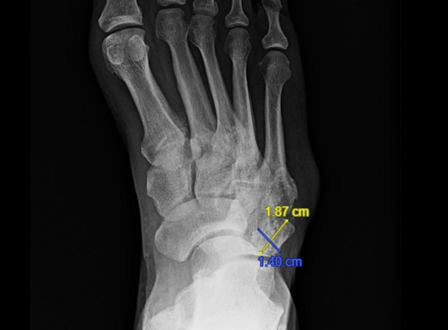
Our team performed a retrospective case-control study evaluating the influence of metatarsal protrusion distance on reulceration rates after partial first ray amputation. The hypothesis was that significant first ray shortening would be a poor prognostic indicator for recurrent ulceration and proximal amputation.6 Thirty-two consecutive patients underwent unilateral partial first ray amputation at a single institution from January 2018 to 2020. Six female and twenty-six male patients with a mean age of 61 (age range 41 to 79) years participated in the study, all with a preoperative diagnosis of osteomyelitis. Preoperative and postoperative radiographs helped calculate the 1-2 metatarsal protrusion distance, measured between the distal dorsal lateral aspect of the residual first metatarsal and second metatarsal head’s articular surface. The surgeon of record determined the amount of bone resected and strategy for closure intraoperatively. Postoperatively, patients bore weight on the heel only in a CAM boot and received broad-spectrum antibiotics, followed by a course of culture-driven antibiotics if indicated. Daily follow-up took place for the first three days, then at one week, two weeks, and monthly, dependent on patient adherence to the treatment plan.6
Fourteen patients developed reulceration to the ipsilateral foot in the postoperative period (43.8 percent), and the mean time to ulcer formation after surgery was 104 days.6 Twenty-eight percent of patients with reulceration underwent additional minor amputations, and four required a more proximal amputation. Active smoking status was the only independent variable associated with increased risk of ulceration, applying to 13/14 (93 percent) patients in the reulceration group versus 8/18 (44 percent) in the ulcer-free group. Radiographic comparison revealed a significantly larger metatarsal protrusion distance of 36.1 + 15.0 mm for patients with reulceration, compared to 25.9 + 12.4 mm for those without new ulcer formation. Further analysis showed that patients with a metatarsal protrusion distance greater than 37 mm were nine times as likely to develop recurrent ulceration following partial first ray amputation, supporting the hypothesis that preserving first metatarsal length can help prevent new ulcer formation.6
Preserving The Length Of The Fifth Ray
Thus far in the literature, there are essentially no studies that examine how the amount of resection and the remaining length of the fifth metatarsal in partial fifth ray resections affect outcomes. These outcomes, such as reulceration rates and reamputation rates, were the primary outcome measures investigated in a multicenter retrospective study involving 117 patients over an eight-year period who underwent partial fifth ray amputations.5 The study included 66 patients from the University of Pittsburgh Medical Center and 51 from Wake Forest Baptist Medical Center. Of the 117 patients in the study, 102 (87.2 percent) patients had a preoperative diagnosis of diabetes with a mean hemoglobin A1c of 8.5 percent, 113 (96.6 percent) presented with a wound, and 108 (92.3 percent) had a diagnosis of osteomyelitis. Regarding additional risk factors, 92 (78.6 percent) patients had peripheral neuropathy, 59 (50.4 percent) had peripheral vascular disease, eight (6.8 percent) had Charcot neuroarthropathy, and 13 (11.1 percent) cavus foot deformity. Radiographic postoperative analysis assessed/ the residual fifth metatarsal length and the level of amputation, categorized into one to four groups including: distal diaphyseal metaphyseal junction; mid-diaphyseal; proximal diaphyseal-metaphyseal junction; and isolated base resections.5
Unfortunately, the study found that most patients reulcerated after fifth metatarsal ray resections.5 Approximately 60 percent (n=71) of the patients developed a postoperative wound, and 40 percent (n=46) remained wound-free through to their final postoperative follow-up. Those with wound recurrence underwent wound classification to better elucidate potential causation. Recurrence of the previous wound accounted for 30 of the wounds, 22 were due to transfer lesions, and 13 involved characteristics of both transfer lesions and recurrence. The follow-up time needed for patients with reulceration was significantly higher at an average of 35 months versus 12 months for those without postop wound recurrence. Of the 117 patients that underwent fifth metatarsal resections, 28 percent required additional, more proximal, ipsilateral fifth ray resection, 12.8 percent underwent transmetatarsal amputations, and 22.2 percent went on to have proximal amputations.5
Ultimately, this study revealed no statistically significant association with the level of fifth ray resection and reulceration or reoperation rates. There is somewhat of a dogma in the surgical field to spare as much viable bone as possible during metatarsal resections. Interestingly, Atway and colleagues examined residual osteomyelitis after partial forefoot amputations and found that 57 percent of patients had residual osteomyelitis.10 The investigators attributed this to inaccurate inspection by the surgeon and fear of over-resection, which would thereby potentially create an unstable foot. Their conclusions stated that wide resection to ensure clean margins is the most important factor in preventing poor outcomes. The data from this fifth metatarsal study posits that bone-sparing resection should perhaps not be the primary goal, as the data from the study suggests that residual fifth ray length may not impact the short-to-medium term viability of the amputation. Instead, it appears that factors such as adequate soft tissue coverage, adequate tissue perfusion, and widely excised margins may be as important, or of greater importance, in successful fifth ray resections with good outcomes.5
Considering Future Directions For Reulceration Prevention
These studies highlight an important issue in our field, being that reulceration after amputations of the fifth ray and first ray are unfortunately a common and persistent issue which requires longer follow-up times and potential further surgical intervention. A systematic review found that reamputation rates at one, three, and five years are 20.1 percent, 29.63 percent, and 45.72 percent, respectively.11 This highlights the importance of persistent follow-up, patient education, and wide resection with adequate soft tissue coverage. It also highlights the need for further research into preventing reulceration and reoperation. Research reviewing the short- to longer-term outcomes of amputations with and without gastrocnemius resection, coupled with partial ray amputations, is an area of potential investigation. Systematic review of literature shows that there are mostly only studies of lower levels of evidence supporting the use of gastrocnemius recession to heal or prevent re-ulceration in the diabetic population.12,13
One combined systematic review and meta-analysis found, in their 11 study analysis with 614 patients, that tendo-Achilles lengthening and gastrocnemius recession were equivalent to the gold standard of total contact casting in regards to time to healing and rate of ulcers healed, while also being significantly more successful in preventing the rate of ulcer recurrence in comparison to total contact casting.13 To our knowledge, there is currently no research investigating the use of gastrocnemius recession in conjunction with ray resections to reduce the rate of reulceration following these surgeries. However, this type of adjunctive surgery could reduce reulcerations by increasing ankle dorsiflexion and reducing the length of plantar pressure in gait. Studies with high qualities of evidence such as randomized controlled trials are necessary to further research in this area. Additionally, investigation into different postoperative protocols and their respective outcomes may influence how surgeons choose to treat their postoperative patients to prevent reulceration and enhance surgical wound healing.
Drs. LeSavage and Leffler are first-year residents with the Podiatric Medicine and Surgery Residency Program in the Department of Orthopaedic Surgery at Wake Forest Baptist Medical Center in Winston-Salem, NC.
Drs. Brackney and Hoffler are third-year residents with the Podiatric Medicine and Surgery Residency Program in the Department of Orthopaedic Surgery at Wake Forest Baptist Medical Center in Winston-Salem, NC.
Dr. Powers is an Assistant Professor at the Podiatric Medicine and Surgery Residency in the Department of Orthopaedic Surgery at Wake Forest Baptist Medical Center in Winston-Salem, NC.
1. Yazdanpanah L, Nasiri M, Adarvishi S. Literature review on the management of diabetic foot ulcer. World J Diabetes. 2015;6(1):37.
2. Adler, AI, Boyko EJ, Ahroni JH, Smith DG. Lower-extremity amputation in diabetes. the independent effects of peripheral vascular disease, sensory neuropathy, and foot ulcers. Diabetes Care. 1999;22(7):1029–1035.
3. Lavery LA, Lavery DC, Quebedeax-Farnham TL. Increased foot pressures after great toe amputation in diabetes. Diabetes Care. 1995;18:1460.
4. Waters RL, Perry J, Antonelli D, Hislop H. Energy cost of walking of amputees. J Bone Joint Surg. 1976;58(1):42–46.
5. Hoffler HL, Honeycutt BJ, Brackney CK, et al. Reulceration and reoperation incidence after isolated partial fifth ray amputations: a multicenter study. [published online ahead of print, 2021 Aug 21]. J Foot Ankle Surg.
6. Hoffler HL, Powers NS, Evans JK, et. al. Metatarsal protrusion distance and its influence on reulceration rates after partial first ray amputations: a retrospective study. J Am Podiatr Med Assoc. 2021. (In Press).
7. Fernando ME, Woelfel SL, Perry D, et al. Dosing activity and returning to pre-ulcer function in diabetic foot remission: patient recommendations and guidance from the Limb Preservation Consortium at USC and The National Rehabilitation Center at Rancho Los Amigos. J Am Podiatr Med Assoc. 2021. doi: 10.7547/20-166.
8. Dalla PL, Faglia E, Caminiti M, et al. Ulcer recurrence following first ray amputation in diabetic patients: a cohort prospective study. Diabetes Care. 2003;26:1874–1878.
9. Molines-Barroso RJ, Lazaro Martinez JL, Aragon Sanchez J, et al. The influence of the length of the first metatarsal on the risk of reulceration in the feet of patients with diabetes. Int J Lower Ext Wounds. 2013;13(1):27–32.
10. Atway VSN, Springer KD, Woodruff DM. Rate of residual osteomyelitis after partial foot amputation in diabetic patients: a standardized method for evaluating bone margins with intraoperative culture. J Foot Ankle Surg. 2012;51:749–752.
11. Zhang P, Lu J, Jing Y, Tang S, Zhu D, Bi Y. Global epidemiology of diabetic foot ulceration: a systematic review and meta-analysis. Ann Med. 2016;49:106–116.
12. Cychosz CC, Phisitkul P, Belatti DA, Glazebrook M, DiGiovanni GW. Gastrocnemius recession for foot and ankle conditions in adults: evidence-based recommendations. Foot Ankle Surg. 2015;21(2):77.
13. Dallimore SM, Kaminski MR. Tendon lengthening and fascia release for healing and preventing diabetic foot ulcers: a systematic review and meta-analysis. J Foot Ankle Res. 2015;8:33.