


Essential Insights In Treating Medial Ankle Sprains
Ankle sprains are one of, if not the most, common injuries in sports. The incidence of injuries appears to be increasing with the higher number of people participating in athletics through all phases of their lives. It is estimated that 30,000 ankle sprains happen each day.1 Lateral ankle sprains are by far the most common with only 5 to 6 percent of ankle sprains occurring medially.2,3 Medial ankle sprains can be more debilitating and have longer recovery times than lateral sprains.
In order to effectively diagnose and treat medial ankle sprains, one must have a strong understanding of the anatomy of the medial aspect of the ankle. The deltoid ligament is made up of two parts, the superficial deltoid and the deep deltoid. The superficial deltoid ligament arises from the anterior portion of the medial malleolus and inserts into the navicular, talar neck and the calcaneus at the sustentaculum tali. Portions of the ligament extend medially and plantarly to the navicular to the level of the spring ligament. These fibers are aligned in the sagittal plane. The superficial deltoid primarily resists hindfoot eversion.
The deep deltoid ligament is a very short, thick, strong ligament, which arises off the posterior portion of the medial malleolus and inserts into the medial aspect of the talus. It is associated with the medial capsule of the ankle joint. The deep deltoid ligament fibers are primarily directed in the transverse plane. These fibers prevent external rotation of the talus and prevent lateral subluxation of the talus and medial gutter widening. Although the deltoid plays a role in preventing lateral displacement of the talus, accompanying injury to the lateral malleolus or lateral ligament complex is usually required for lateral shift of the talus.
Deltoid ligament injuries are the result of an external rotation of the talus that may or may not be associated with a rearfoot eversion. There are a variety of ways in which athletes can suffer injuries to the deltoid complex.
How Athletes Can Be Susceptible To Deltoid Ligament Injuries
Gymnasts may have a deltoid ligament injury when they excessively evert the heel while missing a landing. Ballet dancers in positions one through five are vulnerable as each of these positions has the feet in 180-degree alignment with varying spacing between the feet and can cause a forced heel eversion or talus rotation.
Soccer provides many opportunities for deltoid injuries. Running on uneven ground can cause injury, especially when athletes are playing in early or late season matches when fields may be in less than ideal shape. “Fifty-fifty balls,” in which both players strike opposite sides of the ball at the same time with the inside of the forefoot, result in an external rotation force to the rearfoot and the potential for injury. Repetitively striking balls with the instep may lead to injury. Many deltoid ligament injuries are also caused by an opposing player performing a slide tackle to the lateral ankle, causing eversion of the rearfoot.
Basketball players may land from a jump on another player’s foot, allowing the rearfoot to move excessively into valgus.
Keys To Assessing Deltoid Ligament Issues And Related Injuries
Evaluation of the deltoid ligament begins with a thorough history and physical. Patient recall of the mechanism of injury can lead to clues about the deltoid injury and other associated injuries. However, players often recall nothing more than a collision with another player and subsequently being on the ground.
Palpation of the deltoid ligament and medial ankle will usually elicit pain with direct pressure. The clinician should also palpate the lateral ankle ligaments and fibula for injury. If symptoms allow, one may perform an anterior drawer test. Of special concern with eversion injuries is the palpation of the posterior tibial tendon and the proximal fibula. Medial ankle injuries may be associated with Maisonneuve fractures and the posterior tibial tendon can undergo stretching with rearfoot eversion. Evaluate the syndesmosis for possible injury in association with the deltoid ligament.
Evaluate radiographs of the ankle for associated distal fibular fractures or medial malleolar fractures. Direct attention to the medial ankle joint space. Excessive widening may be present but this usually requires a lateral ankle injury to either the ligamentous structures or distal fibula to allow the talus to translate laterally. An anterior-posterior eversion stress radiograph may show medial widening of the ankle joint space.
Further evaluation via magnetic resonance imaging (MRI) may be warranted. Examine the deep and superficial deltoid as well as possible stress reactions in the distal tibia, fibula and talus. Evaluate the posterior tibial tendon for tears. Proceed to examine the talonavicular joint and pay particular attention to the spring ligament. Rupture of the spring ligament may accompany deltoid ligament tears due to the mechanism of injury.
What You Should Know About Treatment And The Return To Activity
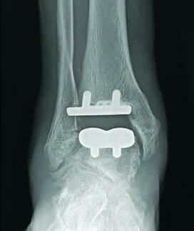
Treatment of the deltoid ligament injury largely depends on the associated injuries. Distal fibular fractures may require open reduction internal fixation (ORIF) to anatomically restore the ankle joint. If medial gutter widening was present prior to the ORIF or closed reduction, it is important to continue to evaluate the medial gutter during the reduction.
Sometimes, the lateral ankle joint appears to be fully reduced but the medial gutter widening persists. If this occurs, there must be a high index of suspicion for the posterior tibial tendon moving up and impinging in the ankle joint. This can be a difficult problem with closed reduction and may require surgical intervention to move the posterior tibial tendon and restore the medial ankle to its anatomic position.
Isolated medial injuries without joint space widening often involve the posterior tibial tendon and the deltoid ligament. Our initial treatment involves early immobilization with a cast (or removable cast boot) with a few days of non-weightbearing that progresses to weightbearing as tolerated for three to four weeks.
The length of time in the cast is dependent upon symptoms, associated injuries and, for upper-level athletes, timing during the season/off-season. After the first week, the athlete may return to activities that do not place significant pressure on the ankle. Easy spinning on an exercise bike is permitted along with core and seated upper body exercise. The patients then progress to a removable boot for an additional two to three weeks. During this time, they are in aggressive physical therapy and may return to modified sport activity.
One should coordinate return to activity with the team trainer and perform modified taping to support the medial ankle in conjunction with a supportive lace-up style ankle brace. Many of the lace-up braces have additional crossing straps and the medial strap can “hold up the arch” to support the posterior tibial tendon and the deltoid ligament.
Athletes gradually progress from the exercise bike to the elliptical machine on a low incline and eventually back to controlled running. Straight ahead running becomes asymptomatic first but cutting and turning can stay somewhat symptomatic for months after the injury. Continued taping and bracing for a few months will help the athlete get back onto the field.
Custom orthotics to address the posterior tibial tendon and medial ankle can be of great value for these patients. These orthotics often include a low medial flange and deep heel cup to support the medial column. Send cleats with the molds to the lab to help ensure a proper athletic fit.
Modifying shoegear is also important. This may involve switching from narrow, firm ground cleats to a wider, more supportive turf shoe. Too often, clinicians ignore the shoes athletes wear off the field or court. Wearing good shoes on the field or court, and then wearing flip-flops or Ugg-type boots (“the winter flip-flop”) off the field can be self-defeating. The fashion arguments of young athletes aside, patients will benefit from an appropriately fitted running shoe worn off the field for at least the first few weeks out of the boot.
Physical therapy is extremely important. This must include proprioceptive and balance exercises, and progress to the complex movement patterns of the athlete’s particular sport. The athlete must then continue these exercises at home for a few months after the conclusion of physical therapy.
Pertinent Pointers On Surgical Intervention
The majority of isolated medial ankle injuries heal well without the need for surgical intervention. However, chronic instability may require reconstruction. This involves direct repair of the deltoid ligament and may involve removal of a fracture fragment or accessory bone from the medial gutter. While these fragments may clearly show on radiographs, they are often encased in scar and the thick fibers of the deltoid ligament. Accordingly, they can be somewhat difficult to find. Intraoperative imaging to localize the fragment can be of great benefit in this case.
When you are having thoughts about deltoid ligament reconstruction, you should also give significant consideration to the posterior tibial tendon, which is often involved in the injury and may become insufficient. Direct repair of the deltoid ligament and posterior tibial tendon may not be enough. Severe valgus deformities may require medial calcaneal slide osteotomies, spring ligament repair or other procedures associated with flatfoot reconstruction to provide long-term successful outcomes.
In Conclusion
While medial ankle injuries are not nearly as common as lateral ankle injuries, they can cause prolonged pain and discomfort. Some high-level soccer players report feeling occasional symptoms while playing for up to nine months after the injury. Most of the time, protection, physical therapy, shoegear modification, orthotics and appropriate bracing can get the athlete back into play without the need for surgical intervention.
Dr. Corwin is an Associate of the American College of Foot and Ankle Surgeons. He is in private practice in Media and Phoenixville, Pa.
Dr. Richie is an Adjunct Associate Professor in the Department of Applied Biomechanics at the California School of Podiatric Medicine at Samuel Merritt University. He is a Fellow and Past President of the American Academy of Podiatric Sports Medicine.
References
1. DiGiovanni BF, Partal G, Baumhauer J. Acute ankle injury and chronic lateral instability in the athlete. Clin Sports Med 2004; 23(1):1-19.
2. Myerson M. Foot and Ankle Disorders. W.B. Saunders Company, Philadelphia, 2000, p. 1459.
3. Mann R, Coughlin M. Surgery of the Foot and Ankle, 6th ed. Mosby, St. Louis, 1993, p. 1139.


