Expecting The Unexpected: When A Small Wound Has Big Implications
Acral lentiginous melanoma only accounts for only two to 10 percent of all reported melanoma types.1 Although the term “lentiginous” often refers to the typical dark coloration of the pathology, the dark coloration is not always a clinical finding, as multiple studies report amelanotic lesions, as well.2,3,4 Therefore, it is pertinent for every practitioner to give due diligence to any suspicious lesion and to know the key findings that may help differentiate a serious lesion from one that is more benign.
What Does The Research Say About Acral/Nodular Malignant Melanoma?
Cutaneous melanoma is the most common type of melanoma subtype by far and accounts for over 90 percent of melanoma cases.5 Melanoma is the nineteenth most common cancer worldwide, with an estimated age-standardized incidence rate of 2.8-3.1 per 100,000.6 There is considerable variation in incidence worldwide, mainly due to skin phenotypes and sun exposure, with the overwhelming majority of incidences in the United States occurring in white-skinned individuals (98.2 percent).5 There are four main subtypes of invasive cutaneous melanoma, with each occurring at various frequencies: superficial spreading (70 percent); nodular (15 to 30 percent); lentigo malignant (10 to 15 percent); and acral lentiginous (less than five percent).
While superficial spreading melanoma is the most common subtype and occurs primarily in sun-exposed areas, nodular melanoma deserves special attention, as it is the most aggressive form of melanoma.7 More specifically, it tends to grow more rapidly in thickness and less in diameter than other forms of melanoma. Presentation varies, but they generally appear dome-shaped, smooth, friable, ulcerated, show bleeding after progression, grow rapidly, and unlike other melanoma, may be symmetrical.7
Although acral lentiginous melanoma is less common than other types of melanoma, it is notable as the most common melanoma in dark-skinned and Asian individuals, who typically have less incidence of other types of melanoma.8 Furthermore, it is particularly important to the podiatric physician as it most commonly appears on plantar, palmar, and subungual surfaces. Subungual melanoma typically presents as a longitudinal hyperpigmented band below the nail plate (melanonychia striata) and may extend into the proximal nail fold (Hutchinson sign). It may present with or without nail destruction, or as a mass with ulceration. Due to the variety of presentations and the fact that multiple pathologies can cause a hyperpigmented band below the nail plate, a dermatological consult is often wise.9
However, one should note that while the risk for malignant melanoma increases drastically as risk factors increase (people with four or five risk factors were 4.4 times more likely to have suspected malignant melanoma than those with one or no risk factors), those with four or five risk factors still only account for 13.6 percent of those diagnosed with suspected melonomas.10 For this reason, it is important to keep in mind that while risk factors are important in assessing the likelihood of melanoma in any given individual, anyone can have a malignancy, and the majority of diagnoses are in patients with no or few risk factors.10
Expected outcomes and five-year survival rates vary greatly based on the extent of the lesion, both in terms of vertical thickness and histological characteristics (ie depth in the dermis or penetration to the subcutaneous tissue).8,11 Two of the most common staging classifications are the Clark Scale and Breslow Scale, although the American Joint Committee on Cancer melanoma staging system (TNM) is also prominent.
|
Clark Scale (level of invasion) |
Histological Tumor Characteristics |
Breslow Scale (vertical thickness) |
Risk for Metastasis |
Five-year survival rate |
|
1 |
Confined to epidermis |
In situ |
None |
100% |
|
2 |
Invasion of the papillary dermis |
Less than 0.75mm |
Minimal |
90% |
|
3 |
Filling of papillary dermis |
0.75-1.5mm |
Significant/Medium |
80% |
|
4 |
Invasion of reticular dermis |
1.51-4.0mm |
High |
50% |
|
5 |
Invasion of deep subcutaneous tissue |
Greater than 4.0mm |
Extremely High |
20% |
It is worth noting how drastically expected patient outcomes worsen as the tumor increases in depth and vertical thickness. While an in situ (confined to the epidermis) tumor has a nearly 100 percent five-year survival rate, this survival decreases to only 20 percent once the lesion invades the subcutaneous tissue.4,5 The fact that outcomes for melanoma are so optimistic when caught quickly and so disheartening when caught later and after extensive tissue infiltration further emphasizes the need for a quick, rapid response when one suspects melanoma.
Treatment typically involves total excision, though the required margins vary based on the lesion's extent. A melanoma still in situ requires a five mm margin and a superficial spreading lesion with one mm thickness or less needs a one cm margin. Thickness of one to two mm requires a margin of one to two cm and a two cm margin is necessary for a lesion thicker than two mm. A patient also needs a sentinel node biopsy if the lesion is thicker than one mm, ulcerative, Clark level 4 or 5, or has a positive deep margin. Other adjuncts to treatment for advanced disease include hyperthermic limb perfusion with melphalan, CO2 laser, intralesional bacillin Calmette-Guerin, dinitrochlorobenzene, ipilimumab, vemurafenib, trameinib, and interferon alpha-2b.11,14,15
When A Patient Presents With A Seemingly Typical Wound
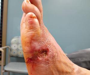
A 68-year-old female with a history of type 2 diabetes, peripheral neuropathy, hypertension, and anemia, presented to the clinic with a concern of a painless wound that she felt stemmed from a dog bite two weeks earlier. Examination revealed a Wagner Stage 1 wound was located on the plantar left foot, between the fourth and fifth digits, and measured 2.7cm x 1.7cm x 0.1cm. During the initial visit, the wound had a granular wound bed with erythematous periwound tissue. As the patient’s history and physical were fairly unremarkable other than that noted above, we initiated a typical wound care protocol consisting of sharp debridement, antibiotic cream, and dry sterile dressings. We scheduled the patient for weekly follow-up visits until wound resolution.
Around one month into treatment, the patient’s wound progress began to stall, despite previous slow improvement. The wound became macerated and did not undergo any changes in size, tissue pattern, or classification. Labs showed a HbA1c of 5.6 percent, C-reactive protein (CRP) was 8.0, and cultures showed moderate Staphylococcus aureus, beta-hemolytic Streptococcus group C and rare Alcaligenes faecalis. At this point, we felt the best course of action was to refer the patient to the hospital’s wound care center.
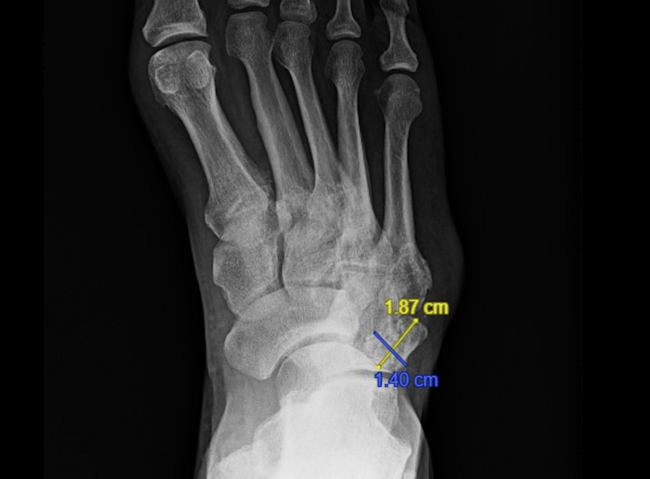
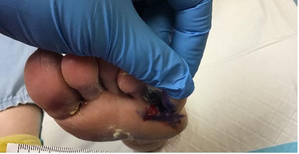
Five months after initial presentation of the wound, we determined that a tissue biopsy was necessary to help determine the etiology of the wound healing issues. Local care consisting of weekly debridements and dry sterile dressings took place while awaiting the biopsy results. One week after performing the initial biopsy, results indicated a Clark Level 4 malignant melanoma, acral/nodular type. After a detailed discussion with the pathologist, we determined that a second opinion by a dermatopathologist at another institution would be beneficial. The next week, that dermatopathologist agreed that the results were indeed consistent with acral lentiginous malignant melanoma. We notified the patient of the results, and placed consults to general surgery and oncology. The patient then scheduled a radical dissection of the lesion and necessary margins of the affected foot and proximal lymph node dissection to prevent metastasis.
A Deeper Dive Into Pertinent Diagnostic And Therapeutic Details
Diagnostic Biopsy Technique. When performing a biopsy on tissue suspected of being cancerous in origin, it is important to remember a few key insights, as misdiagnosis may result from biopsy site selection, techniques, or choice of transport media.12 According to the American Academy of Dermatology, “Important potential sources of error include false-negative direct immunofluorescence results based on poor site selection, uninformative biopsy specimens based on both site selection and technique, and spurious interpretations of pigmented lesions and nonmelanoma skin cancer based on biopsy technique.” In the presence of a suspicious lesion, there are a handful of practices that may help a practitioner perform a successful biopsy:12-15
- Always biopsy any "new" lesions of concern;
- Aim for the darkest and/or most elevated portion of the lesion;
- Handle the biopsy specimen with care and avoid damaging the tissue with instrumentation;
- One, three-mm punch biopsy or two, two-mm punch biopsies are preferable; and
- Allow one to three mm of margin if performing an excisional biopsy.
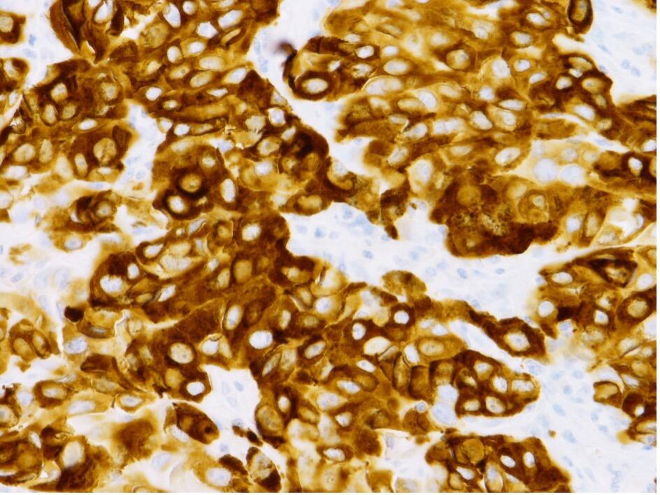
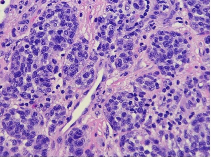
From the Pathologist’s Point Of View. In order to arrive at the final diagnosis of acral/nodular malignant melanoma with 2.0 mm thickness and possible intralymphatic invasion, this case underwent examination of 10 slides in total (four H&E and one each of S-100, HMB-45, Ki-67, Melan-A, D2-40, and p 16). I noted prominent nucleoli, atypical mitotic figures, and a high nuclear-to-cytoplasmic ratio of tumor cells. HMB-45, a monoclonal antibody that reacts against an antigen present in melanocytic tumors such as melanomas, and can be used as a marker in such tumors, was also homogenously expressed throughout the tumor. Additionally, the tumor suppressor genes p 16 are also noticeably absent.
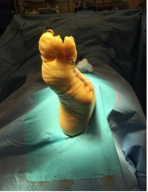
Treatment Protocol And Referral. After notifying and counseling the patient about her diagnosis, we immediately consulted general surgery and oncology. The wound care team developed a treatment plan that involved a radical dissection of the forefoot, primarily partial fourth and fifth ray resections. Dissection extended proximally until our pathologist noted clear margins of all tissue samples delivered to the lab. A general surgeon performed a sentinel node biopsy to remove malignant tissue located within the lymph nodes of the left lower extremity. Definitive resection revealed an end tumor classification of T3bN2bM0. The patient was initially referred to a tertiary care center to treat the melanoma but was unable to continue care due to extenuating circumstances and ultimately decided to seek treatment at a local oncology program.
In Summary
Due to the inherent difficulties one often finds in healing typical neuropathic wounds, biopsy may not always be an obvious initial choice when a wound is healing at a less than ideal pace. It is, in the opinion of the authors that, in general, biopsy is an underutilized tool of the podiatric profession. It is hard to determine exactly why that is the case. It is possible that when a patient does have a suspicious lesion, this lesion may be an incidental finding and not why the patient made an appointment. If that is the case, patients presenting for a musculoskeletal complaint may be reluctant to receive an injection of local anesthetic and undergo a biopsy, especially when the incidence of malignancy is low. Additionally, in our experience, there is little doubt that the eye sees what the mind knows, and young practitioners who have yet to encounter a malignancy may be less likely to aggressively biopsy a lesion that appears borderline between typical and atypical presentations.
However, a biopsy should be a frequent consideration due to its low risk nature and the very high reward of potentially diagnosing a life-threatening illness. Patient education is of utmost importance, as even patients who refuse a biopsy should know their lesion needs monitoring, and immediate follow up with a physician if changes occur. The patient should know that in the case of malignancy, the sooner one obtains a diagnostic biopsy, the quicker one can implement lifesaving treatment, and the better that patient's prognosis will be.
Dr. Shingledecker is a Fellow of the American College of Foot and Ankle Surgeons and the American Society of Podiatric Surgeons, for which he is on the Board of Directors. He practices in Metairie, LA.
Dr. Lamkin practices with Delta Foot and Ankle in Metairie, LA. He is an Associate of the American College of Foot and Ankle Surgeons and a Diplomate of the American Board of Podiatric Medicine.
Dr. Janda is in practice with Gateway Foot and Ankle in Nashville, TN.
Dr. Sartin is the Medical Director of the Department of Pathology at East Jefferson General Hospital in Metairie, LA.
This content was created in partnership with the American Society of Podiatric Surgeons.![]()
- Gerslova A, Pokorna A, Stukavcova A, Veverkova L. Rare cause of non-healing foot wound--acral lentiginous melanoma. Neuro Endocrinol Lett. 2016;37(1):12-17.
- Thomas S, Meng Y-X, Patel VG, Strayhorn G. A rare form of melanoma masquerading as a diabetic foot ulcer: a case report. Case Rep Endocrinol. 2012. Available at: https://doi.org/10.1155/2012/502806. Published April 4, 2012. Accessed November 28, 2021.
- Okhovat JP, Tahan SR, Kim CC. A pink enlarging plaque on the plantar foot: amelanotic acral lentiginous melanoma. Dermatol Online J. 2019;25(1):13030/qt3p91j5db.
- Bradford PT, Goldstein AM, McMaster ML, Tucker MA. Acral lentiginous melanoma: incidence and survival patterns in the United States, 1986-2005. Arch Dermatol. 2009;145(4):427–434.
- Chang AE, Karnell LH, Menck HR. The National Cancer Data Base report on cutaneous and noncutaneous melanoma: a summary of 84,836 cases from the past decade. The American College of Surgeons Commission on Cancer and the American Cancer Society. Cancer. 1998;83:1664–1678.
- Ferlay J, Shin HR, Bray F. Estimates of worldwide burden of cancer in 2008: GLOBOCAN 2008. Int J Cancer. 2010;127:2893–2917.
- Peak SC, Sober AJ, Taso H, et al. Cutaneous melanoma. In: Wolff K, Goldsmith LA, Katz SI, Gilchrest BA, Paller AS, Leffell DJ (eds). Fitzpatrick’s Dermatology in General Medicine, 7th ed. New York:McGraw Hill Medical;2008:1134.
- James W, Berger TG, Elston DM, Newhaus IM. Andrews’ Diseases of the Skin: Clinical Dermatology, 12th ed. Philadelphia:Elsevier;2016:691-694.
- Ruben BS. Pigmented lesions of the nail unit: clinical and histopathologic features. Semin Cutan Med Surg. 2010;29(3);148–158.
- Macneil, JS. New criteria spot melanoma risk, need for total skin exam. Family Practice News. 2007;37(11):27.
- Ali Z, Yousaf N, Larkin J. Melanoma epidemiology, biology and prognosis. EJC Suppl. 2013;11(2):81–91.
- Alguire PC, Mathes BM. Skin biopsy techniques for the internist. J Gen Intern Med. 1998;13(1):46–54.
- Tran KT, Wright NA, Cockerell CJ. Biopsy of the pigmented lesion—when and how. J Am Acad Dermatol. 2008;59(5):852–871.
- Houghton AN, Coit DG, Daud A, National Comprehensive Cancer Network. Melanoma. J Natl Compr Canc Netw. 2006;4(7):666–684.
- Sober AJ, Chuang TY, Duvic M, et al. Guidelines of care for primary cutaneous melanoma. J Am Acad Dermatol. 2001;45(4):579–586.