How To Handle Complications With Minimally Invasive Bunion Surgery
While there has been a strong resurgence of interest for minimally invasive bunion surgery in recent years, a variety of complications can occur regardless of one’s experience with these procedures. Accordingly, this author shares insights from the literature and his experience in addressing common complications ranging from bunion recurrence and hardware breakage to malunion and arthrofibrosis of the first MPJ.
Minimally invasive bunion surgery has regained popularity in recent years after decades of abandonment due to high rates of complications. Advances in technology and newer techniques now allow the performance of minimally invasive bunion surgery to be more predictable with fewer complications.1-5 Proponents of minimally invasive bunion surgery suggest there are less complications in comparison to open bunionectomy.6-8 While minimally invasive bunion surgery is a safe and effective method, complications are still inevitable and minimally invasive techniques open one up to a whole host of new and potentially more challenging complications to manage.8-11
Surgeons perform minimally invasive bunion surgery through much smaller, portal-like incision(s) rather than a typical traditional bunion surgery incision, which extends several inches.12-16 Minimally invasive techniques may involve a single small incision or several tiny incisions.
Several bunion correction methods lend themselves to minimally invasive techniques, particularly bunion shaving and/or a realignment bone cut (distal metatarsal osteotomy). The location of the bone cut differs slightly from a traditional distal osteotomy. A minimally invasive distal first metatarsal osteotomy involves placing the cut slightly more proximal on the metatarsal bone, specifically at the metatarsal neck just proximal to the sesamoids. This allows the surgeon to reposition the entire first metatarsophalangeal joint complex as a unit, allowing for greater correction.
One can perform the osteotomy with a rotary burr, sagittal saw and/or osteotome. The method of bone fixation generally distinguishes one minimally invasive realignment osteotomy technique from another. The three most common methods of fixation are intramedullary percutaneous K-wire, percutaneous screw fixation or intramedullary plate fixation. Each technique has its advantages and disadvantages, however, screw fixation or plate fixation seems advantageous as it allows for stability directly across the osteotomy site.
The entire range of complications associated with traditional bunion surgery continue to exist with minimally invasive techniques. A systematic review of 18 studies (1,594 feet) involving patients who had hallux valgus percutaneous osteotomies identified the following complication rates: K-wire infection (1.6%), recurrence (1.8%), nonunion (0.4%), complex regional pain syndrome (CRPS) (0.9%), transfer metatarsalgia (1.2%), osteonecrosis (0.1%) and joint stiffness (1.9%).12 While newer fixation methods allow one to perform the procedure with more predictability, these methods also open the door for additional complications that have yet to be highlighted.
Minimally invasive techniques are more difficult to perform and there is a steep learning curve, lending to more complications with inexperienced surgeons.
In 2017, a prospective study of a single surgeon’s first 106 consecutive procedures using the percutaneous screw technique identified nearly twice as many complications or postoperative events in the surgeon’s first 53 cases.17 In 2007, Kadakia, Smerek and Myerson abandoned the percutaneous Kirschner wire due to an “unacceptable rate of complications.”18
Should surgeons master a method of minimally invasive bunion surgery, it is important to recognize that complications will still arise for even the most confident surgeons. Determining which patients are good candidates for these procedures will be the best way to avoid postoperative problems.
Assessing The Potential For Bunion Recurrence
Recurrence is possible after any bunionectomy. Some bunions lend themselves to minimally invasive methods and others may be better served with other procedures. Larger bunions with midfoot hypermobility may encourage recurrence, especially if the correction does not re-establish the windlass mechanism of the foot. Narrow metatarsals that limit the amount of the metatarsal head translation may promote a recurrence if one does not fully correct the bunion at the index operation. Even a perfectly executed correction in the “perfect” candidate for minimally invasive bunionectomy may result in a recurrence.
In 2013, Ianno and colleagues reported 16 recurrences in 85 feet using the Bösch technique (percutaneous K-wire fixation).19 Another recent study by Lucattelli and team identified seven recurrences in 195 feet with a percutaneous unfixated osteotomy.6
Raising Awareness Of Possible Nerve Injury And CRPS-Related Complications
Researchers have also reported nerve injury and CRPS with minimally invasive bunion surgery.20 Since inimally invasive surgery does not allow direct visualization of the underlying nerves, these structures are at potential risk for irritation and/or damage.
Depending on the particular minimally invasive method, some nerves are more susceptible than others. The percutaneous metatarsal osteotomy with Kirschner wire fixation is probably least traumatic to surrounding soft tissue neurovascular structures. Methods that involve tunneling and shaving of bony prominences likely create more risk for nerve issues. Protecting these nerves is of utmost importance. Postoperative scar sensitivity may be a sign of neuritis. In a consecutive series of 30 feet, Redfern and colleagues reported two patients who developed CRPS after a minimally invasive chevron osteotomy with rigid screw fixation.20
What About Malunion, Bony Prominences And Hypertrophic Bone?
Malunion is a known occurrence with minimally invasive procedures because surgeons cannot directly visualize bony alignment and mostly rely on intraoperative imaging. Tactile senses may reveal a bony prominence indicating a bony step-off during surgery, suggesting a misalignment. Small misalignments may not become problematic. Depending on the minimally invasive method, malunion may occur in one or all three planes (sagittal, transverse and frontal), leading to additional complexities. Orienting bone cuts in a transverse manner can cause sagittal and frontal plane problems. Elevation of the metatarsal head may occur through plastic deformation and/or bone collapse with weightbearing (despite fixation methods). This is more prevalent with non-adherent patients and larger angular corrections. A plantarflexed malunion may result in a symptomatic dorsal bony step-off prominence and/or altered hallux purchase from the resultant rotated metatarsal head cartilage.
Bony prominences and hypertrophic bone at the osteotomy site are byproducts of secondary bone healing callus formation. These prominences may require repeat operations to smooth out any issues. Minimally invasive techniques seem to be less appealing when one has to shave prominences through an additional incision at a later date. Bone wax application may help prevent regrowth should one need a repeat surgery. Also, leaving the proximal metatarsal shaft intact at the osteotomy site during the index operation may create a pseudobunion (a bony prominence aesthetically resembling a bunion further back on the foot), leaving patients dissatisfied.
Key Considerations With Potential Peri-Implant Fractures And Hardware Breakage
Any bunion surgery is susceptible to peri-implant fracture. Minimally invasive techniques are indeed at risk, especially when one performs them with screw fixation. This specific risk is due to the fixation construct and lack of bone-to-bone contact. Furthermore, with some minimally invasive techniques, surgeons remove additional medial bone so there is also less bone support, lending to peri-implant failure.
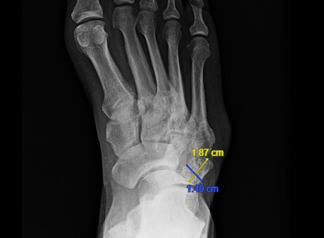
Fracture around the hardware often results in structural instability and loss of position and correction. Some factors that contribute to these fractures are excessive walking, postoperative trauma, multiple points of fixation, stress risers and/or poor bone quality. When it comes to minor fractures that are stable with acceptable alignment, one may treat these fractures without surgery. Displaced fractures often require open reduction with internal fixation and may be challenging to repair due to comminution and bone loss from the index operation. Accordingly, bone grafting may be necessary to repair structural defects (see first set of images at right).
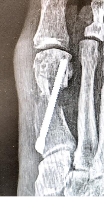
Hardware breakage may also occur. This can result in a loss of position and may be associated with a nonunion (see second set of images at right). Larger shifts of the capital fragment place more stress on the hardware, which may lead to increased strain on the fixation, resulting in breakage. Placing two screws may decrease the loads and lessen the chance of fixation failure.
Is Nonunion A Significant Consideration With Minimally Invasive Bunion Surgery?
Nonunion, or failure of the bones to mend, is a big concern many surgeons have when it comes to minimally invasive bunion surgery. This is because the procedure calls for metatarsal shifts, leaving less bone-to-bone contact than a traditional osteotomy. Making matters more concerning is that the metatarsal bone cut (osteotomy) is at the metatarsal neck within the diaphyseal/metaphyseal junction where healing is more challenging.
However, even with these factors, the overall rate of nonunion appears to be less than one percent among all minimally invasive bunion surgery techniques. Ianno and colleagues had three cases of avascular necrosis in 85 feet using the Bösch technique (percutaneous K-wire fixation).19 A recent multicenter study by Siddiqui and colleagues with percutaneous extra-articular wire fixation resulted in 100 percent union in 217 osteotomies.21 Lucattelli and team’s study of unfixated osteotomies in 195 feet identified two persistent pseudarthroses after bone stimulator treatment.6
Factors that contribute to nonunion are similar to that of the traditional bunionectomy. Nonunions may benefit from bone stimulators. Asymptomatic nonunions may be an acceptable result as long as they are stable. Symptomatic nonunions would likely require a more traditional open revision with plating, bone grafting and non-weightbearing. Any repeat surgery at the metatarsal neck is at risk for nonunion despite best methods given the challenges of bone healing at the diaphyseal/metaphyseal junction.
What You Should Know About Other Potential Complications
Overcorrection resulting in hallux varus is indeed a possibility with minimally invasive bunion surgery. Hallux varus generally occurs from a significant capital fragment translation coupled with adductor release and medial eminence resection. Additionally, performing an Akin osteotomy may increase the risk. In some cases, there is not a particular identifiable cause of the varus but minimally invasive techniques seem to promote a muscular imbalance of the big toe complex, lending itself to this deformity (see third set of photos to right).
It may be beneficial not to perform an adductor release unless it is absolutely necessary. If hallux varus is beginning to develop in the early post-op period, one should initiate splinting of the toe in abduction in the hope of stopping excessive adduction. Treatment of hallux varus is no different than with a traditional bunionectomy, including consideration of early surgical intervention. Hallux varus correction may involve suture tunneling repair with or without a reverse osteotomy. Patients susceptible to varus after a minimally invasive bunionectomy on one side may be more likely to develop a varus on the other extremity.
Forefoot overload and transfer metatarsalgia may occur after both traditional and minimally invasive bunion surgeries. Metatarsal shortening is inherent with any osteotomy and/or fusion (Lapidus), and surgeons should take steps to compensate intraoperatively. Performing an angled osteotomy to lengthen the metatarsal along with translation may be beneficial. One could consider translating or plantarflexing the capital fragment inferiorly but this may inadvertently lead to malunion-related issues. Additionally, when it comes to patients with preoperative lesser metatarsalgia and a long second metatarsal, surgeons may want to consider a shortening osteotomy to balance the metatarsal parabola.
Stiffness of the first MPJ (arthrofibrosis) resulting in decreased range of motion is another known complication after minimally invasive bunion surgery, particularly with the percutaneous Kirschner wire method. Since the Kirschner wire stabilizes by traversing the medial capsule of the first metatarsal, the first MPJ is rendered immobile during the postoperative healing period, resulting in stiffness. Resecting any medial eminence with a rotary burr also creates an opportunity for intra-articular debris and stiffness. A dedicated postoperative physical therapy program is helpful. Intra-articular steroid injections are an option if physical therapy is not producing results.
Infection and wounds may occur at an osteotomy site created with a rotary burr. There may be burning of skin and/or bones with excessive heat leading to wounds and/or osteomyelitis. Wounds overlying an osteotomy may cause contiguous infections. A best practice is to try and avoid damage in the first place with copious irrigation. Excise any portal burns that occur during the index operation rather than risking a postoperative wound. Subsequent deep infections may be complex due to the exposed intramedullary space and hardware. Removing hardware for infection before osteotomy healing creates structural instability that may require external fixation.
In Conclusion
Minimally invasive bunion surgery is more challenging to perform than open traditional bunionectomy. While it has its advantages, surgery performed through small incisions is not without risks. These techniques carry the same potential complications as traditional bunionectomy with some complications being amplified, creating more challenging issues to manage. Understanding how to avoid these complications and manage issues when they occur ultimately results in better outcomes and success with these techniques.
While the majority of complications are experienced by the inexperienced surgeon, one should not underestimate the complexity of these surgeries and every surgery brings its own set of circumstances. Experience and confidence with minimally invasive bunion surgery should not breed complacency, and appropriate patient selection is critical.
Dr. Blitz, the creator of the Bunionplasty®, is in private practice in both Midtown Manhattan, New York and Beverly Hills, Calif. He is board-certified by the American Board of Foot and Ankle Surgery, and is a Fellow of the American College of Foot and Ankle Surgeons. To learn more about minimally invasive bunion surgery, visit www.bunionsurgery.com.
1. Vernois J, Redfern D, the GRECMIP. Percutaneous Chevron: the union of classic stable fixed approach and percutaneous technique. Fuß & Sprunggelenk. 2013;11(2):70–75.
2. Kaufmann G, Dammerer D, Heyenbrock F, Braito M, Moertlbauer L, Liebensteiner M. Minimally invasive versus open chevron osteotomy for hallux valgus correction: a randomized controlled trial. Int Orthop. 2019;43(2):343-350.
3. Magnan B, Pezzè L, Rossi N, Bartolozzi P. Percutaneous distal metatarsal osteotomy for correction of hallux valgus. J Bone Joint Surg Am. 2005; 87(6):1191–1199.
4. Giannini S, Faldini C, Nanni M, Di Martino A, Luciani D, Vannini F. A minimally invasive technique for surgical treatment of hallux valgus: simple, effective, rapid, inexpensive (SERI). Int Orthop. 2013;37(9):1805-1813.
5. Brogan K, Voller T, Gee C, Borbely T, Palmer S. Third-generation minimally invasive correction of hallux valgus: technique and early outcomes. Int Orthop. 2014;38(10):2115-2121.
6. Lucattelli G, Catani O, Sergio F, Cipollaro L, Maffulli N. Preliminary experience with a minimally invasive technique for hallux valgus correction with no fixation. Foot Ankle Int. 2019. doi: 10.1177/1071100719868725.
7. Maffulli N, Longo UG, Oliva F, Denaro V, Coppola C. Bosch osteotomy and scarf osteotomy for hallux valgus correction. Orthop Clin North Am. 2009;40(4):515-524.
8. Radwan YA, Mansour AM. Percutaneous distal metatarsal osteotomy versus distal chevron osteotomy for correction of mild-to-moderate hallux valgus deformity. Arch Orthop Trauma Surg. 2012;132(11):1539-1546.
9. Holme TJ, Sivaloganathan SS, Patel B, Kunasingam K. Third-generation minimally invasive Chevron Akin osteotomy for hallux valgus. Foot Ankle Int. 2019. doi: 10.1177/1071100719874360.
10. Lee M, Walsh J, Smith MM, Ling J, Wines A, Lam P. Hallux valgus correction comparing percutaneous Chevron/Akin (PECA) and open Scarf/Akin osteotomies. Foot Ankle Int. 2017;38(8):838-846.
11. Lai MC, Rikhraj IS, Woo YL, Yeo W, Ng YCS, Koo K. Clinical and radiological outcomes comparing percutaneous Chevron-Akin osteotomies vs open Scarf-Akin osteotomies for hallux valgus. Foot Ankle Int. 2018;39(3):311-317.
12. Bia A, Guerra-Pinto F, Pereira BS, Corte-Real N, Oliva XM. Percutaneous osteotomies in hallux valgus: A systematic review. J Foot Ankle Surg. 2018;57(1):123-130.
13. Roukis TS. Percutaneous and minimum incision metatarsal osteotomies: a systematic review. J Foot Ankle Surg. 2009;48:380–387.
14. Bösch P, Markowski H, Rannicher V. Technik und erste ergebnisse der subkutanen distalen metatarsale-I-osteotomie. Orthop Prax. 1990;26:51–56.
15. Bösch P, Wanke S, Legenstein R. Hallux valgus correction by the method of Bösch: a new technique with a seven-to-ten-year follow-up. Foot Ankle Clin. 2000;5(3):485-498.
16. Magnan B, Samaila E, Viola G, Bartolozzi P. Minimally invasive retrocapital osteotomy of the first metatarsal in hallux valgus deformity. Oper Orthop Traumat. 2008;20:89–96.
17. Jowett CRJ, Bedi HS. Preliminary results and learning curve of the minimally invasive Chevron Akin operation for hallux valgus. J Foot Ankle Surg. 2017;56(3):445-452.
18. Kadakia AR, Smerek JP, Myerson MS. Radiographic results after percutaneous distal metatarsal osteotomy for correction of hallux valgus deformity. Foot Ankle Int. 2007;28(3):355-360.
19. Iannò B, Familiari F, De Gori M, Galasso O, Ranuccio F, Gasparini G. Midterm results and complications after minimally invasive distal metatarsal osteotomy for treatment of hallux valgus. Foot Ankle Int. 2013;34:969–977.
20. Redfern D, Gill I, Harris M. Early experience with a minimally invasive modified chevron and akin osteotomy for correction of hallux valgus. Orthop Proc. 2011;93-B(IV):482.
21. Siddiqui NA, LaPorta G, Walsh AL, Abraham JS, Beauregard S, Gdalevitch M. Radiographic outcomes of a percutaneous, reproducible distal metatarsal osteotomy for mild and moderate bunions: A multicenter study. J Foot Ankle Surg. 2019;58(6):1215-1222.