


Keys To Assessing Peripheral Vascular Disease
 Peripheral vascular disease can be a critical factor with delayed wound healing. Accordingly, these authors offer pertinent diagnostic insights, review current concepts in revascularization and discuss the optimal timing of surgical intervention after revascularization.
Classic risk factors for peripheral vascular disease (PVD) include smoking, diabetes mellitus, hypertension and hyperlipidemia. In patients with PVD, adequate and timely healing of foot wounds depends on glycemic control, blood flow, nutritional status and proper pressure offloading of the site. Whether it is treating a patient with a non-healing foot ulcer or considering elective surgery in a patient with signs and symptoms of PVD, how much blood is enough for healing?
Determining adequate vascular flow for healing begins with a thorough history of earlier vascular interventions and clinical examination of the extremities. Doppler evaluation of pulses and non-invasive vascular testing may also indicate the extent of vascular disease. Depending on the level and extent of vascular impairment based on preliminary evaluation, invasive vascular studies may aid in defining the anatomic pattern of disease to direct treatment.
Rapidly evolving technologies have presented the possibility of improving vascularity for healing while minimizing perioperative morbidity and mortality. However, some have questioned the effectiveness and durability of minimally invasive endovascular techniques and there have been concerns about an endovascular first approach. In this article, we will discuss the key principles and evidence that define our approach in the evaluation of adequate wound healing in the lower extremities of patients with peripheral vascular disease.
What To Look For In The Physical Exam And Non-Invasive Vascular Testing
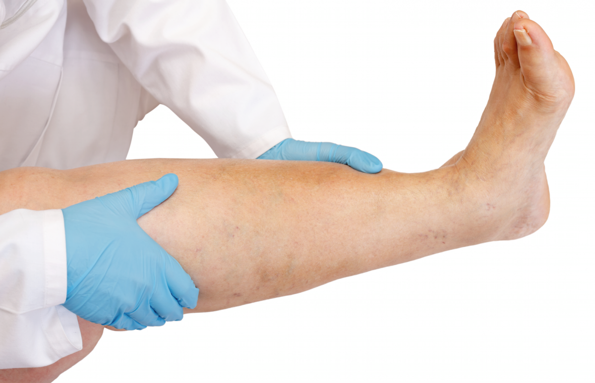
The evaluation of the patient with peripheral vascular disease begins with the physical exam of the lower extremities. Many of us are guilty of focusing solely on the patient’s chief complaint and thus fail to evaluate the lower extremity adequately and thoroughly. An adequate examination can often reveal substantial clues regarding impaired vascularity and the presence of PVD.
It is important to promptly address any local wound sepsis or the treatment of systemic symptoms of infection with surgical intervention (drainage, amputation or debridement) even in the presence of PVD. The surgical treatment of systemic manifestations of infection takes immediate priority with the workup of PVD delayed in order to minimize further tissue loss, limb loss or loss of life due to uncontrolled infection.
Physical examination findings in patients with PVD vary. They may include absent or diminished pulses, abnormal skin color, poor hair growth and cool skin. The most reliable physical findings of PVD are diminished or absent pedal pulses, the presence of femoral artery bruit, abnormal skin color and/or cool skin. However, it is important to note that the absence of these symptoms does not necessarily preclude the presence of PVD.1
 Of these symptoms, the three most sensitive indicators of peripheral vascular disease include abnormal pulses, the presence of a femoral bruit and cool skin. Absence of both the dorsalis pedis artery and posterior tibial artery pulses is 72 percent sensitive and 99 percent specific for PVD.2 If either the dorsalis pedis artery pulse or posterior tibial artery pulse is absent and the other is weak, there is a 63 percent sensitivity and 92 percent specificity for PVD. Less sensitive but highly specific, the presence of a femoral bruit is 29 percent sensitive and 95 percent specific for PVD. Finally, research has found that unilaterally cooler skin in comparison to the contralateral side is 10 percent sensitive and 98 percent specific for PVD.2
Despite the accuracy of the pulse exam for detecting PVD, the reliability of the test itself is unclear. McGee and Boyko evaluated the interobserver reliability of the pulse evaluation based on whether the participating examiners could palpate pulses.1 They found the interobserver coincidence rate of the pulse exam was less than 70 percent. Given the highly sensitive and specific nature of the pulse exam in diagnosing PVD, it should lead the clinician to have a higher index of suspicion for PVD even when a palpable pulse exists.
For any patient with a non-healing (greater than four weeks) foot ulcer, gangrenous changes, cyanosis or rest pain who lacks one or both pedal pulses, one can assume some degree of PVD and perform non-invasive vascular testing. Non-invasive vascular testing includes ankle-brachial index (ABI), pulse volume recording (PVR) and segmental pressures, and provides a baseline of limb perfusion. Additionally, these non-invasive tests can help determine the level of disease and guide further invasive testing when results suggest impaired flow.
Consider PVD to be present when the ABI is less than 0.8 with severe disease present when the ABI is less than 0.5.2 An ABI of < 0.6 corresponds with a significant impairment in wound healing. Peripheral vascular disease specificity with ABI ranges between 83.3 and 99 percent with an accuracy ranging between 72.1 and 89.2 percent for detecting stenosis of greater than 50 percent in patients with ABIs less than 0.9.3 However, the sensitivity of ABI for peripheral vascular disease is wider, reportedly ranging between 15 and 79 percent.
We can attribute the wide-ranging sensitivity of ABI to the frequent presence of extensive calcification of the tibial arteries in patients with diabetes, rendering ankle pressure (and thus ABI) of uncertain value. As a result, a more accurate measure of foot perfusion in the patient with diabetes is toe pressure (and toe-brachial index or TBI).4 Transcutaneous oximetry and transtarsal pulse volume recordings can also be of value in the assessment of PVD with the caveat that they may be poorly predictive and thus require clinical correlation.5
Segmental pressure values have also been helpful in predicting wound healing. Researchers have shown that patients with diabetes with toe pressures greater than 55 mm Hg have a greater healing potential.6 Values of less than 45 mmHg in patients with diabetes are associated with poor wound healing potential with a minimum of 30 mmHg required to heal digital wounds.
Essential Considerations With Invasive Vascular Testing
Based on non-invasive testing and the physical exam, the physician can determine if there is need for more invasive testing to define the anatomic distribution of disease. Imaging studies such as computed tomographic angiography, magnetic resonance angiography (MRA), duplex ultrasound and catheter-based angiography can be useful in both a diagnostic and therapeutic fashion to improve wound healing potential.
 Digital subtraction angiography remains the gold standard imaging modality for evaluating the distribution of PVD when one plans revascularization. Angiography has the advantage of allowing simultaneous endovascular intervention. Due to the need for contrast, patients with diabetes and renal insufficiency are at risk for contrast-induced nephropathy. As a precaution, patients with renal impairment should receive intravenous fluids and stop using metformin prior to digital subtraction angiography as it may cause lactic acidosis.7
When it comes to assessing PVD in patients with known renal impairment, one should refer patients for magnetic resonance angiography, which is minimally invasive and capable of clearly imaging calcified vessels while avoiding the need for iodinated contrast. Disadvantages of magnetic resonance imaging include limited spatial resolution and a relative contraindication for the use of gadolinium in patients with severe renal insufficiency (creatinine clearance < 30 mL/min).
Vascular surgeons can perform computed tomography angiography (CTA) when magnetic resonance angiography is contraindicated.8 It offers improved spatial resolution as well as the speed of examination. Limitations include poor imaging capabilities in the presence of calcified vessels. A systematic review comparing the sensitivity and specificity of duplex ultrasound, contrast-enhanced magnetic resonance angiography and multidetector-row computed tomography angiography reported similar accuracies across these modalities for detecting high-grade stenosis above and below the knee.9
After defining the distribution of disease, one must determine the most appropriate intervention to improve flow for adequate healing. This decision must integrate several factors including patient risk, the severity of ischemia, arterial anatomy and the availability of vein graft for bypass. Only when all of this information is available can one decide on the optimal revascularization strategy.
Which Revascularization Option Is Best For Patients With PVD?
Restoration of direct, pulsatile flow to the foot should be the goal of any revascularization attempts.
The two major options for the treatment of PVD are endovascular treatment or open bypass surgery. The debate continues over whether an endovascular first approach provides sufficient revascularization or whether bypass surgery is necessary for wound healing.10 The belief is that with endovascular intervention, there is the chance that the surgeon can establish arterial flow and tissue perfusion with lower morbidity and mortality in comparison to open bypass. This is especially true in patients who are not candidates for open bypass due to comorbidities, a lack of bypass target vessels or a lack of vein conduit.
Initial comparable levels of limb salvage with endovascular and bypass techniques fueled enthusiasm for open bypass. Taha and colleagues found that technical success, defined as a palpable pulse or Doppler-able biphasic pulse, occurred in 90.7 percent of the open bypass group in comparison to 79.9 percent in the endovascular group.10 The amputation rate was 10 percent in the bypass group and 7.2 percent within the endovascular group at 30 days. At one year, the amputation rate was 16.3 percent within the bypass group and 13 percent within the endovascular group. They concluded that the overall amputation rate was similar but open bypass was statistically more effective in achieving technical success.
 Mashaki and coworkers also compared the effectiveness of bypass versus endovascular intervention for infrapopliteal lesions in patients with critical limb ischemia.11 Endpoints included the patency of the target vessel at three years as well as the limb salvage rate. They found the patency rate was statistically more significant in the bypass group at 72 percent in comparison to the endovascular group at 54 percent. The limb salvage rate at three years was similar with the bypass group at 86 percent and the endovascular group at 82 percent. These findings were similar to the Taha study with bypass creating more successful flow but ultimately similar limb salvage rates.
A landmark level 1 study compared bypass first or endovascular first treatment for critical limb ischemia.12 The Bypass versus Angioplasty in Severe Ischaemia of the Leg (BASIL) study examined 452 patients with critical limb ischemia, assessing limb salvage rates and overall survival. There was no difference in limb salvage rates or overall survival at the one-year evaluation period. However, at the two-year mark, there was a survival advantage associated with the bypass first group and a trend toward improved limb salvage.
Most interestingly, patients who underwent bypass surgery after a failed initial angioplasty fared much more poorly than patients who had an initial bypass, suggesting that the “free shot” with an endovascular first approach may worsen subsequent revascularization with bypass.12 Finally, the authors concluded that vein bypass surgery offered the best long-term outcomes.
How Much Blood Is Enough For Surgery?
Following revascularization, how long should the physician wait before performing a surgical procedure? When is perfusion going to be the most optimal?
To answer these questions, Arroyo and colleagues evaluated TcPO2 levels before bypass and one, two and three days post-bypass.13 Immediately after the bypass surgery, TcPO2 levels increased to the target tissue and continued to rise, reaching maximal levels at day three post-bypass. As a result, the authors concluded that one should perform surgical intervention three days after surgery to maximize healing potential.
An additional concept to consider after revascularization is the principle of angiosomes in wound healing. An angiosome is an anatomic region that a specific artery supplies. On the surface, the notion that revascularizing an artery that supplies an ischemic region may create a greater chance of healing a wound seems to make sense rather than relying on collateral circulation forming.
Neville and colleagues performed a retrospective analysis assessing if targeting an artery to a specific angiosome matters in wound healing.14 They divided patients into two groups: direct revascularization or bypass to the artery supplying ischemic angiosome, and indirect revascularization or bypass to an unrelated artery of ischemic angiosome. They found that 91 percent of wounds healed and 9 percent went on to amputation with direct revascularization in comparison to 62 percent healed and a 38 percent amputation rate with indirect revascularization. This demonstrates that targeting the vessel that feeds the ischemic region of interest is preferable when possible in revascularization procedures.
In Conclusion
Peripheral vascular disease can have detrimental effects on healing. Without proper blood flow, wounds will fail to heal. Evaluation for peripheral vascular disease begins with a thorough clinical exam with subsequent invasive testing. These simple tests can be very valuable in predicting and defining disease.
When treating PVD, endovascular intervention can be successful to increase blood flow and perfusion with wound salvage rates similar to bypass in the short term. Endovascular intervention is associated with lower morbidity but a higher incidence of re-intervention and the need for close follow up due to technical failures.
Achieving pulsatile flow via bypass surgery still remains the gold standard but imposes a higher incidence or morbidity due to the invasive nature of the procedure. Bypass also has a higher incidence of limb survival if success remains to the five-year mark postoperatively.
To increase the healing potential of an ischemic area, targeting the artery that supplies the specific angiosome site creates the greatest potential to heal the region. Also consider the timing to take a patient to surgery following revascularization. Currently, waiting three days or longer gives the patient the greatest chance of healing as oxygenation to the tissue reaches maximum levels at this time.
Dr. Kaminsky is a first-year podiatric surgical resident at the Beth Israel Deaconess Medical Center in Boston.
Dr. Dinh is an Assistant Professor in Surgery at Harvard Medical School. She is also affiliated with the Beth Israel Deaconess Medical Center in Boston.
References
- McGee SR, Boyko EJ. Physical examination and chronic lower-extremity ischemia: a critical review. Arch Intern Med. 1998. 22;158(12):1357-64.
- Bakker K, Apelqvist J, Schaper NC. Practical guidelines on the management and prevention of the diabetic foot 2011. Diabetes Metab Res Rev. 2012;28(Suppl. 1):225e31.
- Xu D, Li J, Zou L, et al. Sensitivity and specificity of the ankle–brachial index to diagnose peripheral artery disease: a structured review. Vascular Medicine. 2010;15(5)361–369
- Cao P, Eckstein HH, De Rango P, et al. Chapter II: diagnostic methods. Eur J Vasc Endovasc Surg. 2011; 42(Suppl 2):S13-S32.
- Andersen CA. Noninvasive assessment of lower extremity hemodynamicsin individuals with diabetes mellitus. J Vasc Surg. 2010; 52(supl):76S-80S.
- Arsenault KA, McDonald J, Devereaux PJ, et al. The use of transcutaneous oximetry to predict complications of chronic wound healing: a systematic review and meta-analysis. Wound Repair Regen. 2011; 19(6):657-63.
- Heikkinen M, Salmenpera M, Lepantalo A, Lepantalo M. Diabetes care for patients with peripheral arterial disease. Eur J Vasc Endovasc Surg 2007;33(5):583-91.
- National Institute for Health and Clinical Excellence. Lower limb peripheral arterial disease: diagnosis and management. Clinical guideline 147. Available at https://guidance.nice.org.uk/CG119 . Published 2012.
- Collins R, Burch J, Cranny G, Aguiar-Ibáñez R, Craig D, Wright K, et al. Duplex ultrasonography, magnetic resonance angiography, and computed tomography angiography for diagnosis and assessment of symptomatic, lower limb peripheral arterial disease: a systematic review. Br Med J 2007;334(7606):1257.
- Taha AG, Byrne RM, Avgerinos EG, et al. Comparative effectiveness of endovascular versus surgical revascularization for acute lower extremity ischemia. J Vasc Surg. 2015; 61(1):147–154.
- Masaki H, Tabuchi A, Yunoki Y, et al. Bypass vs. endovascular therapy of infrapopliteal lesions for critical limb ischemia. Ann Vasc Dis. 2014;7(3):227-31.
- Adam DJ, Beard JD, Cleveland T, et al. Bypass versus angioplasty in severe ischaemia of the leg (BASIL): multicentre, randomised controlled trial. Lancet. 2005;3;366(9501):1925-34
- Arroyo Cl, Tritto VG, Buchbinder D, et al. Optimal waiting period for foot salvage surgery following limb revascularization. J Foot Ankle Surg. 2002;41(4):228-32.
- Neville RF, Attinger CE, Bulan EJ, et al. Revascularization of a specific angiosome for limb salvage: does the target artery matter? Ann Vasc Surg. 2009;23(3):367-73.
For further reading, see “PAD Testing: Methods, Myths And Medicare Requirements” in the June 2014 issue of Podiatry Today or “Current Insights On ABI And Diagnosing PAD” in the September 2013 issue.
For an enhanced reading experience, check out Podiatry Today on your iPad or Android tablet.

