


Rethinking Our Approach To Jones Fractures To Facilitate Shorter Post-Op Recovery
If there was a surgical technique that could abbreviate the time it took to achieve clinical and radiographic healing of first metatarsal base osteotomies by three weeks, podiatric physicians would be obligated to investigate. Further, if this new technique afforded superior outcomes in comparison to the existing surgical standard, word would spread quickly to foot surgeons everywhere. Imagine how much more rapidly athletes could bear weight, exercise, go to work or return to their sport. Alas, I am not aware of any “new technique” that affords us this luxury in the instance of first metatarsal osteotomies. However, in the case of Jones fractures of the fifth metatarsal, there appears to be a novel and perhaps better method to address this enigmatic fracture. In fact, this method does appear to facilitate accelerated radiographic and clinical healing in comparison to the more commonly employed surgical practices. Like so many other surgical improvements, this new technique is not revolutionary. Rather, it is evolutionary, taking some of the desirable attributes from existing techniques and combining them in an effort to improve outcomes. It employs the idea presented by Torg and colleagues, who advocated using autogenous bone graft, especially in cases with medullary sclerosis.1 This technique also implements rigid internal fixation as advocated by many authors but primarily credited to Delee and co-authors.2 By combining these two fundamental concepts, this technique appears to have significantly accelerated the time to union of these fractures.
Weighing Surgical Versus Conservative Treatment For Jones Fractures
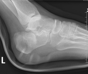
Before delving into a unilateral surgical discussion, it is important to acknowledge the persistent debate about conservative and surgical care of these fractures. Many true Jones fractures do heal with conservative management. In instances in which patients are poor surgical candidates due to comorbidities, certainly non-invasive treatment is justified. Even in healthy populations, many practitioners anecdotally maintain that non-weightbearing with or without external bone stimulators has facilitated consistent healing. However, even the most ardent supporters of conservative management must agree that healing is slow, unpredictable and re-fractures are relatively common. Clapper and colleagues found that even in cases with no medullary sclerosis, the average time to union was 21.2 weeks with a 28 percent failure rate with conservative treatment.3 Suffice it to say, this is an eternity for an active patient even if he or she is lucky enough to heal. In my practice, I encourage early surgical intervention. I believe a thorough inspection of the literature validates this contention. Before examining this technique, it is critical to establish the terminology of proximal fifth metatarsal fractures. As a general rule, there are three types of fifth metatarsal base fractures: the tuberosity avulsion fracture, the true Jones fracture and the diaphyseal stress fracture.4 A true Jones fracture involves the metaphyseal-diaphyseal junction and often enters the fourth and fifth intermetatarsal articulation. The diaphyseal stress fracture is distal to the fourth and fifth intermetatarsal articulation, and often displays significant cortical hypertrophy and medullary sclerosis. Most authors contend that the diaphyseal stress fracture is more commonly a true pathologic stress fracture due to repetitive use.5 This is in contrast to a Jones fracture, which is most commonly considered an acute injury due to a sudden axial and adduction force to the fifth metatarsal.6 However, it is my contention that these two types of fractures are more readily associated with repetitive stress overload and subsequent microfractures, and ultimately progress to macrofracture in lieu of an acute trauma. To this point, I would encourage you to pull radiographs of patients who have had Jones fractures. Take a moment to evaluate their foot type. Do they have a cavus foot or metatarsus adductus? Chances are, patients had one or the other, whether you were dealing with a diaphyseal stress fracture or a true Jones fracture. Furthermore, is there any sclerosis or cortical hypertrophy near the fracture? If so, chances are the fracture was a result of repetitive stress, usually in a higher arched foot, with or without some acute insult. This serves as the proverbial “straw that broke the camel’s back.” To summarize, I believe the majority of Jones fractures are true stress fractures in a vascularly compromised portion of bone found in a consistent foot type.7 This philosophy inspired the application of a different surgical technique in an attempt to address many of these observations. More than an academic exercise, it is an effort to improve and abbreviate your patient’s recovery time.
A Guide To The Surgical Technique
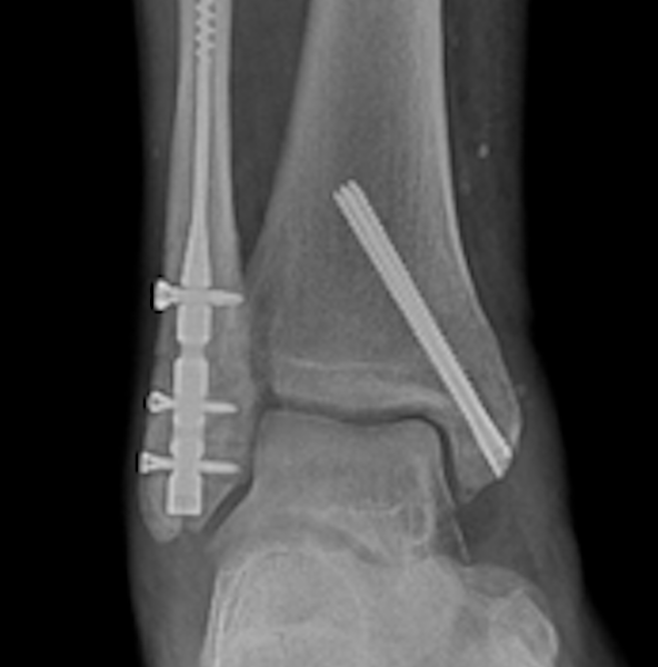
The technique is designed to address the factors that we know facilitate osseous healing in Jones fractures. There are three fundamental components to the procedure: • the evacuation of sclerosed bone around the fracture and in the medullary canal; • the introduction of healthy, autogenous bone graft; and • the implantation of rigid internal fixation. One would make a longitudinal incision over the fifth metatarsal shaft to the level of the fifth metatarsocuboid joint. Proceed to perform careful blunt dissection through the subcutaneous layer in an effort to protect the terminal branch/branches of the sural nerve. Identify the fracture plane. Then drive a cylindrical reamer (in this instance, a cylindrical reamer from an anterior cruciate ligament reamer system used in knee reconstruction) from lateral to medial to evacuate the bone on either side of the fracture line. Typically, the reamer is between 7 and 9 mm in diameter, and one does not drive it through the medial cortex. Remove the plug and take care to ensure the medullary canal is open distally and proximally. When it comes to extremely hard bone, it may be necessary to drill out the canal to facilitate endosteal healing. At this point, obtain a cylindrical autogenous graft from the calcaneus through a separate incision. The diameter of this graft is typically 1 mm larger than the evacuated bone on the fifth metatarsal to ensure a snug fit in the recipient site. Take some small cancellous bone fragments from the heel and pack them up and down the medullary canal. Proceed to place the autogenous plug into the recipient site with the lateral calcaneal cortex serving as the new fifth metatarsal cortex. A bone tamp may be necessary to accomplish a flush graft site. Finally, place a straight plate to bridge the fracture. Typically, there are two screws distal and proximal to the plug. Furthermore, drill the initial screws on either side of the graft eccentrically to compress the graft. Currently, I am using a six-hole plate from the Synthes Mini Fragment Set (Synthes). However, there are numerous small plates from various companies that I am sure would serve this purpose effectively. Currently, we are in the process of designing a “Jones Fracture Kit” complete with reamers, harvest cylinders and a locking plate in an attempt to standardize the necessary equipment.
In Conclusion
While it appears there are numerous steps to this technique, after a few cases, surgeons can execute it quite efficiently. In my practice, I have found that this technique clearly improves and accelerates the recovery from this fracture. To date, I have been employing this technique for roughly nine years and my partner (Richard Bouché, DPM) has used it for over five years. We have consistently seen osseous healing within four to five weeks. Originally, this technique was dedicated to those fractures that demonstrated sclerosis or presented with a prodrome of symptoms. However, as confidence in the technique evolved with excellent, predictable success in athletes, we are now recommending the procedure even in acute fractures devoid of sclerosis. To be clear, it is not my assertion that one should address all Jones fractures in this manner. In fact, most foot surgeons have been content with the results utilizing intramedullary screw fixation. If something works in your hands, by all means, stick to your guns. In my experience, the intermetatarsal screw fixation has been less than gratifying. Admittedly, most Jones fractures heal consistently with a well placed intermetatarsal screw. That said, the average time to union is well over seven weeks.8 This is not to mention some of the potential issues including: hardware irritation; plastic deformation; re-breakage after screw removal; and nerve entrapment with percutaneous screw fixation.5,9 Furthermore, what a luxury to be able to tell your active patients that you can honestly predict their return to activity will be two to three weeks earlier with this technique. Currently, our Sports Medicine Clinic in Seattle is beginning a formal, prospective, clinical trial to test the assertions in this article in a more objective, scientific manner. Observation, time and experience will be the true judges of this new technique. That said, after having addressed the Jones fracture in this manner for the better part of a decade, I can honestly contend that it is a superior procedure. I would be surprised if future research contradicts this assertion. Dr. Blahous is in private practice at the Sports Medicine Clinic in Seattle. He is a Fellow of the American College of Foot and Ankle Surgeons. Dr. Richie is an Adjunct Associate Professor in the Department of Applied Biomechanics at the California School of Podiatric Medicine at Samuel Merritt University. He is a Past President of the American Academy of Podiatric Sports Medicine. Dr. Richie is in private practice in Seal Beach, Ca. References 1. Torg J, Balduini F, et al. Fractures of the base of the fifth metatarsal distal to the tuberosity. J Bone Joint Surg. 1984; 66A: 209-214. 2. Delee JC, Evans JP, Julian J. Stress fracture of the fifth metatarsal. Am J Sports Med. 1983; 11(5):349-353. 3. Clapper MF, O’Brien TJ, Lyons PM. Fractures of the fifth metatarsal. Clin Ortho Rel Research. 1995; 315:238-241. 4. Lawrence SJ, Botte MJ. Jones fractures and related fractures of the proximal fifth metatarsal. Foot Ankle. 1993; 14(6):358-365. 5. Porter DA, Duncan M, et al. Fifth metatarsal Jones fracture fixation with a 4.5 mm cannulated stainless steel screw in the competitive recreational athelete: a clinical and radiographic evaluation. Am J Sports Med. 2005; 33(5):726-733. 6. Jones R. Fracture of the fifth metatarsal bone. Liverpool Med Chir J. 1902; 22:103-107. 7. Draves D. Lower Extremity Anatomy. Williams and Wilkins, Baltimore, 1986. 8. Mologne TS, Lundeen JM, et al. Early screw fixation versus casting in the treatment of acute Jones fractures. Am J Sports Med. 2005; 33(7):970-975. 9. Larson CM, et al. Intramedullary screw fixation of Jones fractures: analysis of failure. Am J Sports Med. 2002; 30(1):55-60.


