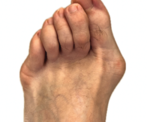
Roundtable Insights On Minimally Invasive Bunion Surgery
What approaches provide optimal outcomes? How do interested surgeons overcome the steep learning curve? When and how should one train in these techniques? What are the relevant indications and contraindications? Also, what does the future hold for minimally invasive surgery for hallux valgus? The panelists address these questions and more, sharing their insights on and experience with this evolving surgical pathway.
Q: Which minimally invasive (MIS) or percutaneous approach for surgical correction of bunion deformities do you routinely perform, and why? Furthermore, describe your typical postoperative course.
A:
Bradley Abicht, DPM, FACFAS, shares that, in a majority of mild-to-moderate hallux valgus deformities, he employs a percutaneous transverse distal first metatarsal osteotomy, with or without percutaneous Akin hallux osteotomy, as previously described by Vernois and Redfern.1 He says similar approaches have been coined minimum incision chevron Akin (MICA) or percutaneous chevron Akin (PECA). According to Dr. Abicht, patient benefits include small, cosmetically pleasing scars, immediate weight-bearing, less postoperative pain and swelling, less great toe joint stiffness and decreased overall complications compared to open approaches.
“After the outpatient same-day procedure, patients may immediately weight-bear in a surgical shoe,” he says. “I then see them in one week for a dressing change, and a week later for suture removal. Patients then use a compression garment, interdigital spacer and surgical shoe, fully weight-bearing from two to four weeks postop, at which time I obtain weight-bearing radiographs.”
He shares that his patients then transition to a supportive shoe with continued weight-bearing activity as tolerated. He permits athletic activity at three months, obtaining additional radiographs at three and six months.
Brian Loder, DPM, FACFAS, currently uses the percutaneous chevron Akin approach (PECA). This technique utilizes a low-speed, high-torque Shannon burr for the osteotomy and fixates with 4-0 cannulated chamfered screws.
“I have had the privilege of using most, if not all of the percutaneous systems on the market, and I find the PECA approach to be the most user friendly,” he says. “The increased stiffness of the guide wire allows for a more controlled insertion of the guide pins. Also, this technique utilizes the Stryker core power supply, which many hospitals already have.”
Dr. Loder’s typical postoperative protocol allows immediate weight bearing in a surgical shoe for four weeks. Beginning at two weeks, he starts range-of-motion exercises of the first MPJ to prepare for advancement to athletic shoes at five weeks. He relates that patients progressively increase their activity as tolerated.
Thomas Roukis, DPM, PhD, FACFAS, stresses that when discussing minimum-incision and percutaneous surgery it is critical to understand the nomenclature. He goes on to explain that minimum-incision surgery refers to the smallest incision necessary to perform the procedure under direct visualization. For first ray minimum-incision surgery, he says his typical incision length is between one and 2.5cm long.
“One performs the osseous work with a power saw blade under direct visualization of the intended structures, with or without intraoperative image intensification,” he adds.
Differentiating from “minimum-incision,” Dr. Roukis shares that percutaneous surgery refers to the smallest possible working incision without direct visualization of the underlying target structures. For first ray percutaneous surgery, his typical incision length is between one and three mm long. A power rotary burr using tactile sense accomplishes the osseous work, most commonly under intraoperative image intensification.
“Percutaneous surgery is also known as “blind” or “closed” surgery,” he says. “After 20 years in practice, countless surgical skills courses and direct observation of skilled international surgeons, I have not been able to convert to percutaneous 1st ray surgery. To be clear, this is not because the technique is a poor one, but rather that I am fearful of entering into another learning curve. So, I prefer minimum incision approaches for first ray surgery whenever feasible.”
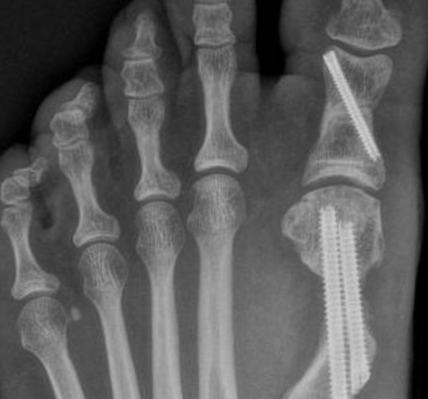
Dr. Roukis says his specific technique involves a transverse guided osteotomy fixated with an intra-medullary locked nail/plate (Centrolock®, Novastep). The osteotomy is a modified Hohmann fixed with an intra-medullary device as popularized by Bösch/ Giannini/Kramer and colleagues depending on the incision description (percutaneous, MIS, open, respectively).2-4
“The system I employ allows for precise manipulation of the first metatarsal head in the transverse plane (lateral translation ± shortening and PASA/DMAA rotational correction), sagittal plane (plantar displacement) and frontal plane (derotation) in addition to axial compression,” he says. “This is the so-called ‘3D+’ correction since in my experience there is no such thing as ‘4D’ correction.”
Dr. Roukis adds that his typical postop course involves a compression dressing and immediate flat-foot, non-propulsive weight-bearing in a postoperative shoe for two weeks. After suture removal, he advises a large toe spacer for the first webspace to support the soft-tissue balancing and a compression anklet for use at all times other than bathing. At this time, he says the patient then ambulates in an athletic, lace-up shoe to their tolerance with no ballistic activities until pain and swelling are totally absent, usually at the four-month mark.
Patrick DeHeer, DPM, FACFAS, FASPS, says he uses the SERI/Bösch technique.2,3 A minimally invasive Hohmann osteotomy, he adds that it is the only osteotomy that provides for triplanar correction.3 He cites fixation technique as critical to lock in all three planes of correction.
“I use a 2.0 mm Steinmann pin placed along the medial aspect of the distal phalanx, proximal phalanx and metatarsal head,” he shares. “I then redirect it from extra-osseous to intra-osseous by deflecting the pin with a hemostat within the canal of the metatarsal shaft. Either by hand or mallet, I advance the pin into the metatarsal base. The next step is critical to lock in the frontal plane correction. By not releasing any capsular or ligamentous structures, if you rotate the hallux so the toenail is directly straight up, this will result in correction of rotation at the osteotomy site. The pin is then driven past the metatarsal base, through the first cuneiform, into the navicular.”
He adds that crossing multiple cortices is important for stability and reducing pin tract infection risk.
“After doing several with just one pin, I noticed some sagittal plane displacement of the distal capital fragment, so I added a second point of fixation to stabilize the sagittal plane,” says Dr. DeHeer. “I use a 0.062-inch K-wire from the dorsal distal lateral metatarsal head to plantar proximal medial metatarsal shaft. This pin is a little tricky to throw, but very doable.”
He stresses the importance of verifying placement of both pins on AP and lateral fluoroscopy views. Postop, he says he allows immediate weight-bearing in a cast boot, removing the pins at six weeks and allowing transition into an athletic shoe at that time as well. He says he does not allow any high-impact activity until about 10 weeks postop.
Jonathan Sharpe, DPM, FACFAS, uses both minimal-incision and percutaneous approaches for bunionectomies.
“I have not abandoned more traditional practices of bunion correction such as the chevron osteotomy or Lapidus arthrodesis, but I perform increasingly more MIS and percutaneous approaches,” he clarifies. “Patients are becoming aware of the option and come for surgical consultation seeking it. We then discuss the approaches available and the associated benefits.”
He shares he typically recommends partial weight-bearing with crutches and a surgical shoe for seven to 10 days postop, followed by full weight-bearing in a protective surgical sandal or boot until radiographic evidence of bone union, on average at six to eight weeks.
Noman Siddiqui, DPM, MHA, FACFAS, relates he performs the same procedure he described in Podiatry Today in 2014,5 since then, incorporating screw placement and an adjunct Akin osteotomy
“I read Magnan and team’s article from 20056 and it made a lot of sense to me,” he says. “I knew I would incorporate this into my practice. My typical postoperative course is very simple; weight-bearing as tolerated in flat surgical shoe for four weeks. I transition to sneakers at four weeks postop. Impact activities take place with radiographic and clinical signs of healing, typically eight to 10 weeks in adults and six to eight weeks in pediatrics.”
Tea Nguyen, DPM, MPH, relates that she learned traditional MIS technique through cadaver labs and private training.
“I prefer either the exostectomy or Reverdin-Isham bunionectomy, with or without proximal phalanx osteotomy, all without internal fixation and three to five mm incisions,” she shares. This method, described by de Prado and team7, has consistently provided exceptional results and is extensively used in other countries. I have no reason to use fixation anymore.”
She continues on to say that she feels previous iterations of MIS fell out of favor for many possible reasons, including unstable osteotomies, short after-care and technical challenges with the drill system at that time.
“I follow Dr. de Prado’s technique7 and pioneers of the “traditional” MIS including Drs. Isham8 and La Bourde,” she adds. “The actual cut (Reverdin-Isham) is very stable, allowing immediate weight-bearing.”
Q: The literature illustrates a steep learning curve associated with mastering MIS and percutaneous bunion surgery approaches. No matter your preferred MIS or percutaneous surgical technique, what pearls can you provide other surgeons to overcome this learning curve?
A:
Dr. Abicht feels that when approaching any new surgical technique, it requires an understanding of the history or evolution of the technique, as well as knowledge of the current literature, including surgical pearls and best practices.
“Most importantly, the surgical technique requires practice,” he says. “Whether through sawbones, cadaver labs or other hands-on repetition/habituation techniques, practicing prior to performing the actual procedure on a live patient is imperative to master the stereokinetic skills required for reproducible success.”
He advocates for seeking training and labs with expert surgeons that routinely perform high volumes of percutaneous and MIS techniques. Finally, he recommends selecting a first patient with a mild bunion deformity and performing a thorough consent process regarding goals and potential to conversion to an open approach if necessary.
Dr. Loder agrees that patient and deformity selection is key, along with considering the OR space. He feels it is initially best to choose a patient with a moderate intermetatarsal angle, around 13 to 15 degrees.
“This allows an easier angle of entry for the guide pins and still leaves plenty of room for translation,” he explains. “In the beginning I try to teach surgeons to throw the guide wires first before performing the osteotomy so an escape plan can be easier. The best advice I can give is to train with a surgeon that that already mastered the technique, take your time and go slow. The MIS hallux valgus technique was designed to lead to quicker and less painful recovery for the patient, not necessarily to be easier for the surgeon.”
He adds that the beginning MIS surgeon needs to become comfortable with capital fragment translocation of 80 to 100 percent as this is where he feels one can find the best outcomes.
Being true to one’s skill set and knowing one’s limitations are key in Dr. Roukis’ opinion.
“Attend multiple training courses, digest every bit of information available, ask/spend time with experts, and ultimately, take a look in the mirror to make sure that what you are doing benefits patients over other techniques you have already mastered,” he relates.
Dr. DeHeer advises surgeons go slow and initially use fluoroscopy abundantly to assure the correct location for the osteotomy and appropriate fixation.
“The SERI/Bösch tricky point is redirecting the 2.0 mm pin to inside the bone,” he says. “I use a mallet to tap the pin distally and deflect it off the hemostat down the shaft of the metatarsal.”
Dr. Sharpe agrees that the learning curve poses a challenge and strongly advocates for cadaveric training on multiple occasions, including one that is spatially close to your first “real” procedure. He, too, agrees that frequent fluoroscopy usage, along with optimizing the OR set up, is important. Specifically, he recommends that the surgical foot rest on the fluoroscopic imager with the screen situated on the opposite side.
“Earlier in my MIS career I would translate the capital fragment and temporarily pin it to the second metatarsal with a K-wire to stablize the bone,” he adds. “This helped me maintain correction while delivering the guidewires for the cannulated implants. Also, placing these guidewires prior to making the osteotomy can be a very helpful practice.”
Dr. Siddiqui stresses that one should choose an approach that he or she likes and stick to it, not modifying the technique.
“Just pick a technique and do it that way without deviation until you understand the various nuances of positioning, radiographic tolerance and see the outcomes over many months,” he advises. “The tendency to deviate too soon can be a recipe for disaster.”
Dr. Siddiqui clarifies that he has only performed a transverse cut whereas Vernois and Redfern describe a chevron.1
“In my opinion, that is not the easiest to initially execute, so start with transverse as it is easier, reproducible and and I find no difference in healing or outcomes,” he says.
Dr. Nguyen shares the opinion that traditional MIS mastery requires more than just one lab. She relates she participated in private coaching with T.J. Ahn, DPM, to learn proper technique and after-care.
“For me, it was essential to get one-on-one training by a mentor who could guide me for at least one year, through the learning and healing phases,” she explains. “I highly recommend that interested parties, especially in methods without fixation, obtain a private mentor with vast experience who can show you all of the small, but important nuances of this procedure.”
Q: Should medical schools, residencies and fellowships teach MIS and percutaneous techniques alongside more traditional open approaches for surgical correction of bunion deformities? Why or why not?
A:
All of the panelists agree that these techniques should be a part of medical education curriculums, but have different viewpoints as to how and when.
Dr. Abicht advocates to include these concepts early in a learner’s career, alongside traditional open approaches. Due to pros and cons for both techniques, he feels some will excel with one over another.
He draws similar comparisons to open versus arthroscopic ankle arthrodesis, or open reduction with internal fixation of a calcaneal fracture through an open lateral extensile approach versus closed reduction with percutaneous fixation.
“As advances in technology and mastery of procedural efficiency takes place, we can bestow significant benefits to our patients, which should remain the ultimate goal,” says Dr. Abicht. “After practicing for a decade and previously training exclusively on open approaches, converting to MIS/percutaneous bunion correction has been the single biggest “game-changer” and patient pleaser I’ve witnessed in my own surgical practice.”
Due to his belief that MIS procedures will soon be an integral part of podiatric surgeons’ tool boxes, Dr. Loder agrees it is necessary to begin this education early in one’s career. Specifically, he opines that residencies and fellowships will need to offer MIS training to stay in-demand and competitive.
Dr. Roukis generally agrees, but adds that he feels learners need self-awareness and must train with true expert surgeons. Ultimately, he proposes that the role of the schools, residency and fellowship programs is to help learners obtain self-awareness, after which, they can best undergo training with masters, following the concepts of the “Conscious Competence Ladder.”9
Perspective on the role of MIS in podiatric surgical practice is important, according to Dr. DeHeer. He does support inclusion in the surgery curriculum in schools, but more importantly as part of residency training.
“Unfortunately, not a lot of foot and ankle surgeons do MIS. I do some, but it is only about 10 to 20 percent of my surgical cases,” he shares. “Allowing evidence-based research to guide us on the role of MIS in foot and ankle surgery will be critical.”
While supporting individuals’ decisions as to whether or not to adopt MIS, Dr. Sharpe says he believes that all trainees should have introduction to the technique.
“If the attendings in that program do not perform or subscribe to MIS bunion procedures that is perfectly okay,” he says. “They should explain why, but not withhold training or cadaveric labs from their students/ residents/felllows.”
Dr. Siddiqui concurs with open communication within training programs, especially regarding data, outcomes and prior biases against MIS.
“From a surgical standpoint, residents need to focus on open procedures,” he adds. “MIS is an advanced surgical technique, in my opinion, and a poor open surgeon will be poor MIS surgeon. Therefore, focus should be on good principles, performed open.”
Full exposure to comprehensive training for future surgeons is key, according to Dr. Nguyen, who feels that MIS is the way of the future. With the high cost of hospital surgeries and inconveniences of leaving the office, she maintains that it is important for foot and ankle specialists to know what is available so they can decide what works for them.
“I have been doing office-based bunion surgery almost exclusively in my practice in the past 13 months, she shares. “You definitely need the foundation of open surgery to understand the intricate anatomy of the foot. This helped me convert to MIS, knowing exactly where my incisions can safely go and if complications arise, I can still open if needed. I have not had to do this so far.”
Q: In your opinion, what are the relevant indications and contraindications of your preferred MIS or percutaneous approach? When would you consider an alternate MIS/ percutaneous or other procedure instead of your preferred approach?
A:
Regarding percutaneous distal first metatarsal osteotomy and percutaneous Akin osteotomy for surgical hallux valgus correction, Dr. Abicht relates that the main indications are a mild-to-moderate symptomatic bunion deformity in a surgically-appropriate patient for which conservative care has failed. He adds that these are typically primary hallux valgus procedures, but may apply in certain revision situations. Contraindications are similar to open bunion procedures, he says, but would include active acute or chronic infection, skeletally immature patients with open epiphyses and various revision cases best addressed through an open approach.
Dr. Loder agrees regarding the contraindications, and adds that one should avoid both MIS and open procedures for hallux valgus in moderate-to-severe osteopenia and peripheral vascular disease.
“The indication for MIS has more to do with the room present between the first and second metatarsal heads and less to do with the intermetatarsal angle,” he says. “When the space between the first and second metatarsal heads is greater than the width of the first metatarsal head, MIS will not fully correct the deformity. In these cases, I will use a Lapidus to get correction.”
Both Drs. Abicht and Loder comment that new Lapidus techniques could emerge that may allow for successful and reproducible results through MIS approaches.
Dr. Sharpe notes that the Lapidus is very valuable for an unstable medial column with a bunion deformity. He also says he gravitates to first MPJ arthrodesis for very large deformities in patients of advanced age.
He comments that for a patient with vascular concerns or who smokes, there might be some advantage to not opening the soft tissues. Otherwise, he agrees with the other panelists who feel the indications and contraindications for MIS parallels that of open bunion procedures.
Dr. DeHeer says he chooses an MIS approach where he previously used an Austin bunionectomy, essentially for a mild-to-moderate bunion deformity.
Focusing specifically on hallux valgus, Dr. Siddiqui says that if a patient has contraindications for open surgery, he does not perform MIS. Otherwise, he shares that he prefers MIS for all hallux valgus procedures.
Dr. Nguyen offers patients both open and MIS options, including discussing the risks and benefits, allowing patients to decide from there. She shares that the vast majority choose MIS under local anesthesia.
“For office-based surgery, patients should be relatively healthy, adhere to medical advice and able to sit in the exam chair for 45 minutes under local anesthesia,” she explains.
Acknowledging some patients prefer monitored anesthesia care in the surgery center, she will provide that as well. Conversely, she notes she has had success performing office surgery for higher-risk patients with non-healing foot ulcers due to underlying biomechanics, or for those who don’t want sedation or general anesthesia.
“You have to know and trust what will work for your patient,” she adds. “To date, I have no reported infections from office-based surgery and I see these patients within three to four days after MIS and continuously until they heal.”
“As the American College of Foot and Ankle Surgeons tag line states, we are all lifelong learners,”10 says Dr. Roukis. “In this context, we need to be self-critical of our outcomes and adjust the technique to improve the patient’s experience without perpetually being in a learning curve. I will leave expanding the indications for MIS and percutaneous surgery for those more skillful than I and follow their lead.”
Q: In the future, do you think MIS and percutaneous approaches will be more common than traditional open approaches for primary bunion surgeries? Why or why not?
A:
Most of the panelists see a significant role for MIS bunion surgery in the future. Dr. Abicht believes open and MIS or percutaneous approaches will continue, but more surgeons will learn MIS techniques and witness the resultant outcomes.
“Foot and ankle surgeons do need more high-quality long-term comparative prospective studies analyzing the superiority of both approaches, as the current literature lacks a high volume of studies involving the MIS or percutaneous approaches,” adds Dr. Abicht. “This is especially true of the American literature. Nevertheless, patients also seek out surgeons that can perform hallux valgus correction through MIS and percutaneous approaches over open traditional techniques. This factor more than anything may drive the continued evolution of MIS.”
Dr. Loder concurs that patient interest will drive selection of MIS procedures, and sees it as the procedure of choice for hallux valgus in the future. He cites shortened recovery time, less joint contracture, great correction and less pain as key considerations.
“I find that patients are willing to live with the bunion deformity rather than undergo an open procedure and are content to wait for the MIS technique to be offered in their community,” he says. “We, as a profession, have a long way to go in the process of developing a nearly perfect bunion correction procedure. I truly believe the current MIS techniques will continue to advance and outcomes will get better and better with each improvement.”
Contending that foot and ankle surgeons will need to settle into a balance between open and MIS/percutaneous techniques for first ray surgery, Dr. Roukis encourages review of “Scott’s Parabola”11 to determine where MIS and percutaneous first ray surgery are on the curve.
“I fear we are at the “peak of inflated expectations,” and we would all do well to minimize an eventual, unpleasant tumble down the curve into the “trough of disillusionment.”10
Dr. DeHeer feels the role of MIS surgery must align with a shift in the paradigm of understanding of hallux valgus.
“There is a bit of trend to the concept of MIS Lapidus,” he notes. “I have difficulty understanding how one can accomplish triplanar correction without releasing any of the dorsal, plantar or lateral ligamentous structures of the first tarsometatarsal joint. Frankly, the length of the incisions for debridement and fixation are not that much smaller than a traditional open approach.”
He cites three core principles he feels are important in HAV surgery: triplanar correction; being extra-articular to the first MPJ, preventing arthrofibrosis if possible; and preserving the metatarsal head blood supply.
Dr. Sharpe believes that, regardless of trends, results and long term outcomes will determine the future of MIS bunion surgery.
“Simply put, if patients are equally or more satisfied, then I believe it may become the norm in the future,” he adds.
Although he feels those who learn the methods will increasingly transition to more MIS procedures, Dr. Siddiqui says there will still be those with bias against it. Therefore, he feels MIS will not be the only approach. Instead, he comments that it could replace open distal osteotomies for hallux valgus.
Dr. Nguyen is hopeful that MIS will become more common than open bunionectomies, however she points out that it takes a different mindset when performing MIS.
“We are used to seeing everything opened up and using hardware, so I don’t think … MIS will be for everyone,”she says. “But I do hope more and more people adopt it, as I believe it could alleviate some of the burnout physicians face with our very convoluted U.S. healthcare system.
Noting that she has been very happy, even throughout the COVID-19 pandemic, performing office-based MIS bunion surgery, Dr. Nguyen also comments that patients express satisfaction with cost savings and transparency this situation affords. Although she stresses that she does not use hardware in her MIS procedures, due to the reliability she has experienced, she suggests that surgeons interested in MIS could potentially take advantage of opportunities to learn techniques with fixation systems as an introduction.
“(Those using MIS systems with fixation) are also getting great results,” she says. It ends up being whatever works best for the surgeon that will produce the best results for patients.”
Dr. Abicht is the Section Chair of the Podiatric Medicine and Surgery Department at Gundersen Health System in La Crosse, Wis. He is a Fellow of the American College of Foot and Ankle Surgeons. He discloses that he is a consultant for Novastep.
Dr. DeHeer is the Residency Director of the St. Vincent Hospital Podiatry Program in Indianapolis. He is a Fellow of the American College of Foot and Ankle Surgeons, the American Society of Podiatric Surgeons, the American College of Foot and Ankle Pediatrics and the Royal College of Physicians and Surgeons of Glasgow.
Dr. Loder is a Fellow of the American College of Foot and Ankle Surgeons and practices in Clinton Township, Mich. He is the Director of the Podiatric Medicine and surgery Residency at Henry Ford Macomb Hospital in Clinton Township, Mich.
Dr. Nguyen is a fellowship-trained podiatrist and was an International Visiting Scholar in the division of Plastic Reconstructive Microsurgery at Chang Gung Memorial Hospital in Taiwan. She is in private practice in Freedom, Calif. Dr. Tea Nguyen can be reached at teadpm.com.
Dr. Roukis is a Fellow and Past President of the American College of Foot and Ankle Surgeons. He is a Professor in the Department of Orthopaedic Surgery and Rehabilitation at the University of Florida Health System in Jacksonville, Fla. Dr. Roukis discloses that is he a developer of Centrolock for Novastep.
Dr. Sharpe is a Fellow of the American College of Foot and Ankle Surgeons and Director of Fellowship Training at Orthopedic Associates of Lake County in Concord, Ohio. Dr. Sharpe discloses that he was a member of the surgeon design team for Centrolock for Novastep, and is a consultant for Stryker/Wright and Novastep.
Dr. Siddiqui is a Fellow of the American College of Foot and Ankle Surgeons and the Director of Podiatric Surgery and the Deformity Correction and Orthoplastics Fellowship at the Rubin Institute for Advanced Orthopedics/ International Center for Limb Lengthening in Baltimore. He discloses that he is a consultant for Arthrex.
1. Redfern D, Vernois J, Legré BP. Percutaneous surgery of the forefoot. Clin Podiatr Med Surg. 2015;32(3):291-322.
2. Bösch P, Markowski HP, Rannicher V. Technique and first results of the subcutaneous distal osteotomy of the first metatarsal bone. Orthop Praxis. 1990;26:51-56.
3. Giannini S, Faldini C, Nanni M, Di Martino A, Luciani D, Vannini F. A miniminally invasive technique for surgical treatment of hallux valgus: simple, effective, rapid, inexpensive (SERI). Int Orthop. 2013;37(9):1805-1813.
4. Lamprecht E, Kramer J. Die metatarsale -i- osteotomie nach behandlung des hallux valgus. Orthop Praxis. 1982;8:636–645.
5. Siddiqui NA, LaPorta G. Emerging insights on minimally invasive hallux valgus correction. Podiatry Today. 2014;27(9):26-32.
6. Magnan B, Pezzé L, Rossi N, Bartolozzi P. Percutaneous distal metatarsal osteotomy for correction of hallux valgus. J Bone Joint Surg Am. 2005;87(6):1191-1199.
7. de Prado M, Golanó P, Ripoli PL. Minimally invasive foot surgery. 2009:About Your Health Publishers.
8. Bauer T, de Lavigne C, Biau D, De Prado M, Isham S, Laffenetre O. Percutaneous hallux valgus surgery: a prospective multicenter study of 189 cases. Orthop Clin North Am. 2009;40(4):505-514
9. MindTools. The conscious competence ladder. Available at: https://www.mindtools.com/pages/ article/newISS_96.htm . Accessed April 9, 2021.
10. American College of Foot and Ankle Surgeons. Available at: www.acfas.org. Accessed April 9, 2021.
11. Scott JW. Scott’s parabola. BMJ. 2001;323(7327):1477.