


Treating Secondary Infection In A Patient With Severe Tinea Pedis
These authors offer insights on the management of a 43-year-old patient with tinea pedis complicated by cellulitis and multiple chronic ulcers. The most common causative microorganisms of the dermatophyte tinea pedis are Trichophyton rubrum, Trichophyton mentagrophytes and Epidermophyton floccosum with T. rubrum being the most common cause worldwide. A hot, humid, tropical environment and prolonged use of occlusive footwear are the most common risk factors for all types of tinea pedis. Swimming and communal bathing may also increase the risk of infection.1,2 Tinea pedis is the most common dermatophytosis worldwide with 70 percent of the population infected at some time.3 Tinea pedis is not associated with significant mortality or morbidity. Tinea pedis affects every racial and ethnic group. The disease more commonly affects males. The prevalence of tinea pedis increases with age and it is rare during childhood.4 There are different clinical variations of tinea pedis. They include interdigital, chronic hyperkeratotic, inflammatory/vesicular and ulcerative tinea pedis. Interdigital. This is one of the most characteristic types of tinea pedis with erythema, maceration, fissuring and scaling. It is often associated with pruritus. Chronic hyperkeratotic. This presentation is characterized by chronic plantar erythema with slight scaling to diffuse hyperkeratosis. Inflammatory/vesicular. This presentation is marked by painful, pruritic vesicles or bullae, most often on the instep or anterior plantar surface. The lesions can contain either clear or purulent fluid. Ulcerative. This is characterized by rapidly spreading vesiculopustular lesions, ulcers and erosions, typically in the interdigital spaces, and is usually associated with a secondary bacterial infection. Cellulitis, lymphangitis, pyrexia and malaise are usually associated with this infection. This type is commonly present in immunocompromised patients and those with diabetes. One can diagnose tinea pedis using potassium hydroxide (KOH) staining for fungal elements. The infection is easily visible under the microscope. A fungal culture may confirm the diagnosis. Treatment usually consists of topical antifungals, oral antifungals or a combination of both. One would use topical agents for one to six weeks. Untreated tinea pedis can lead to secondary cellulitis, lymphangitis, pyoderma and osteomyelitis. The type of tinea pedis infection and underlying conditions affect the prognosis but with appropriate treatment, the prognosis is good.
A Closer Look At The Initial Patient Presentation
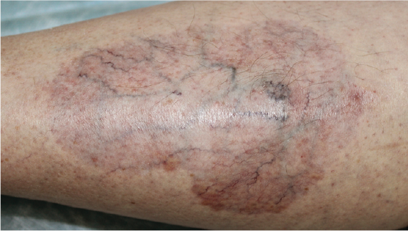
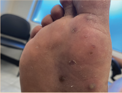
A 43-year-old male presented with a chief complaint of a chronic fungal infection of the right foot. He says the first symptoms of the infection appeared about three years prior when he noticed itching, burning and pain on ambulation in the third and fourth interdigital spaces of the right foot and developed acute cellulitis of the right foot. His primary care physician treated him with an antibiotic for acute cellulitis followed by a third month’s treatment with a topical antifungal cream. After prolonged treatment, the patient developed chronic multiple ulcers of the third and fourth interdigital spaces of the right foot. The patient was subsequently admitted to a local wound care center with a chief complaint of burning, throbbing, sharp pain and a non-healing, post-fungal wound of the third and fourth interdigital spaces of the right foot with serosanguinous exudate. The initial treatment was minor debridement of the wound with application of gentian violet and prescription terbinafine (Lamisil, Novartis) PO. He also received topical econazole nitrate (Spectazole cream, Janssen Pharmaceuticals) and hydrocodone (Vicodin, Abbott Laboratories) for pain management. At a fourth week of follow-up, he had decreases in edema, exudate, pain and the size of the wound. The physician recommended he continue topical antifungal therapy for three more weeks. At the two month follow-up, the patient had an increase in the width and length of the wound with edema and erythema of skin of the right foot and serous exudates from the wound. The patient received aseptic debridement of the wound, topical gentian violet application and prescribed amoxicillin clavulanate (Augmentin, GlaxoSmithKline) PO and topical Betadine ointment (Purdue Pharma) for two weeks with no relief. Four months after the original admission to the wound care center, the patient was admitted again to the center with tinea pedis complicated with cellulitis and chronic ulcers of the third and fourth interdigital space of the right foot. He received antifungal treatment with terbinafine PO, topical econazole nitrate cream, gentian violet and a Xeroform petrolatum dressing (Covidien), and pain management with hydrocodone. After four weeks with no improvement, physicians added griseofulvin (Gris-PEG, Pedinol) PO and naftifine gel (Naftin, Merz Pharmaceuticals) to the treatment regimen. Physicians discontinued terbinafine PO and econazole nitrate cream, and continued hydrocodone for pain management. Two months later, the patient had complaints of a painful, swollen right foot and an open, tender pruritic lesion of the third and fourth interspace and dorsal aspect of the second through the fifth toes of the right foot. His condition was getting progressively worse with severe pain on ambulation and pain in all type of shoes, causing him to miss work. The patient was taking griseofulvin 250 mg TID, hydrocodone, and topical naftifine gel, He was also using a Xeroform petrolatum dressing and a controlled ankle motion walker. His vascular, neurologic and orthopedic exams were normal. X-rays showed no radiographic evidence of osteomyelitis. We obtained wound cultures. Initial treatment consisted of a therapeutic injection with lidocaine (Xylocaine, Astra Zeneca) and bupivicaine (Marcaine) for pain relief, application of gentian violet and prescribing Pedi-Dry, Betadine soak gauze and lidocaine patches with weekly follow-up.
How The Patient Fared During Follow-Up
On the first follow-up, we added empiric antibiotic therapy with amoxicillin clavulanate PO and piroxicam (Feldene, Pfizer) PO for pain management to the treatment regimen. The culture came back positive for gram-negative Pseudomonas aeruginosa. We added levofloxacin (Levaquin, Ortho-McNeil) PO with amoxicillin clavulanate d/c and topical silver sulfdiazine (Silvadene, Monarch Pharmaceuticals) to his treatment. At the two week follow-up visit, treating the affected area with levofloxacin PO showed much success with decreased edema, erythema, width, length and depth of the wound and a 100 percent granular base of the lesion. However, in the next three weeks, the patient started feeling severe pain on ambulation, itching and a burning sensation in interdigital spaces three to four on the right foot. He also had increased swelling and hyperemia of the right foot. In regard to the physical exam, the Wood’s lamp was positive. We suspected an erythrasma superinfection and prescribed erythromycin with continued Betadine soak gauze and daily local wound care. We saw the patient on a weekly basis and provided local wound care. Due to persistent severe pain, we added hydrocodone for pain management. His condition significantly improved, his skin showed excellent signs of healing and the Wood’s lamp test showed resolution of erythrasma. We subsequently referred the patient to physical therapy to help reduce pain and increase the range of motion of the right foot. After one week of physical therapy, the patient went on vacation. During this time, he swam and walked barefoot. After vacation, the patient came back to the clinic with the complaints of severe pain, swelling and drainage from the wound of the right foot. The Wood’s lamp test was negative and we obtained a wound culture, which was positive for Pseudomonas aeruginosa. To rule out an abscess of the right foot, we sent the patient for magnetic resonance imaging (MRI) of the right foot. To rule out neoplasm, we recommended a periodic acid Schiff (PAS) stain with histopathology. The PAS results were positive for orthokeratotic hyperkeratosis and negative for fungus. The MRI demonstrated chronic phlegmon with no signs of abscess. The treatment consisted of moxifloxacin (Avelox, Bayer) PO for P. aeruginosa, local wound care with application of Burow's solution to reduce inflammation and piroxicam for pain management. We successfully treated the patient for severe foot infection but due to his non-adherence to follow-up, it was hard to obtain the final results of our treatment.
Understanding The Prevalence And Impact Of Pseudomonas
Pseudomonas is a strictly aerobic gram negative bacterium. Pseudomonal species have been found in water, plants, soil and animals. P. aeruginosa colonization occurs in more than 50 percent of humans and Pseudomonas aeruginosa are considered the most common pseudomonal species.5 Pseudomonas is a clinically serious and opportunistic pathogen, usually causing nosocomial infections.6 These organisms also exhibit innate resistance to many antibiotics and can develop new resistance after exposure to antimicrobial agents. P. aeruginosa rarely causes disease in healthy people. Most infections occur in compromised hosts, such as those with disrupted physical barriers to bacterial invasion like macerated skin, ulcers, burn injuries, intravenous lines, urinary catheters, dialysis catheters, endotracheal tubes and impaired immune systems that one may encounter in patients with. HIV, hypogammaglobulinemia, cystic fibrosis, complement deficiency and iatrogenic immunosuppression.7 According to data from the Centers for Disease Control and Prevention (CDC) National Nosocomial Infections Surveillance System of 2004, P. aeruginosa is the leading cause of intensive care unit-related pneumonia and osteochondritis.8 It is the second most common gram-negative organism, responsible for 9 percent of all nosocomial bacterial and fungal isolates. P. aeruginosa is the fourth most common cause of surgical site infections and hospital-acquired gram-negative rod bacteremia.8
What You Should Know About Secondary Infections Caused By Pseudomonas
Children have a higher risk than adults to pseudomonal osteochondritis infections following puncture wounds of the foot.9 Older patients are more susceptible to pseudomonal bone and joint infections. Children have a higher incidence of developing pseudomonal folliculitis than adults.9 Skin and soft tissue infections caused by Pseudomonas include burn wound sepsis, dermatitis, ecthyma gangrenosum, pyoderma, surgical and wound infections, including cellulitis, hot tub folliculitis, necrotizing fasciitis and chronic paronychia. Ecthyma gangrenosum is a rare but pathognomonic form of pseudomonal infection. Although ecthyma gangrenosum is often due to Pseudomonas, case reports indicate that the same frequency is associated with non-Pseudomonas ecthyma gangrenosum (i.e., Fusarium, Klebsiella). Ecthyma gangrenosum in patients without neutropenia is very rare as are non-bacteremic ecthyma gangrenosum cases. However, non-septicemic cases occur in patients without immunosuppression or neutropenia, or following antibiotic therapy. Pseudomonal burn wound infections occur with systemic involvement (e.g., bacteremia, fever, hypothermia, disorientation, obtundation, hypotension, oliguria, ileus and leukopenia). Chronic paronychia slowly evolves and initially presents with tenderness and mild swelling about the proximal and lateral nail folds. Pseudomonal toe web infections are distinctive clinical entities that are often misdiagnosed as tinea pedis. Dried exudate or a green stain (caused by pyoverdin) may be present on socks or toenails. Pseudomonas folliculitis presents as cutaneous lesions any time from eight hours to five days or more (mean incubation period is 48 hours) after using a contaminated whirlpool, home hot tub, water slide, physiotherapy pool or contaminated loofah sponge. Malaise and fatigue may occur during initial days of the eruption. Fever (uncommon) is low grade when present. Localized cellulitis can occur as a secondary infection of tinea of the toe webs or groin. It may also be triggered by bedsores, stasis ulcers or burns. Localized cellulitis can also occur in grafted areas or under the foreskin of the penis. Maceration or occlusion of these skin lesions leads to secondary infection. Deep erosions and tissue necrosis may occur before one correctly diagnoses the condition. Severe pain is highly characteristic of an evolving infection. The hallmark lesions resulting from pseudomonal wound infections are multifocal with dark-brown to black or violaceous discoloration of the burn eschar. This is accompanied by edema and hemorrhagic necrosis. Ecthyma gangrenosum lesions are multiple noncontiguous ulcers or solitary ulcers. These lesions begin as isolated, red, purpuric macules that become vesicular, indurated and eventually, bullous or pustular. The bullae may be hemorrhagic but contain little if any pus. Lesions can remain localized or more often can extend over several centimeters. The central area of these lesions becomes hemorrhagic and necrotic. Then the lesion denudes to form a gangrenous ulcer with a gray-black eschar and erythematous halo. Although lesions can occur anywhere, they occur mainly in the gluteal and perineal regions (57 percent), the extremities (30 percent), the trunk (6 percent) and the face (6 percent).5 These skin lesions heal slowly. Patients with septicemia have associated signs that include elevated temperatures, chills, hypotension, tachycardia and tachypnea. In chronic paronychia, the skin around the nail becomes pale, red, painful and swollen. A small amount of pus may occasionally emanate from beneath the proximal nail fold. The nail plate turns green-black, which is characteristic of pseudomonal infections. The condition causes little discomfort or inflammation. This presentation is often confused with subungual hematoma. A Pseudomonas-infected toe web presents as a thick, white, macerated scale with a green discoloration. The most consistent clinical feature is soggy, wet toe webs and adjacent skin. In the mildest form of pseudomonal infection in the toe web, the affected tissue is damp, softened, boggy and white. The second, third and fourth toe webs are the most common sites of initial involvement. Severe forms may progress to denuded skin and profuse, serous or purulent material. Pseudomonas folliculitis presents with a few to more than 50 urticarial plaques that measure 0.5 to 3 cm in diameter with a central papule or pustule on all skin surfaces other than the head. The rash can be a polymorphous eruption or a mixture of follicular, maculopapular, vesicular or pustular lesions. These lesions often are pruritic. Most clear in seven to 10 days, leaving round spots of red-brown post-inflammatory hyperpigmentation. However, some patients may have recurrent crops of lesions over an extended period of three months. Pseudomonas cellulitis presents with a dusky red to bluish or green skin discoloration and purulent discharge. The typical fruity or mouse-like odor has been linked to pseudomonal infection. Vesicles and pustules may occur as satellite lesions. The eruption may spread to cover wide areas and cause systemic manifestations.
Current Insights On Lab Tests And Pseudomonas Infections
The following laboratory results are helpful to confirm a pseudomonal infection. * Complete blood cell counts reveal leukocytosis with a left shift and bandemia, which indicates the possible presence of toxic granulations or vacuoles. * Erythrocyte sedimentation rate (ESR) and C-reactive protein (CRP) levels may be elevated in infection. * Metabolic profile reveals any electrolyte abnormalities, the degree of dehydration and worsening renal function. * Wound and burn cultures can be helpful to identify pseudomonal infections. * A Wood light examination of a pseudomonal toe web reveals a green-white fluorescence from the elaboration of pyoverdin. * A computed tomography scan of soft tissue and bone of skin infections may be required to exclude an abscess formation or osteomyelitis.
Key Treatment Considerations
Treatment of Pseudomonas infections entails the use of antimicrobial agents.10 Anti-pseudomonal drug combination therapy (such as a beta-lactam antibiotic with an aminoglycoside) is usually recommended for the initial empiric treatment of a pseudomonal infection, especially for patients with neutropenia, bacteremia, sepsis, severe upper respiratory infection or abscess formation. The choice of antibiotic also depends on the site and extent of infection, and on local antibiotic resistance patterns. Reports of more resistant strains of Pseudomonas organisms to the currently used antimicrobials are causing much concern.11 B. cepacia has grown resistant to aminoglycosides, anti-pseudomonal penicillins and most beta-lactam agents. Some strains are variably susceptible to third-generation cephalosporins, ciprofloxacin (Cipro, Bayer), trimethoprim-sulfamethoxazole (Septra, GlaxoSmithKline), ampicillin-sulbactam (Unasyn), chloramphenicol (Neogen) or meropenem (Merrem, AstraZeneca).12 Since human cases of glanders are rare, limited information is available about antibiotic treatment of the organism in humans. Sulfadiazine has been effective in experimental treatments of animals and humans. B. mallei organisms are usually sensitive to tetracyclines, ciprofloxacin, streptomycin, novobiocin, gentamicin, imipenem (Primaxin, Merck), ceftazidime (Fortaz, GlaxoSmithKline) and sulfonamides. Researchers have reported resistance to chloramphenicol.12 Treatment duration is often prolonged, from one to two months, and clinicians often combine this with surgical drainage. Ceftazidime alone or in combination with either trimethoprim-sulfamethoxazole or amoxicillin clavulanate is the therapy of choice for B. pseudomallei.12 The organism is usually sensitive to imipenem, penicillin, doxycycline (Adoxa, PharmaDerm), azlocillin, ceftazidime, ticarcillin-clavulanic acid (Timentin, GlaxoSmithKline) and ceftriaxone (Rocephin, Hoffmann-La Roche). Initiate treatment early in the course of the disease. The organism is resistant to ciprofloxacin and aztreonam (Azactam, Bristol-Myers Squibb). Treatment is often prolonged, from three to 12 months, with the longest duration of therapy used for chronic extrapulmonary disease. Management of pseudomonal cellulitis includes the use of a PO antibiotic for seven to 10 days. This often resolves a localized infection. Pseudomonal toe web infections require initial debridement with applications of silver nitrate or 5% acetic acid to the toe webs and the dorsal and plantar areas. Following this initial treatment, apply a topical antibiotic, silver sulfadiazine cream or Castellani paint until infection resolves. A PO quinolone effectively reduces the duration of infection. Pseudomonal folliculitis is often "self-limiting." Treatment may require only application of silver sulfadiazine cream or 5% acetic acid wet compresses for 20 minutes two to four times daily with topical antibiotics.
In Conclusion
Interdigital toe web space is a warm, moist, protected environment that predisposes to the proliferation of both dermatophytes and gram-negative organisms. Tinea pedis is by far the most common fungal infection. Maceration, scaling and fissures result, allowing for overgrowth of bacteria that normally inhabit this interspace. Proliferation of these organisms, which include, most prominently, Pseudomonas aeruginosa, other gram-negative bacteria (e.g., Escherichia coli and Proteus mirabilis) and gram-positive bacteria, leads to an aggressive, painful infection. In our case, we used all appropriate diagnostic, therapeutic and prophylactic tools recommended by many authors to succeed in treatment of the patient. Some authors also recommend treatment with topical amikacin 5% gel (Amikin, Sopharma), Castellani paint and compresses with 2% to 5% acetic acid, which we might add to our treatment approach. In terms of antibiotic treatment, in our opinion, a first-generation quinolone such as ciprofloxacin might be more effective versus the second-generation levofloxacin or third-generation moxifloxacin that cover gram-negative organisms but are more active against gram-positive flora. In addition, the patient was getting treatment in different wound care facilities for long periods of time with medications such as amoxicillin-clavulanate and antifungal creams that were not covering P. aeruginosa infection. This might lead to antibiotic resistance. Dr. Moharan is the Former Chief Podiatry Resident at St. John’s Episcopal Hospital in Far Rockaway, N.Y. and Senior Resident at Kennedy University Hospital in Turnersville, N.J. Dr. Ahmed is a Former Chief Podiatry Resident at Mount Sinai Hospital of Queens, N.Y. and currently a Wound Care Fellow at St. John’s Episcopal Hospital. Dr. Karpenko is Former Chief Resident at Kingsbrook Jewish Medical Center in Brooklyn, N.Y. and is currently practicing in Brooklyn. References 1. Gentles JC. The isolation of dermatophytes from the floors of communal bathing places. J Clin Pathol. 1956; 9(4):374-7. 2. Resnik SS, Lewis LA, Cohen BH. The athlete's foot. Cutis. 1977; 20(3):351-3, 3. Malcolm B. Tinea pedis. Practitioner. 1998; 242(1584):225. 4. Elewski BE. Cutaneous mycoses in children. Br J Dermatol. 1996; 134(Suppl 46):7-11; discussion 37-8. 5. Ng W, Tan CL, Yeow V, Yeo M, Teo SH. Ecthyma gangrenosum in a patient with hypogammaglobulinemia. J Infect 1998; 36(3):331-5. 6. Pollack M. The virulence of Pseudomonas aeruginosa. Rev Infect Dis. 1984; 6(S6):17-26. 7. Kielhofner M, Atmar RL, Hamill RJ, Musher DM. Life-threatening Pseudomonas aeruginosa infections in patients with human immunodeficiency virus infection. Clin Infect Dis. 1992; 14(2):403-11. 8. Centers for Disease Control and Prevention. National Nosocomial Infections Surveillance Systems Report. Am J Infect Control. 2004; 32(4):470-85. 9. Jarvis JG, SkipperJ. Pseudomonas osteochondritis complicating puncture wounds in Children. J Pediatr Orthop. 1994; 14(6):755-9. 10. Giamarellou H, Antoniadou A. Antipseudomonal antibiotics. Med Clin North Am. 2001; 85(1):19-42, v. 11. Carmeli Y, Troillet N, Eliopoulos GM, Samore MH. Emergence of antibiotic-resistant Pseudomonas aeruginosa: comparison of risks associated with different antipseudomonal agents. Antimicrob Agents Chemother. 1999; 43(6):1379-82 12. Byrne S, Maddison J, Connor P, et al. Clinical evaluation of meropenem versus ceftazidime for the treatment of Pseudomonas spp. infections in cystic fibrosis patients. J Antimicrob Chemother. 1995; 36(Suppl A):135-43. Additional References 13. Baltch AL, Griffin PE. Pseudomonas aeruginosa bacteremia: a clinical study of 75 patients. Am J Med Sci. 1977; 274(2):119-29. 14. Brewer SC. Clinical investigations in critical care: ventilator-associated pneumonia due to Pseudomonas aeruginosa. Chest. 1996; 109(4):1020-30. 15. Gupta AK, Baran R, Summerbell R. Onychomycosis: strategies to improve efficacy and reduce recurrence. J Eur Acad Dermatol Venereol. 2002; 16(6):579-86. 16. Pellizzari C. Recherche sur Trichophyton tonsurans. G Ital Mal Veneree. 1888; 29:8. 17. Douidar SM, Snodgrass WR. Potential role of fluoroquinolones in pediatric infections. Rev Infect Dis. 1989; 11(6):878-89.

