What Caused These Multiple Hyperpigmented Macules of the Mouth?
© 2023 HMP Global. All Rights Reserved.
Any views and opinions expressed are those of the author(s) and/or participants and do not necessarily reflect the views, policy, or position of The Dermatologist or HMP Global, their employees, and affiliates.
Case Report
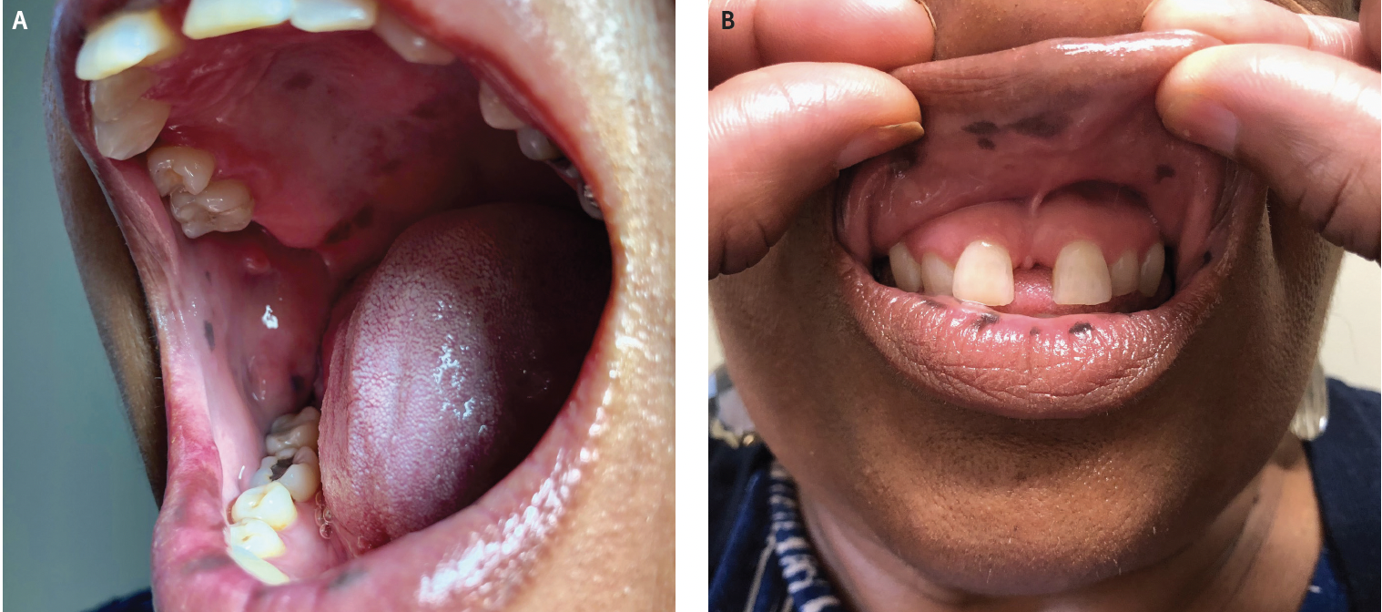
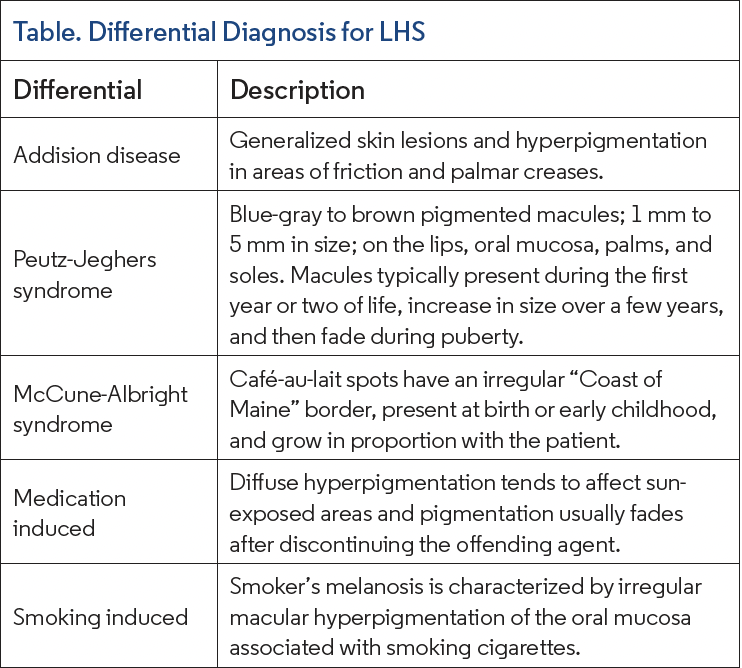
A 62-year-old Black woman presented to the outpatient dermatology office at the recommendation of her dentist for evaluation of many asymptomatic, small, dark brown hyperpigmented macules of the mouth with onset during the past 7 years. The lesions were located on the hard palate, buccal mucosa, and mucosal lips (Figure). Further evaluation identified four 1- to 3-mm macules of the distal fingers and a single, uniform, 1.5-mm longitudinal brown stripe of the right fifth digit but no other areas of melanonychia. The patient denied any changing lesions, ulceration, or genital involvement and declined a genital examination. There was no family or personal history of colon cancer, and a previous screening colonoscopy was normal. No other family members had similar oral lesions. A 3-mm punch biopsy of a macular pigmented area of the right lower mucosal lip was taken. Histopathology revealed no increase in melanocytes, consistent with a melanotic macule.
What Is The Diagnosis?
Check your answer below!
Diagnosis
Laugier-Hunziker syndrome
Laugier-Hunziker syndrome (LHS) is described as a benign macular hyperpigmentation disorder without concomitant somatic alterations or risk of malignant transformation. This syndrome primarily affects women in the third to fifth decade of life and there is typically no family history of LHS.1
Clinical Presentation and Histology
The lesions are primarily characterized by hyperpigmented macules on the lips, oral and genital mucosa, palms, soles, and nails. The macules tend to be characterized as well-defined and light brown to black, usually 0.1 cm to 0.5 cm in size. Nail involvement is common, with pigmentation in the nail fold called pseudo- Hutchinson sign.2
Histologically, these lesions commonly have epidermal melanosis without melanocytosis and frequently increased basal layer pigmentation with normal numbers of melanocytes. However, there have been a few cases reported with histopathology findings of increased intra-epidermal melanocytes and one report describing cellular atypia of intraepithelial melanocytes from a lesion on a sun-exposed area of the lip.3
Pathogenesis
Currently, the etiology and pathogenesis of LHS have not been elucidated. It has been reported as occurring both sporadically without a genetic association and as an autosomal dominant condition in families. It has been postulated that an alteration of the melanocytes leads to an increased synthesis of melanosomes and deposition within the basal cell layer, but specific mutations have yet to be discovered.4
Differential Diagnosis
The diagnosis of LHS is one of exclusion. Because this syndrome may mimic more serious diseases, certain conditions must be ruled out before the diagnosis of LHS can be given. The differential diagnosis for LHS includes Addison disease, Peutz-Jeghers syndrome, McCune-Albright syndrome, and adverse effects of medications and smoking (Table).
LHS can be differentiated from Addison disease by checking the cortisol level, which is decreased in Addison disease and within normal limits in LHS. The skin lesions seen in Addison disease tend to be generalized. Most commonly, these pigmentary changes are seen in sun-exposed areas, areas exposed to chronic friction, and palmar creases. The hyperpigmentation in Addison disease will fade within a few days of starting glucocorticoid therapy.5
Peutz-Jeghers syndrome can present with similar pigmented macules; however, these tend to be blue-gray to brown; 1 mm to 5 mm in size; and occur on the lips, oral mucosa, palms, and soles. However, the macules of Peutz-Jeghers syndrome typically present during the first year or two of life, increase in size over a few years, and then fade during puberty. LHS typically presents during the third to fifth decade of life.6
McCune-Albright syndrome is defined as a triad of precocious puberty, café-au-lait lesions, and fibrous dysplasia of bone. Café-au-lait spots in McCune-Albright syndrome tend to have an irregular “Coast of Maine” border, present at birth or early childhood, and grow in proportion with the patient.7
There are several drugs that can cause diffuse hyperpigmentation, such as chemotherapeutic agents, antimalarials, oral contraceptives, heavy metals, prostaglandin agonists, amiodarone, diltiazem, minocycline, zidovudine, and psychotropic drugs. Drug-associated hyperpigmentation tends to affect sun-exposed areas, and the pigmentation usually fades after discontinuing the offending agent.8 Smoking cigarettes can cause smoker’s melanosis, which is characterized by irregular macular hyperpigmentation of the oral mucosa.9
Treatment and Complications
Because LHS is a benign disorder, treatment is primarily cosmetic. Options for therapy include cryotherapy, CO2 lasers, and avoiding sun exposure. Although LHS is classically benign, there have been several cases reported with associated esophageal melanocytosis, actinic lichen planus, hypocellular bone marrow, and thrombocytopenia.10
Our Patient
In the absence of childhood onset of polyposis of the colon and no family history of colon cancer or similar cutaneous findings, the diagnosis of LHS was made. The patient was counseled on the benign nature of the lesions and elected for continued observation rather than cosmetically aimed treatment.
Conclusion
LHS is an acquired, benign hyperpigmentation condition of the skin characterized by hyperpigmented macules on the lips, oral and genital mucosa, palms, soles, and nails. Clinical diagnosis is made via the exclusion of other pathologies. The benign nature of the disorder does not necessitate treatment unless the patient has cosmetic concerns for which utilization of CO2 lasers may be considered.
References
1. Laugier P, Hunziker N. Pigmentation mélanique lenticulaire, essentielle, de la muqueuse jugale et des lèvres. Arch Belg Dermatol Syphiligr. 1970;26(3):391-399.
2. Koch SE, LeBoit PE, Odom RB. Laugier–Hunziker syndrome. J Am Acad Dermatol. 1987;16(2 Pt 2):431-434. doi:10.1016/s0190-9622(87)70055-3
3. Moore RT, Chae KA, Rhodes AR. Laugier and Hunziker pigmentation: a lentiginous proliferation of melanocytes. J Am Acad Dermatol. 2004;50(5 Suppl):S70- S74. doi:10.1016/j.jaad.2003.09.016
4. Nayak RS, Kotrashetti VS, Hosmani JV. Laugier-Hunziker syndrome. J Oral Maxillofac Pathol. 2012;16(2):245-250. doi:10.4103/0973-029X.99079
5. Dunlop D. Eighty-six cases of Addison’s disease. Br Med J. 1963;2(5362):887-891. doi:10.1136/bmj.2.5362.887
6. Utsunomiya J, Goncho H, Miyanaga T, Hamaguchi E, Kashimure A. Peutz- Jeghers syndrome: it’s natural course and management. Johns Hopkins Med J. 1975;136(2):71-82.
7. Dumitrescu CE, Collins MT. McCune-Albright syndrome. Orphanet J Rare Dis. 2008;3:12. doi:10.1186/1750-1172-3-12
8. Kang S, Lerner EA, Sober AJ, Levine N. Pigmentary disorders from exogenous causes. In: Levine N (ed). Pigmentation and Pigmentary Disorders. CRC Press; 1993. Updated with data from Krause W. Drug-induced hyperpigmentation: a systemic review. J Dtsch Dermatol Ges. 2013;11(7):644-651. doi:10.1111/ddg.12042
9. Axéll T, Hedin CA. Epidemiologic study of excessive oral melanin pigmentation with special reference to the influence of tobacco habits. Scand J Dent Res. 1982;90(6):434-442. doi:10.1111/j.1600-0722.1982.tb00760.x
10. Montebugnoli L, Grelli I, Cervellati F, Misciali C, Raone B. Laugier-Hunziker syndrome: an uncommon cause of oral pigmentation and a review of the literature. Int J Dent. 2010;2010:525404. doi:10.1155/2010/525404