Reducing Unnecessary Anxiety for Patients With Clinically Atypical Moles
Rarely does a day go by that I do not have a new patient who explains to me they need to be seen every 6 months because they have “precancerous moles.” I have a few questions about this situation. First, who is telling their patients they have “precancerous moles”? Second, which clinicians are telling these patients they need to be seen every 6 months, especially when they only have a handful of clinically atypical moles?
What Does “Precancerous Moles” Really Mean?
Let’s start with the concept of “precancerous moles.” Not only is the concept erroneous, but it creates an unnecessary level of anxiety in patients who receive this diagnosis. It simply sounds ominous. I suppose this nomenclature is referring to dysplastic nevi or clinically atypical moles. But these should certainly never be referred to as “precancerous.” The fact of the matter is that without a genomic analysis of the melanocytes comprising clinically atypical moles, it is impossible to know the true malignant potential of a dysplastic nevus.1,2
To be fair, I completely understand the previous clinicians’ intention in conveying such a concept as a “precancerous mole,” however inaccurate it may be. We know that patients with numerous dysplastic nevi are at increased risk for developing melanoma, but by no means does having multiple dysplastic nevi make getting melanoma inevitable.3 And we also know with certainty that some melanomas arise from a pre-existing nevus. Therefore, I agree with other clinicians who have a heightened concern for patients with multiple dysplastic nevi, especially nevi that are particularly atypical either by the unaided eye or under dermoscopy. But I do not condone the practice of telling patients they have “precancerous moles” because it leads to overdiagnosis, unnecessary procedures, and just plain misinformation.4
Management of Dysplastic Nevi
Now that we are talking about dysplastic nevi and clinically atypical moles, I would like to add that it is high time we come to some sort of a consensus on the most appropriate management of dysplastic nevi.5 The management of dysplastic nevi varies widely among clinicians throughout the country, and even within the same practice. Some clinicians frequently biopsy multiple lesions at a time with a suspicion for melanoma. Others choose to monitor the atypical moles over time, usually with photo documentation, which is my strong preference. The former approach of performing multiple biopsies at a visit leads to the patient who presents with multiple scars throughout the body, usually on the trunk. It is this patient who explains to me, “I used to see Dr X, and every visit he/she took something off me. I was told the lesions were ‘precancerous,’ and some of them required another procedure. They had to keep going back, but they finally got it.”
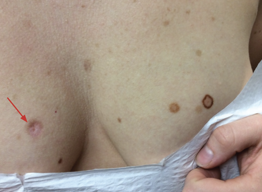
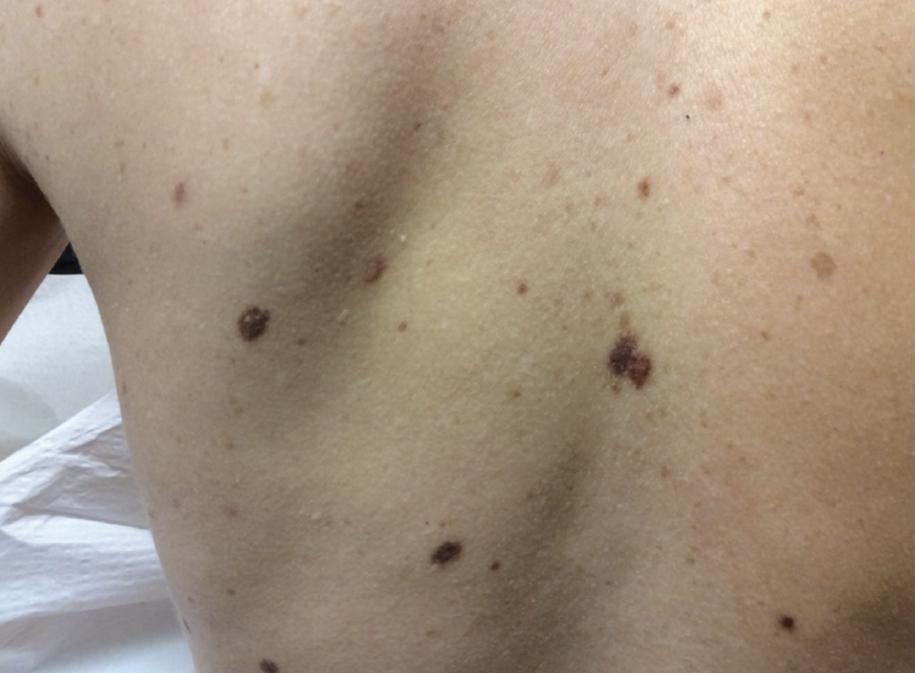
I often check the previous record to find that the original lesion was a 3-mm pigmented nevus diagnosed histopathologically as a “moderately atypical mole.” This is frustrating because I am quite certain that the overwhelming majority, if not all, of these and similar lesions could have been safely watched, avoiding a scarring biopsy procedure, and certainly did not require an additional follow-up procedure.6,7 Not only is this burdensome for the patient, but it also creates an unnecessary utilization of health care resources. Figure 1 demonstrates a woman who presented to me more than 6 years ago with a large hypertrophic scar on her right breast that was the result of the removal of a “mildly dysplastic nevus.” She had numerous scars on her body from previous procedures, and I have yet to perform a biopsy on her. But I do follow her many clinically atypical moles. (Figure 2)
Controversy exists as to the most likely origin of cutaneous melanoma. We know that melanoma can arise from a variety of melanocytic nevoid precursor lesions. It can also derive from other lesions or from skin not suspected to develop into melanoma.8 Finally, and debatably, the most dangerous melanomas emerge de novo from seemingly normal skin.1 The difficulty then lies in determining which nevi have a high likelihood for melanoma and which should be left alone and monitored. Although it is important to identify and remove melanomas as soon as possible, it should also be reassuring to know that the vast majority of melanomas (whether in situ or invasive) will not result in a metastatic event or death. I find it extremely useful to utilize digital photography, and I would submit that any dermatologic clinician who does not document with photography in 2022 is not practicing the most optimal form of dermatology.9
Conclusion
Because we do not have a crystal ball that can reliably predict whether a dysplastic nevus or atypical mole has the potential to develop into melanoma without some form of ancillary sampling, it is probably best to never use the term “precancerous” when referring to these lesions. And we need to ensure that we properly train our medical assistants who may be communicating the diagnosis of dysplastic nevus (or atypical mole) to our patients to also never use the term “precancerous.” On the other hand, if a lesion is sampled and determined to be “severely atypical” by histology or “likely malignant” by genomic analysis, then the clinician can explain to the patient that to the best of our understanding, this particular lesion seems to have the capability of becoming a melanoma. But even then, the lesion could be in the early stages of bona fide malignant melanoma, and we still do not know that with 100% certainty. It is best to acknowledge to the patient that we simply do not know. A good way of explaining this concept to patients with clinically atypical nevi is to explicitly state that having multiple atypical moles puts them at increased risk for melanoma, but with routine checks and self-performed skin examinations, we will likely catch any malignancies in their earliest stages and prevent an untoward outcome.
References
1. Shain AH, Bastian BC. From melanocytes to melanomas. Nat Rev Cancer. 2016;16(6):345-358. doi:10.1038/nrc.2016.37
2. Shain AH, Joseph NM, Yu R, et al. Genomic and transcriptomic analysis reveals incremental disruption of key signaling pathways during melanoma evolution. Cancer Cell. 2018;34(1):45-55.e4. doi:10.1016/j.ccell.2018.06.005
3. Li WQ, Cho E, Weinstock MA, Li S, Stampfer MJ, Qureshi AA. Cutaneous nevi and risk of melanoma death in women and men: a prospective study. J Am Acad Dermatol. 2019;80(5):1284-1291. doi:10.1016/j.jaad.2018.12.058
4. Nault A, Zhang C, Kim K, Saha S, Bennett DD, Xu YG. Biopsy use in skin cancer diagnosis: comparing dermatology physicians and advanced practice professionals. JAMA Dermatol. 2015;151(8):899-902. doi:10.1001/jamadermatol.2015.0173
5. Strazzula L, Vedak P, Hoang MP, Sober A, Tsao H, Kroshinsky D. The utility of re-excising mildly and moderately dysplastic nevi: a retrospective analysis. J Am Acad Dermatol. 2014;71(6):1071-1076. doi:10.1016/j.jaad.2014.08.025
6. Argenziano G, Cerroni L, Zalaudek I, et al. Accuracy in melanoma detection: a 10-year multicenter survey. J Am Acad Dermatol. 2012;67(1):54-59. doi:10.1016/j.jaad.2011.07.019
7. Hocker TL, Alikhan A, Comfere NI, Peters MS. Favorable long-term outcomes in patients with histologically dysplastic nevi that approach a specimen border. J Am Acad Dermatol. 2013;68(4):545-551. doi:10.1016/j.jaad.2012.09.031
8. Cabrera R, Recule F. Unusual clinical presentations of malignant melanoma: a review of clinical and histologic features with special emphasis on dermatoscopic findings. Am J Clin Dermatol. 2018;19(Suppl 1):15-23. doi:10.1007/s40257-018-0373-6
9. Hartge P, Holly EA, Halpern A, et al. Recognition and classification of clinically dysplastic nevi from photographs: a study of interobserver variation. Cancer Epidemiol Biomarkers Prev. 1995;4(1):37-40.
Dr Jarell is an adjunct clinical professor of dermatology at the Geisel School of Medicine at Dartmouth and practicing dermatologist, dermatopathologist, and clinical trials investigator in Portsmouth, NH.
Disclosure: The author serves as a consultant for Castle Biosciences, Inc. Photos printed with patient's permission.