Seborrheic Keratosis: The Most Common Reason Patients See Me
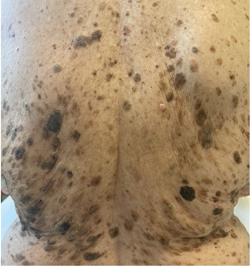
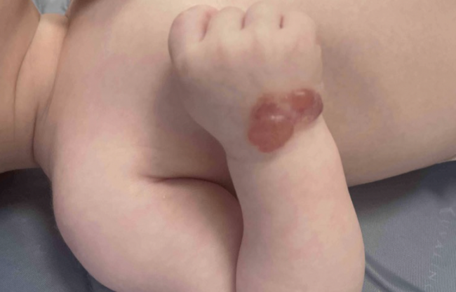
Clinical photograph printed with patient permission.
One of the most satisfying and rewarding aspects of practicing dermatology is being able to provide calming reassur
When patients present to us with a suspicious lesion that we know is an SK, it should be a pleasure to pause and confidently provide reassurance that the concerning lesion is nothing dangerous and incredibly common. I often tell the patient, “This is the most common reason people come to see me.” I believe that providing this information serves multiple purposes. It reassures the patient that the concern they are presenting with is not unusual because many people have this issue. And if patients know I see this entity with tremendous frequency, then they can be assured that I am someone they can trust with this benign diagnosis. Providing that reassurance with confidence is quite gratifying, and I will regularly receive a response such as, “Oh, thank goodness! You just made my day. I was so worried.” I have no doubt similar responses are abundant in medicine, and particularly in dermatology.
As dermatologists, we need to remember that although we can easily take for granted the appearance of most SKs, we need to respect our nondermatologist colleagues who have not had the vast experience of examining thousands of them. Often, patients are referred to us by other medical providers to “rule out a suspicious lesion.” If it is an obvious SK, then we need to patiently and confidently convince the patient that they have nothing to be concerned about even if another provider was uncertain of the diagnosis. This requires deft communication skills because we want to convey our confidence without any hint of arrogance, condescension, or patronization to both the patient and the referring provider. Finally, I strongly recommend against the practice of doing a biopsy to prove what we already know. I feel this way for nearly all dermatologic conditions, but particularly so regarding SK. Performing an unnecessary biopsy not only creates a false sense of heightened suspicion, but is also adds great expense to the visit in an already constrained medical system of limited resources.1 One might argue that performing a biopsy is simple and may alleviate a patient’s anxiety, but I maintain that alleviating anxiety should be accomplished with expertise, and the right course of action is to verbally convince the patient that their lesion is normal and of no medical significance. This is not to say that we should never treat SKs from a cosmetic standpoint, but cosmetic removal needs to be clearly distinguished from medical necessity. Cosmetic removal of SKs should be handled in a manner that is standard for the practice, but their treatment should not be billed to a third-party payor.
On the other hand, unless we are absolutely certain of the benign diagnosis, we must consider that sometimes a banal appearing SK can actually be a malignancy masquerading as an SK. In 2002, Izikson et al showed that although rare, sometimes a melanoma can be found when the suspected diagnosis is SK.2 I still recall verbatim those frightening, yet hallowed, words from my Andrews’ Diseases of the Skin textbook as a first-year resident many years ago, “Even the most seasoned dermatologist has been humbled by the occasional diagnosis of melanoma in low-suspect lesions.”3 If there is any suspicion whatsoever, we need to take the time to more thoroughly evaluate the concerning lesion, and this should be done by dermoscopy.4 There are several dermatoscopic clues to the diagnosis of SK that have been well described.1 If after rigorous analysis there is still suspicion for the possibility of malignancy, then a biopsy should be performed. Before any biopsy, I recommend high-resolution photography.
Although it is possible for an SK to undergo a malignant transformation, the chance of this occurring is extremely low. I generally provide reassurance to my patients that a typical SK will likely remain an SK and may even possibly grow. But there is almost no chance that an SK can evolve into a more ominous entity.
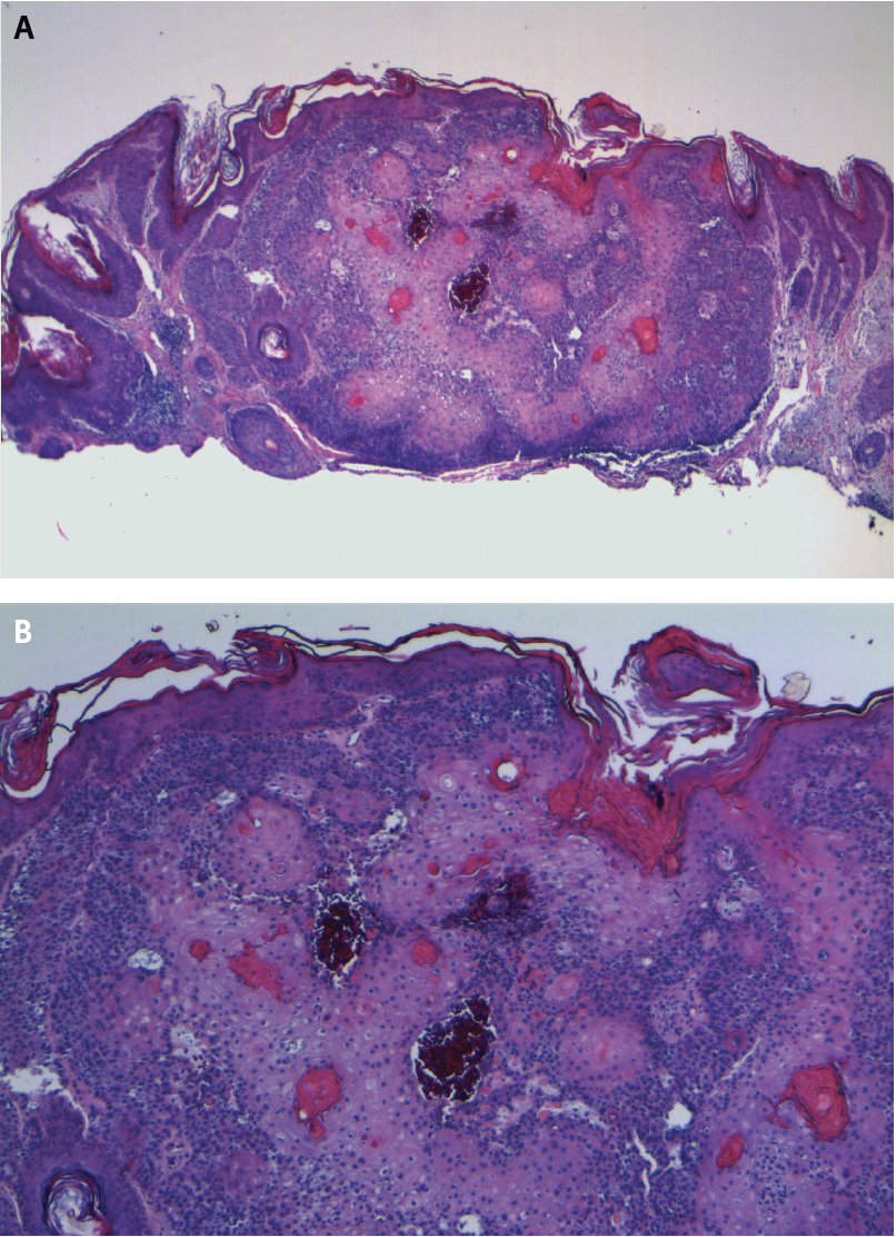
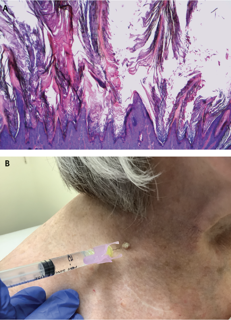
However, to be sure, many atypical squamous proliferations, actinic keratoses, and frank squamous cell carcinomas begin their existence as banal SKs. And this is where it sometimes becomes tricky, especially for the dermatopathologist. In many cases, it is not easy to confidently distinguish histologically an irritated SK (Figure 2) vs actinic keratosis versus verruca vulgaris—many pathologists will simply hedge the diagnosis and fi nalize with “verrucous keratosis” (Figure 3). It warrants repeating that the likelihood of dysplastic or malignant transformation for any given SK is extraordinarily low.
Because SK is so incredibly common, it is incumbent upon the dermatology provider to thoroughly understand this entity, its various manifestations, and when to suspect a more ominous masquerading process. I am grateful that I have acquired the ability to confidently put my patients' minds at ease about their SKs and send them on their way satisfied with our visits.
References
1. Braun RP, Ludwig S, Marghoob AA. Differential diagnosis of seborrheic keratosis: clinical and dermoscopic features. J Drugs Dermatol. 2017;16(9):835-842.
2. Izikson L, Sober AJ, Mihm Jr MC, Zembowicz A. Prevalence of melanoma clinically resembling seborrheic keratosis: analysis of 9204 cases. Arch Dermatol. 2002;138(12):1562-1566. doi:10.1001/archderm.138.12.1562
3. James WD, Elston D, Treat JR, Rosenbach MA. Andrews’ Diseases of the Skin: Clinical Dermatology. 13th ed. Elsevier; 2019.
4. Minagawa A. Dermoscopy-pathology relationship in seborrheic keratosis. J Dermatol. 2017;44(5):518-524. doi:10.1111/1346-8138.13657