A V-Y flap is a modified advancement flap that is used for the repair of small and medium size cutaneous defects. It has the advantage of a robust vascular supply and a reliable healing pattern. Its most common application includes the repair of facial wounds after removal of skin cancers often on the cutaneous lip, but also on the eyebrow and forehead, though it can also be applied in a variety of additional facial and non-facial reconstructive settings.
Previously, this flap was also commonly referred to as the island pedicle flap due to its two main requirements: (1) the separation of epidermal and dermal connections of the flap, resulting in an “island” of skin, and (2) the preservation of a subcutaneous pedicle (consisting of subcutaneous fat, muscle, and, occasionally, superficial musculoaponeurotic system), which provides the flap with its blood supply through perforating vessels. Thus, unlike other random-pattern flaps, the dimensions of a V-Y flap are not limited by decreasing perfusion of the flap at the periphery, as the blood supply is derived from under the flap. In fact, this flap is perfused equally in all directions and tip necrosis is uncommon.
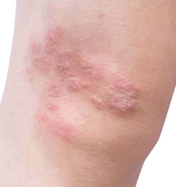
 Figure 1, above: Figure 1a: This patient underwent excision of melanoma in situ. Figure 1b: Planning reconstruction. Figure 1c: Patient post construction. Figure 1d: Patient 1 month after the procedure. Photos courtesy of Dr. Alexander Berlin
When performing a V-Y flap, a triangular incision is made from the edges of the wound and carried down to subcutaneous fat. The height of the triangle is usually 1.5 to 2 times the length of the original base or the “leading-edge” of the flap, though it may be considerably longer, especially on the upper lip. The direction or orientation of the distal point is selected based on adjacent skin laxity and mobility as well as the location of natural or evolving skin creases. In many sites, such as the upper lip, the sides of the triangle are not designed in a straight fashion, but rather in a curvilinear manner to follow the natural landmarks, such as the melolabial fold. In addition, a bilateral V-Y flap can be used to cover a larger defect, with two triangular flaps pointing in opposite directions and the two flaps being brought together in the middle. This technique is also often utilized in the areas of limited skin mobility, such as the shin, where the two triangular areas that would have been removed in a typical elliptical closure are, instead, utilized as double V-Y flaps, thereby reducing wound tension.
Undermining is carried out laterally (away from the flap), while undermining of the actual pedicle is largely avoided (aside from areas of tethering) to provide for the blood supply of the flap. Some undermining of the tail end and the leading edge may be necessary to enhance flap mobility and to avoid tethering of the base of the flap or significant wound tension. Variations in undermining are sometimes used for greater mobilization and include the formation of a muscular sling as the basis for flap movement, laterally based pedicle formation and sub-pedicle undermining. Moreover, the original defect should usually be deepened by removing additional subcutaneous tissue to accommodate the thickness of the incoming pedicle, which may also help to avoid subsequent pin-cushioning of the flap. Once mobilized, the flap is brought into position using a skin hook and a dissolvable key suture is placed in the middle of the leading edge of the advancing triangular flap, followed by additional dissolvable sutures at the corners of the leading edge.
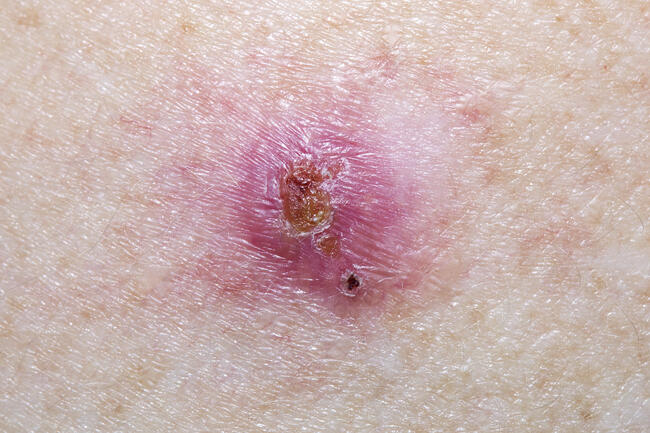
 Figure 2a-c: This patient underwent Mohs surgery with surgical repair with a V-Y advancement flap. Figure 2d: Patient 8 days post surgery. Early follow-up at 2 months shows some mild pincushioning (that could be improved with intra-lesional steroid) and some prominence of the suture line including from the leading edge of the flap (that could be blended with a fractional ablative laser). Photos courtesy of Dr. Joel Cohen.
A somewhat common complication of V-Y flaps is trap-door deformity, also known as “pin-cushioning,” where the flap bulges out of its original position. This process is poorly understood, but its risk appears to be reduced by burying the pedicle deeper in the original defect (as described above), resulting in a more depressed-appearing flap initially, as well as slightly undersizing the flap by 1 mm to 2 mm. If present, trap-door deformity usually resolves spontaneously or with the help of manual massage or intralesional corticosteroids.
Dr. Berlin is President of DFW Skin Surgery Center, PLLC, in Arlington, TX. He is also Clinical Assistant Professor of Dermatology, New Jersey Medical School in Newark, NJ.
Dr. Cohen is the Director of AboutSkin Dermatology and DermSurgery in Colorado. His practice focuses on Mohs surgery and cosmetic dermatology.
Disclosure: Drs. Cohen and Berlin report that they have no conflicts of interest or financial disclosures to report.
A V-Y flap is a modified advancement flap that is used for the repair of small and medium size cutaneous defects. It has the advantage of a robust vascular supply and a reliable healing pattern. Its most common application includes the repair of facial wounds after removal of skin cancers often on the cutaneous lip, but also on the eyebrow and forehead, though it can also be applied in a variety of additional facial and non-facial reconstructive settings.
Previously, this flap was also commonly referred to as the island pedicle flap due to its two main requirements: (1) the separation of epidermal and dermal connections of the flap, resulting in an “island” of skin, and (2) the preservation of a subcutaneous pedicle (consisting of subcutaneous fat, muscle, and, occasionally, superficial musculoaponeurotic system), which provides the flap with its blood supply through perforating vessels. Thus, unlike other random-pattern flaps, the dimensions of a V-Y flap are not limited by decreasing perfusion of the flap at the periphery, as the blood supply is derived from under the flap. In fact, this flap is perfused equally in all directions and tip necrosis is uncommon.
 Figure 1, above: Figure 1a: This patient underwent excision of melanoma in situ. Figure 1b: Planning reconstruction. Figure 1c: Patient post construction. Figure 1d: Patient 1 month after the procedure. Photos courtesy of Dr. Alexander Berlin
When performing a V-Y flap, a triangular incision is made from the edges of the wound and carried down to subcutaneous fat. The height of the triangle is usually 1.5 to 2 times the length of the original base or the “leading-edge” of the flap, though it may be considerably longer, especially on the upper lip. The direction or orientation of the distal point is selected based on adjacent skin laxity and mobility as well as the location of natural or evolving skin creases. In many sites, such as the upper lip, the sides of the triangle are not designed in a straight fashion, but rather in a curvilinear manner to follow the natural landmarks, such as the melolabial fold. In addition, a bilateral V-Y flap can be used to cover a larger defect, with two triangular flaps pointing in opposite directions and the two flaps being brought together in the middle. This technique is also often utilized in the areas of limited skin mobility, such as the shin, where the two triangular areas that would have been removed in a typical elliptical closure are, instead, utilized as double V-Y flaps, thereby reducing wound tension.
Undermining is carried out laterally (away from the flap), while undermining of the actual pedicle is largely avoided (aside from areas of tethering) to provide for the blood supply of the flap. Some undermining of the tail end and the leading edge may be necessary to enhance flap mobility and to avoid tethering of the base of the flap or significant wound tension. Variations in undermining are sometimes used for greater mobilization and include the formation of a muscular sling as the basis for flap movement, laterally based pedicle formation and sub-pedicle undermining. Moreover, the original defect should usually be deepened by removing additional subcutaneous tissue to accommodate the thickness of the incoming pedicle, which may also help to avoid subsequent pin-cushioning of the flap. Once mobilized, the flap is brought into position using a skin hook and a dissolvable key suture is placed in the middle of the leading edge of the advancing triangular flap, followed by additional dissolvable sutures at the corners of the leading edge.
 Figure 2a-c: This patient underwent Mohs surgery with surgical repair with a V-Y advancement flap. Figure 2d: Patient 8 days post surgery. Early follow-up at 2 months shows some mild pincushioning (that could be improved with intra-lesional steroid) and some prominence of the suture line including from the leading edge of the flap (that could be blended with a fractional ablative laser). Photos courtesy of Dr. Joel Cohen.
A somewhat common complication of V-Y flaps is trap-door deformity, also known as “pin-cushioning,” where the flap bulges out of its original position. This process is poorly understood, but its risk appears to be reduced by burying the pedicle deeper in the original defect (as described above), resulting in a more depressed-appearing flap initially, as well as slightly undersizing the flap by 1 mm to 2 mm. If present, trap-door deformity usually resolves spontaneously or with the help of manual massage or intralesional corticosteroids.
Dr. Berlin is President of DFW Skin Surgery Center, PLLC, in Arlington, TX. He is also Clinical Assistant Professor of Dermatology, New Jersey Medical School in Newark, NJ.
Dr. Cohen is the Director of AboutSkin Dermatology and DermSurgery in Colorado. His practice focuses on Mohs surgery and cosmetic dermatology.
Disclosure: Drs. Cohen and Berlin report that they have no conflicts of interest or financial disclosures to report.
A V-Y flap is a modified advancement flap that is used for the repair of small and medium size cutaneous defects. It has the advantage of a robust vascular supply and a reliable healing pattern. Its most common application includes the repair of facial wounds after removal of skin cancers often on the cutaneous lip, but also on the eyebrow and forehead, though it can also be applied in a variety of additional facial and non-facial reconstructive settings.
Previously, this flap was also commonly referred to as the island pedicle flap due to its two main requirements: (1) the separation of epidermal and dermal connections of the flap, resulting in an “island” of skin, and (2) the preservation of a subcutaneous pedicle (consisting of subcutaneous fat, muscle, and, occasionally, superficial musculoaponeurotic system), which provides the flap with its blood supply through perforating vessels. Thus, unlike other random-pattern flaps, the dimensions of a V-Y flap are not limited by decreasing perfusion of the flap at the periphery, as the blood supply is derived from under the flap. In fact, this flap is perfused equally in all directions and tip necrosis is uncommon.
Figure 1, above: Figure 1a: This patient underwent excision of melanoma in situ. Figure 1b: Planning reconstruction. Figure 1c: Patient post construction. Figure 1d: Patient 1 month after the procedure. Photos courtesy of Dr. Alexander Berlin
When performing a V-Y flap, a triangular incision is made from the edges of the wound and carried down to subcutaneous fat. The height of the triangle is usually 1.5 to 2 times the length of the original base or the “leading-edge” of the flap, though it may be considerably longer, especially on the upper lip. The direction or orientation of the distal point is selected based on adjacent skin laxity and mobility as well as the location of natural or evolving skin creases. In many sites, such as the upper lip, the sides of the triangle are not designed in a straight fashion, but rather in a curvilinear manner to follow the natural landmarks, such as the melolabial fold. In addition, a bilateral V-Y flap can be used to cover a larger defect, with two triangular flaps pointing in opposite directions and the two flaps being brought together in the middle. This technique is also often utilized in the areas of limited skin mobility, such as the shin, where the two triangular areas that would have been removed in a typical elliptical closure are, instead, utilized as double V-Y flaps, thereby reducing wound tension.
Undermining is carried out laterally (away from the flap), while undermining of the actual pedicle is largely avoided (aside from areas of tethering) to provide for the blood supply of the flap. Some undermining of the tail end and the leading edge may be necessary to enhance flap mobility and to avoid tethering of the base of the flap or significant wound tension. Variations in undermining are sometimes used for greater mobilization and include the formation of a muscular sling as the basis for flap movement, laterally based pedicle formation and sub-pedicle undermining. Moreover, the original defect should usually be deepened by removing additional subcutaneous tissue to accommodate the thickness of the incoming pedicle, which may also help to avoid subsequent pin-cushioning of the flap. Once mobilized, the flap is brought into position using a skin hook and a dissolvable key suture is placed in the middle of the leading edge of the advancing triangular flap, followed by additional dissolvable sutures at the corners of the leading edge.
Figure 2a-c: This patient underwent Mohs surgery with surgical repair with a V-Y advancement flap. Figure 2d: Patient 8 days post surgery. Early follow-up at 2 months shows some mild pincushioning (that could be improved with intra-lesional steroid) and some prominence of the suture line including from the leading edge of the flap (that could be blended with a fractional ablative laser). Photos courtesy of Dr. Joel Cohen.
A somewhat common complication of V-Y flaps is trap-door deformity, also known as “pin-cushioning,” where the flap bulges out of its original position. This process is poorly understood, but its risk appears to be reduced by burying the pedicle deeper in the original defect (as described above), resulting in a more depressed-appearing flap initially, as well as slightly undersizing the flap by 1 mm to 2 mm. If present, trap-door deformity usually resolves spontaneously or with the help of manual massage or intralesional corticosteroids.
Dr. Berlin is President of DFW Skin Surgery Center, PLLC, in Arlington, TX. He is also Clinical Assistant Professor of Dermatology, New Jersey Medical School in Newark, NJ.
Dr. Cohen is the Director of AboutSkin Dermatology and DermSurgery in Colorado. His practice focuses on Mohs surgery and cosmetic dermatology.
Disclosure: Drs. Cohen and Berlin report that they have no conflicts of interest or financial disclosures to report.


 Figure 1, above: Figure 1a: This patient underwent excision of melanoma in situ. Figure 1b: Planning reconstruction. Figure 1c: Patient post construction. Figure 1d: Patient 1 month after the procedure. Photos courtesy of Dr. Alexander Berlin
 Figure 2a-c: This patient underwent Mohs surgery with surgical repair with a V-Y advancement flap. Figure 2d: Patient 8 days post surgery. Early follow-up at 2 months shows some mild pincushioning (that could be improved with intra-lesional steroid) and some prominence of the suture line including from the leading edge of the flap (that could be blended with a fractional ablative laser). Photos courtesy of Dr. Joel Cohen.