

Treating Calcified PAD With the New Shockwave M5+ IVL Catheter
Vascular Surgeon
Sanger Heart and Vascular Institute
Atrium Health
Charlotte, North Carolina
Vascular Disease Management speaks with vascular surgeon Halim Yammine, MD, from Sanger Heart and Vascular Institute in Charlotte, North Carolina, about treating calcified PAD with the Shockwave M5+ intravascular lithotripsy (IVL) catheter. Dr. Yammine’s case report follows the interview.
What is your current calcified superficial femoral artery/popliteal artery treatment algorithm?
Whenever you’re dealing with a calcified superficial femoral artery (SFA)/popliteal artery, the first thing to think about is crossing the lesion, which is the most important step. Sometimes, you might need pedal access to work in a retrograde fashion to be able to cross it. Once I cross the lesion, I use intravascular ultrasound (IVUS) to investigate the plaque morphology and perform accurate sizing of the blood vessel. It also tells me if I crossed the lesion in true or false lumen. Once I cross the lesion and use IVUS to size the vessel, I’ll typically use IVL and then drug-coated balloon (DCB) angioplasty instead of stenting, depending on the lesion length and the result of the DCB after IVL.
What challenges does calcium pose in getting good results in SFA/popliteal artery treatment?
Calcific lesions are very challenging to cross. You feel like you’re pushing against a rock. Sometimes you must go subintimal to be able to cross the lesion, which is a big challenge. Even if you’re able to cross the lesion and you do balloon angioplasty or DCB, the vessels tend to recoil and there’s a higher rate of dissection because that calcific area acts as a lead point for dissection. And then, even if you’re able to cross the lesion, sometimes a stent cannot track; even if it tracks and you’re able to deploy it, it won’t expand very well because of the hard nature of the calcific plaque, which would lead to well-documented loss of long-term patency in those patients.
What has been your experience with IVL, and what is your perception of the new Shockwave M5+? What value does the faster pulsing, larger size, and further reach of Shockwave M5+ bring to your patients?
It has been a very positive experience. Patients have a better response to DCB angioplasty after IVL. There is a decreased need for stenting and less dissection rate. I believe that IVL will lead to higher patency rates, even long term. The Shockwave M5+ has been a breath of fresh air. It’s much nicer and faster than the Shockwave M5 catheter. It saves time, especially when you’re treating a long SFA lesion. You’re cutting the IVL treatment time in half with 2 times faster pulsing, less radiation, less catheterization time, and less fatigue for the physician and the team.
I really love it, to be honest. You can treat more patients because of the larger sizes the M5+ has. You can go up to 8mm now. The longer catheter allows us to reach lesions just below the knee (BTK) or at the knee level, even in tall patients. You can also treat more vascular beds as you can go into the iliac arteries or the CFA with the 8mm size.
Do you have any tips to share on optimizing your results with IVL?
The most important thing when dealing with a calcific lesion is after you cross the lesion, you need to be able to track the balloons and stents across that lesion. Even with the Shockwave balloon for IVL, you need to be able to track. Sometimes it might be good to try to dilate with the 3mm to 4mm regular balloon to improve trackability through that lesion and be able to deliver the Shockwave balloon to perform IVL. This is helpful, especially in very tight, highly calcific lesions, sometimes in chronic total occlusions (CTOs) as well. I’m also a big fan of IVUS for plaque evaluation and sizing.
What are your thoughts regarding the PAD III randomized control trial 1- and 2-year results that were released at SCAI 2022?
I was very impressed with the results of that study. It did show excellent outcomes at 1 and 2 years in terms of patency and a decreased need for reinterventions on those target lesions. It compared well to studies done for all the DCBs present on the market. Also, it compared well to the drug-eluting stents (DES) present on the market.
When you look at the 2-year patency rates of the DES present on the market now, it drops into the 70% range for patency at 2 years. This is similar to IVL and DCB that we’ve seen in the PAD III study, which tells me I can achieve similar patency rates at 2 years without having to place the stent and leave metal in place. Those, in my opinion, are very favorable outcomes. Patient populations that we typically treat were well represented also in this study, which I think means that it would translate well to real-world practices.
What is your perception of the safety profile of IVL based on your experience and the PAD data?
It has an excellent safety profile. There are very, very low dissection rates. You rarely need a stent to help you in this patient population. There’s a significantly less need for stenting, and in almost all my experience, I didn’t have a single embolic event. When you treat those lesions, you don’t have to use high inflation pressures. You’re sticking with low inflation pressures and letting the shockwaves do their part. IVL has an excellent safety profile; I haven’t had any major issues with it so far, and we use it quite a bit.
What is your advice to your peers if they’re hesitant to try IVL?
I would give them the same advice I got when I was hesitant to try IVL, which is to go ahead, try it once, and you’ll be impressed with the results. You can look with IVUS before and after and you’ll see the lumen expansion and that there is no dissection. You’ll see that you are able to get optimal results in very challenging patients. Treating a patient with IVL and DCB makes a big difference instead of giving them an open bypass. I tell colleagues to try it once and you’ll be impressed with the results, and you’ll be able to treat more patients with this technology.
Case Report
Treating Calcified PAD With Shockwave IVL
Halim Yammine, MD
Sanger Heart and Vascular Institute, Charlotte, North Carolina
A 59-year-old male with medical comorbidities that included longstanding diabetes mellitus type 2, hypertension, coronary artery disease, obesity, ongoing tobacco abuse, end-stage renal disease (ESRD) on hemodialysis (HD), and known severe PAD presented to our clinic for evaluation. He had a small blister on his left heel that progressed into a large ulcer over the previous month. The patient’s duplex ultrasound showed diffuse calcific plaque, which made it difficult to visualize any significant stenoses or potential subtotal occlusion(s) within the left lower extremity (LLE). Monophasic waveforms were noted in the left ankle, which may suggest tibial disease. The left ankle-brachial index was noncompressible with a toe-brachial index of 0.36.
Despite the ultrasound not showing any evidence of stenosis, it was clear that the patient had severe vascular insufficiency contributing to the ulcer not healing. We decided to proceed with a diagnostic angiogram with possible intervention.
Procedures
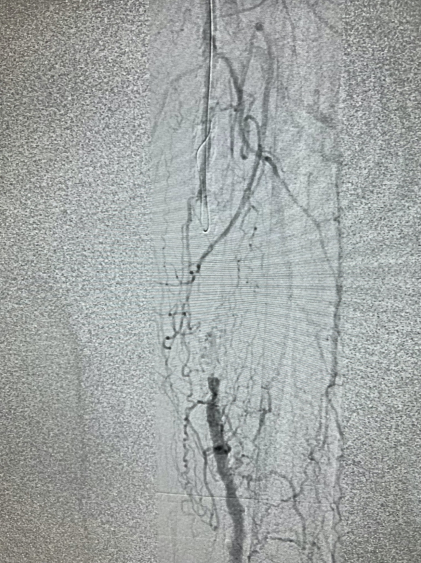
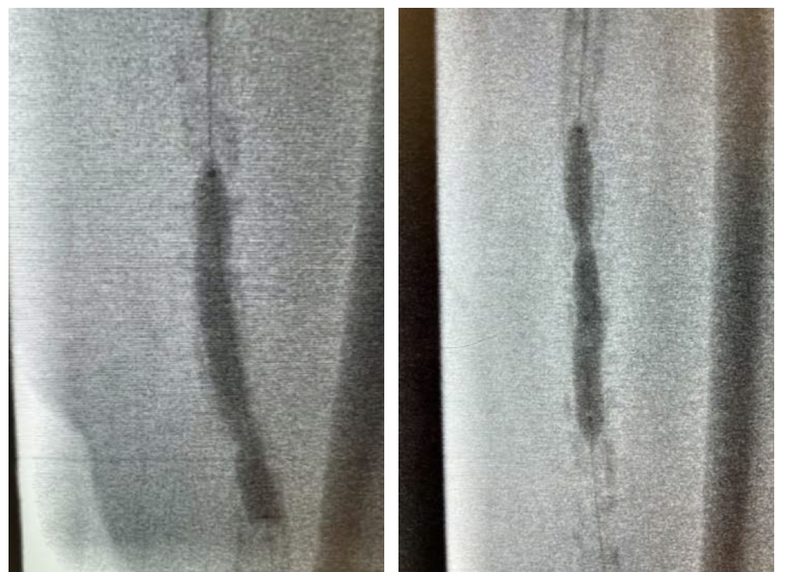
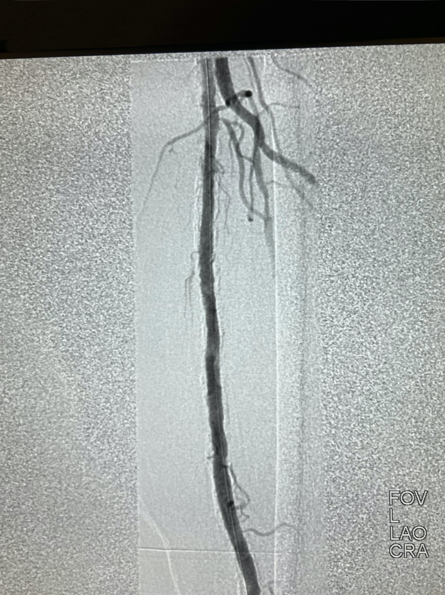
We obtained access of the right common femoral artery (CFA) under ultrasound guidance. We proceeded to perform an aortogram that showed that the inflow vessels were patent. Then a left lower extremity angiogram showed a chronic total occlusion in the left SFA (Figure 1). We started by placing a 7Fr x 45cm sheath up and over into the left CFA. We were able to cross the CTO with moderate difficulty. IVUS was then used to evaluate the plaque morphology and appropriately size the vessel. Given the extent of calcification we decided to use a 7mm x 60mm Shockwave M5+ balloon to perform IVL (Figure 2 and Figure 3). We followed this with DCB angioplasty with a 7mm x200mm Ranger (Boston Scientific). Follow-up angiography showed excellent flow with no residual stenosis (Figure 4). We were satisfied with the results and terminated the procedure. The patient did very well during and after the procedure. He was seen in follow-up and his ulcer is almost completely healed.
Discussion
Critical limb ischemia is a severe form of PAD associated with high rates of limb loss and even mortality. Diabetes mellitus and ESRD are associated with a high degree of calcification in the blood vessels. Calcified plaques can present multiple challenges during endovascular interventions. First, it can be very difficult to cross a highly calcific lesion, and a lot of times it might require additional retrograde access. Fortunately, in our patient the lesion was crossed with only moderate difficulty, and we were able to do it with an antegrade approach only.
Second, calcified lesions notoriously do not respond well to traditional balloon angioplasty, and it is very difficult to achieve any durable luminal gain. Stenting these lesions can also be very challenging, and it can be difficult to even deliver the stent across a highly calcific, tight stenosis. They can also lead to suboptimal expansion of stents, which often lead to aggressive ballooning and running the risk of rupturing the vessel or causing stent fractures.
IVL presents an excellent treatment option for this very challenging patient population. The luminal gain that can be obtained with this technology in highly calcific lesions is unparalleled. It makes delivering DCBs or stents much easier and leads to more optimal results with either. It has an excellent safety profile with very low rates of dissection and distal embolization. In our patient, we were able to treat a long CTO with IVL and DCB alone with excellent results without the need for stenting or any other adjunctive therapies.
Conclusion
Highly calcific lesions in the SFA can be very challenging to treat at times. IVL presents an excellent treatment option for this patient population with excellent outcomes and very low risk of complications.
This interview was supported by Shockwave Medical.
Dr. Yammine is a paid consultant for Shockwave Medical and opinions expressed are those of the speaker and not necessarily those of Shockwave Medical.
In the United States: Rx only.
Indications for Use. The Shockwave Medical Intravascular Lithotripsy (IVL) System is intended for lithotripsy-enhanced balloon dilatation of lesions, including calcified lesions, in the peripheral vasculature, including the iliac, femoral, ilio-femoral, popliteal, infra-popliteal, and renal arteries. Not for use in the coronary or cerebral vasculature.
Contraindications. Do not use if unable to pass 0.014 guidewire across the lesion. Not intended for treatment of in-stent restenosis or in coronary, carotid, or cerebrovascular arteries.
Warnings. Only to be used by physicians who are familiar with interventional vascular procedures. Physicians must be trained prior to use of the device. Use the generator in accordance with recommended settings as stated in the Operator’s Manual.
Precautions. Use only the recommended balloon inflation medium. Appropriate anticoagulant therapy should be administered by the physician. Decision regarding use of distal protection should be made based on physician assessment of treatment lesion morphology.
Adverse effects. Possible adverse effects consistent with standard angioplasty include access site complications; allergy to contrast or blood thinner; arterial bypass surgery; bleeding complications; death; fracture of guidewire or device; hypertension/hypotension; infection/sepsis; placement of a stent; renal failure; shock/pulmonary edema; target vessel stenosis or occlusion; vascular complications. Risks unique to the device and its use: allergy to catheter material(s); device malfunction or failure; excess heat at target site.
Prior to use, please reference the Instructions for Use for more information on indications, contraindications, warnings, precautions, and adverse events. www.shockwavemedical.com
Please contact your local Shockwave representative for specific country availability and refer to the Shockwave S4, Shockwave M5, and Shockwave M5+ instructions for use containing important safety information.
