


Safety and Efficacy of Periorbital Sclerotherapy
© 2023 HMP Global. All Rights Reserved.
Any views and opinions expressed are those of the author(s) and/or participants and do not necessarily reflect the views, policy, or position of Vascular Disease Management or HMP Global, their employees, and affiliates.
VASCULAR DISEASE MANAGEMENT 2023;20(11):E197-E200
Abstract
Periorbital telangiectasias and reticular veins are dilated blood vessels found near the periorbital region, which are often a cosmetic concern. Traditional treatments include laser therapy and microphlebectomy. A more novel approach is the use of sclerotherapy, for which we will share our experience in this case report. An 82-year-old woman presented with bilateral periorbital cosmetic reticular veins. After a comprehensive evaluation, a decision was made to treat with sclerotherapy. The injection of the sclerosant, 0.5% polidocanol, was uneventful without any adverse effects reported. After the procedure, cosmetic satisfaction was assessed using standardized tools, revealing a substantial improvement in the patient’s self-esteem. The success of this case demonstrates that although there is hesitancy to use sclerotherapy for the management of reticular veins due to neurologic and ophthalmic complications, sclerotherapy can be a minimally invasive treatment option. However, additional research is necessary to develop a standardized method for using sclerotherapy for facial veins, and consideration for the potential risk of blindness is still noted.
Introduction
Telangiectasias are dilated and tortuous veins often associated with normal aging or veno-congestive pathology.1 Telangiectasias can occur anywhere in the body but are often found on the legs, chest, hands, and face.1 Facial telangiectasias are often considered part of normal aging; however, pale skin tone, smoking history, UV exposure, and body mass index may further contribute to an increase in incidence.2 There are no specific genetic linkages to the development of facial telangiectasias; however, it is suggested that there may be tentative correlations between single nucleotide polymorphisms of the vascular system and telangiectasia formation.3 Common locations of facial telangiectasias are around the nose, cheeks, and chin.4 Facial telangiectasias may also be present around the orbit.
The management of vascular anomalies, such as venous and lymphatic malformations, as well as telangiectasias and varicose and reticular veins, consists of phlebectomy, ablation, and sclerotherapy, among other techniques.5-7 Sclerotherapy is a novel treatment option for these pathologies that comes with remarkable cosmetic results. After injecting a sclerosant, it works by inducing intimal inflammation, which initiates the intrinsic clotting cascade and creates a thrombus that will subsequently occlude the vessel. Thereafter, the vessel becomes fibrotic and obliterates.6,8 Conventionally, sclerotherapy has been used in the lower extremities4, and it was only recently adopted for usage in periorbital veins with great success.6-11 There are several sclerosant agents, such as glycerin, hypertonic saline, morrhuate sodium, sodium tetradecyl sulfate (STS), and polidocanol. The most used and FDA-approved agents for sclerotherapy are STS and polidocanol.7,12,13
Although sclerotherapy is a promising treatment option, many vascular surgeons have not regularly adopted sclerotherapy for facial use due to the fear of neurologic and ophthalmic complications.4 Studies have mentioned that patients undergoing sclerotherapy run the risk of minor and transient complications, such as edema, ecchymosis, pain, hyperpigmentation, and even telangiectasias from inflammation-induced angiogenesis.7,8,10,11 In several studies, only minor complications were observed. Green et al reported injection site purpura in 6 patients and erythema and edema for 2 to 7 days among all patients who underwent sclerotherapy for the removal of periocular veins.8 Faiz et al observed effects ranging from hyperpigmentation, edema, ecchymosis, subcutaneous atrophy, and pain among 18.1% of their patients who underwent sclerotherapy for venous malformations.11 Major complications that patients are susceptible to are blindness in 1 eye and skin necrosis from an incidental arterial injection, thrombosis, anaphylaxis, and neurological defects.5-7,11,13,14 However, none of these were observed in the aforementioned studies. Theoretical reasons for sclerosants to reach the ophthalmic circulation include retrograde passage of material, high injection pressure, and enough material within the vessel lumen.14 Green et al mentioned that the likelihood of these agents causing thrombosis in orbital and cerebral veins is slim to none as the residual agents will flow in an anterograde direction, the path of least resistance, and will be diluted by the blood in larger veins.8 It is also important to consider what sclerosant to use; for periorbital veins, hypertonic saline is not recommended as it has been associated with pain, burning, and cramps during injection.7
There are many inherent benefits to sclerotherapy. First, it is associated with low recurrence rates. In a study conducted by Razavi et al of 11 patients who underwent sclerotherapy for venous-lymphatic malformation, there was no recurrence observed.6 Secondly, the cost of sclerotherapy when compared with conventional phlebectomy is lower.15,16
Case Report
Our patient, an 82-year-old woman with dark reticular veins on both sides of the forehead, takes daily aspirin for heart health. She has had previous falls in which she fractured her ribs and is obtaining physical therapy for her knee. She is active and has played the piano professionally. She has no pain or medical pathology related to her veins. Her medical and surgical history is unremarkable in relation to her facial cosmetic telangiectasia. All patients undergoing periorbital sclerotherapy first are required to see ophthalmology and oculo-plastics. The patient had a lateral periocular vein that was easily compressible and nontender.
Injection Technique and Follow-up
During initial consultation, the required ophthalmologic evaluation was discussed, along with other treatment options including laser and microphlebectomy. The FDA-approved drug polidocanol was discussed as the injectable treatment option. The risks were explained, which include bruising, swelling, less than optimal results, matting, and visual disturbance.
Treatment was performed in the semi-recumbent position. No more than 1 cc of 0.5% polidocanol was used at a time. After appropriately prepping the skin, the solution was injected via a 30-gauge 0.5" needle in 3 cc syringe. First, cannulation was verified by using negative pressure and blood within the needle. Next, with minimal pressure and slow infusion, a small amount of sclerosant was injected, just enough to displace the blood (normally less than 0.5 cc). Pressure was then directly held over the vein. A 5-minute timed pressure hold was performed. At the end, ice was placed over the area to reduce bruising. The area was inspected for the absence of blood. The patient was asked to refrain from activity for 24 hours post procedure.
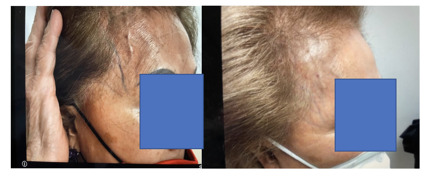
Follow-up included a phone call the next day by office staff, and a 1-, 4-, and 6-week follow-up. If the patient remained unsatisfied at the 6-week follow-up, consideration for repeat injection was performed. Also, if there was an additional contralateral vein, it was treated at this point. The follow-up visit included checking for complications including matting, staining, reported visual disturbance, or unsatisfactory results. There were no such complications, and no bruising remained by the 1-week mark. At the 6-week mark, the patient was satisfied with the aesthetic results (Figure), which in some cases results in improvement but not 100% resolution.
Conclusion
The results of this case report, as well as that of previously reported studies using sclerotherapy for periorbital veins, suggest that sclerotherapy for periorbital veins is indeed a safe and effective technique. There is, however, a major risk for ophthalmic, thrombotic, and neurologic complications due to forceful injection and/or intra-arterial injection, although none have been reported and can be prevented with proper technique and caution. n
The authors have completed and returned the ICMJE Form for Disclosure of Potential Conflicts of Interest. The authors report no financial relationships or conflicts of interest regarding the content herein.
Manuscript accepted October 31, 2023.
Address for Correspondence: Dr. Alissa Brotman, Rowan-Virtua School of Osteopathic Medicine, 42 E Laurel Road, Suite 1300, Mt. Laurel, NJ 08084. Email: brotmaal@rowan.edu
REFERENCES
1. Bowes LE, Goldman MP. Sclerotherapy of reticular and telangiectatic veins of the face, hands, and chest. Dermatol Surg. 2002;28(1):46-51. doi:10.1046/j.1524-4725.2002.01154.x
2. Mekić S, Hamer MA, Wigmann C, et al. Epidemiology and determinants of facial telangiectasia: a cross-sectional study. J Eur Acad Dermatol Venereol. 2020;34(4):821-826. doi:10.1111/jdv.15996
3. Mekić S, Wigmann C, Gunn DA, et al. Genetics of facial telangiectasia in the Rotterdam Study: a genome-wide association study and candidate gene approach. J Eur Acad Dermatol Venereol. 2021;35(3):749-754. doi:10.1111/jdv.17014
4. Gao L, Qu H, Gao N, et al. A retrospective analysis for facial telangiectasia treatment using pulsed dye laser and intense pulsed light configured with different wavelength bands. J Cosmet Dermatol. 2020;19(1):88-92. doi:10.1111/jocd.13179
5. Smith V, Whiteley M. Facial veins – diagnosis and treatment options. The PMFA Journal. 2019;6(2). https://www.thepmfajournal.com/features/post/facial-veins-diagnosis-and-treatment-options
6. Razavi ME, Rajabi MT, Hassanpoor N, Mohammadi SS. Sclerotherapy for eyelid and anterior orbital venous-lymphatic malformation. J Curr Ophthalmol. 2019;31(2):214-217. doi:10.1016/j.joco.2019.01.002
7. Yiannakopoulou E. Safety concerns for sclerotherapy oftTelangiectases, reticular and varicose veins. Pharmacology. 2016;98(1-2):62-69. doi:10.1159/000445436
8. Green D. Removal of periocular veins by sclerotherapy. Ophthalmology. 2001;108(3):442-448. doi:10.1016/s0161-6420(00)00384-5
9. De Maria L, De Sanctis P, Tollefson M, et al. Sclerotherapy for low-flow vascular malformations of the orbital and periocular regions: systematic review and meta-analysis. Surv Ophthalmol. 2020;65(1):41-47. doi:10.1016/j.survophthal.2019.08.003
10. Das S, Agrawal A, Burathoki SK, Nandolia KK, Juneja A, Samanta R. Orbital venolymphatic malformation treated with sodium tetradecyl sulfate: a case report. Cureus. 2022;14(9):e29173. doi:10.7759/cureus.29173
11. Faiz K, Finitsis S, Linton J, Shankar JJS. Bleomycin for orbital and peri-orbital veno-lymphatic malformations—a systematic review. Interv Neuroradiol. 2021;27(2):291-297. doi:10.1177/1591019920972514
12. Andrews RH, Dixon RG. Ambulatory phlebectomy and sclerotherapy as tools for the treatment of varicose veins and telangiectasias. Semin Intervent Radiol. 2021;38(2):160-166. doi:10.1055/s-0041-1727151
13. Lee CH, Chen SG. Direct percutaneous ethanol instillation for treatment of venous malformation in the face and neck. Br J Plast Surg. 2005;58(8):1073-1078. doi:10.1016/j.bjps.2005.04.014
14. Dehghani A, Rezaei L, Ghanbari H, Nasrollahi K, Tavakoli M. ophthalmic artery occlusion following facial sclerosing therapy. J Ophthalmic Vis Res. 2018;13(3):351-354. doi:10.4103/jovr.jovr_29_16
15. Nakano LC, Cacione DG, Baptista-Silva JC, Flumignan RL. Treatment for telangiectasias and reticular veins. Cochrane Database Syst Rev. 2021;10(10):CD012723. doi:10.1002/14651858.CD012723.pub2
16. Lattimer CR, Azzam M, Kalodiki E, Shawish E, Trueman P, Geroulakos G. Cost and effectiveness of laser with phlebectomies compared with foam sclerotherapy in superficial venous insufficiency. Early results of a randomised controlled trial. Eur J Vasc Endovasc Surg. 2012;43(5):594-600. doi:10.1016/j.ejvs.2012.01.032
