

Atherectomy in the Infrainguinal Arteries
© 2023 HMP Global. All Rights Reserved.
Any views and opinions expressed are those of the author(s) and/or participants and do not necessarily reflect the views, policy, or position of Vascular Disease Management or HMP Global, their employees, and affiliates.
VASCULAR DISEASE MANAGEMENT 2023;20(10):E179-E182
Hello and welcome to the October edition of Vascular Disease Management. This month we are tackling the hotly debated topic of atherectomy and plaque modification. Atherectomy/plaque modification technologies deploy a variety of device types to alter the arterial vessel wall with several possible goals: 1) improve device delivery (usually balloon or stent) to a target lesion; 2) increase luminal gain after angioplasty or stenting; 3) prevent stent under-expansion; and 4) avoid the need for stenting.
In light of recent reports alleging overuse of atherectomy devices, we would like to revisit the guidelines and clinical data as to inform the overall discourse surrounding the appropriate use of these devices.
Case Example
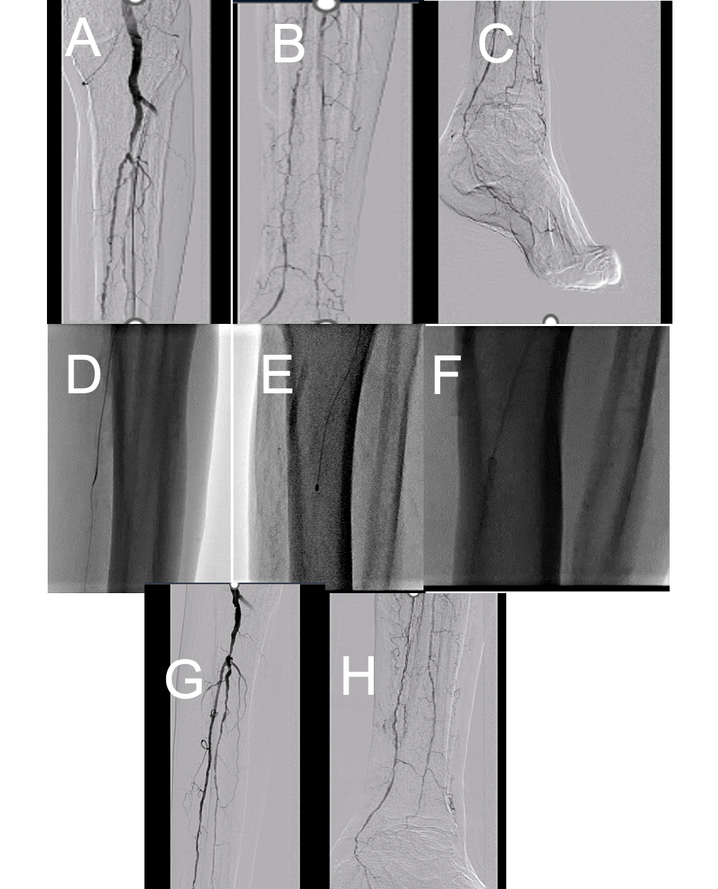
We would like to begin with a case in which atherectomy enabled us to treat a balloon uncrossable lesion (Figure) for limb salvage. A successful restoration of inline flow would not have been possible in this case without atherectomy.
Societal Guidelines
The various major guideline writing committees (Society for Vascular Surgery, American College of Cardiology, and the Trans-Atlantic Inter-Society Consensus II) hold atherectomy as an acceptable adjuvant for endovascular procedures but do not offer specific guidance for use.1-3 The Society for Cardiovascular Angiography and Interventions statement on device selection in femoropopliteal disease maintains that atherectomy treatment is adjunctive prior to definitive balloon or stent treatment4,5 and is generally not meant to be used in isolation. The Appropriate Use Criteria for Peripheral Artery Intervention labels atherectomy as “may be appropriate” in femoropopliteal and below-the-knee lesions and “rarely appropriate” in suprainguinal lesions.4
Safety of Atherectomy
A 2023 paper assessed the safety of atherectomy by propensity matching a large Medicare cohort who underwent atherectomy for femoropopliteal segment arterial lesions during their procedure to a cohort treated with percutaneous transluminal angioplasty (PTA) alone. In the study, 46.7% of patients had critical limb-threatening ischemia (CLTI). Over a median follow-up of nearly 3 years, atherectomy use resulted in no difference in the major safety outcomes of composite death or amputation, as well as major adverse limb events. Patients who underwent atherectomy had a modest reduction in future amputation or surgical revascularization but had an increase in the risk of undergoing a subsequent peripheral intervention.6
Randomized Controlled Trials
It is commonly misstated that randomized data for atherectomy does not exist. The EXCITE trial demonstrated superiority with laser atherectomy plus PTA compared with PTA alone.7 In the trial, 250 patients undergoing treatment of femoropopliteal in-stent restenosis (ISR) were randomized to laser + PTA vs PTA alone. The study showed that in the laser group, the 6-month primary patency was 71.1% vs 56.4% in favor of laser + PTA, P=.004.
Compliance 360° was a multicenter study of 50 patients with calcified femoropopliteal disease randomizing Diamondback 360 orbital atherectomy (Cardiovascular Systems, Inc.) plus PTA vs PTA alone.8 The primary endpoint, freedom from target lesion revascularization (TLR) or restenosis, was observed at 12 months in 81.2% in the orbital atherectomy plus PTA arm vs 78.3% in the PTA arm, P>.99. Despite the lack of a difference in the primary outcome (the study was underpowered as it was meant to be a pilot study), there was less need for stenting with orbital plus PTA when compared with PTA alone (5.3% vs. 77.8%, P<.001).
Highlighted Registry Updates
A recent study of state Medicare data demonstrated that centers that used atherectomy in more than 56% of cases had a significantly lower amputation rate when compared with centers that did not (<10% vs >30%).9 Additionally, the LIBERTY 360 3-year registry results, in which the majority of patients were treated with atherectomy, demonstrated excellent patency across Rutherford groups: the Kaplan-Meier estimates of freedom from major amputation at 36 months were 98.5% in Rutherford class 2 to 3, 94.0% in Rutherford class 4 to 5, and 79.9% in Rutherford class 6.10
Highlighted Recent Publications and Conference Presentation Based on Device Type
Intravascular Lithotripsy
Shockwave balloons (Shockwave Medical) employ pulsatile sound energy to modify calcified plaque in the wall of the vessel. DISRUPT PAD II was a multicenter, single-arm registry assessing intravascular lithotripsy (IVL) in 60 patients with moderate-to-severe femoropopliteal calcified stenosis. The study’s primary safety endpoint, 30-day major adverse events, was 1.7% with only 1 serious dissection and no distal embolization. The primary effectiveness endpoint, patency at 12 months, was 54.5%, and clinically driven TLR was 20.7%. This was followed by DISRUPT PAD III, examining IVL in patients with femoropopliteal calcified lesions followed by treatment with drug-coated balloons (DCB). The 2-year follow-up data showed freedom from TLR rate after Shockwave treatment + DCB of 70.3% compared with 51.3% with PTA alone, P=.003. This study was followed by the larger DISRUPT PAD III observational study (OS), which included 1,373 patients and demonstrated results consistent with DISRUPT III. The OS study also importantly confirmed a very low complication rate with IVL, with 0.7% significant dissection and no instances of embolization, no reflow or abrupt closure.11 Lastly, the ongoing DISRUPT BTK II trial will further elucidate efficacy and safety of these devices in the below-the-knee vascular beds.
Rotational Devices with Aspiration Capabilities
The JetStream for the Treatment of In-Stent Restenosis (JET-ISR) study examined 60 patients with femoropopliteal ISR. Freedom from TLR at 6 months and 1 year were 79.3% and 60.7%.12
The Rotarex device (BD) was evaluated in a single-center registry of 74 high-risk patients with chronic total occlusions and ISR. Patients were treated with Rotarex followed by DCB; at 1 year, the restenosis rate was 20.5%.13
Directional Atherectomy
The Hawk (Medtronic) and Pantheris (Avinger) are currently approved directional atherectomy devices that use rotating cutting discs to remove the atherosclerotic plaque from the vessel wall. The recent multicenter, single-arm evaluation of Hawk in complex femoropopliteal disease, called the VIVA REALITY study, showed a 1-year primary patency and freedom from major adverse events among patients treated with Hawk directional atherectomy plus DCB of 76.7% with a freedom from TLR of 92.6%.14 In the study, only 8.8% of treated lesions required bailout stenting.
A single-center, head-to-head study of directional vs. orbital atherectomy in femoropopliteal lesions was recently performed, with directional atherectomy resulting in greater luminal gain and less stent placement acutely.15 The extended follow-up of this study is slated to be presented in the coming weeks.
Orbital/Rotational Atherectomy
The Rotablator system (Boston Scientific) is available for use in infrainguinal disease, particularly below the knee. Limited contemporary studies of Rotablator exist in the peripheral vasculature. The Diamondback 360 orbital atherectomy system has a crown that orbits inside the vessel via a 360-degree arc, allowing for differential atherectomy for varying vessel sizes. Recent results with Diamondback 360 are discussed above.
Conclusion
With a large volume of clinical registry data available in a variety of clinical settings and 1 positive randomized trial in ISR, it is hard to argue against atherectomy in selected clinical scenarios such as severe calcific disease. Concerns do arise, particularly in patients with wounds, whether microembolization leading to no reflow could worsen outcomes in individuals where this risk may be elevated. This question represents an interesting area of future research. While randomized trials certainly would be beneficial in advancing knowledge, there is little impetus for device companies to perform them; therefore, government-funded studies may be required to develop this level of evidence. The current controversies in the press regarding appropriate use of atherectomy may be sufficient to spark funding for additional trials in the field. These devices remain an important part of the clinical armamentarium for the treatment of infrainguinal peripheral arterial disease. n
REFERENCES
1. Conte MS, Bradbury AW, Kolh P, et al. Global vascular guidelines on the management of chronic limb-threatening ischemia. Eur J Vasc Endovasc Surg. 2019;58(1S)S1-S doi:10.1016/j.ejvs.2019.05.006.109.e33
2. Hirsch AT, Haskal ZJ, Hertzer NR, et al. ACC/AHA 2005 guidelines for the management of patients with peripheral arterial disease (lower extremity, renal, mesenteric, and abdominal aortic): a collaborative report from the American Association for Vascular Surgery/Society for Vascular Surgery, Society for Cardiovascular Angiography and Interventions, Society for Vascular Medicine and Biology, Society of Interventional Radiology, and the ACC/AHA Task Force on Practice Guidelines (Writing Committee to Develop Guidelines for the Management of Patients With Peripheral Arterial Disease) endorsed by the American Association of Cardiovascular and Pulmonary Rehabilitation; National Heart, Lung, and Blood institute; Society for Vascular Nursing; TransAtlantic Inter-Society Consensus; and Vascular Disease Foundation. J Am Coll Cardiol. 2006;47(6):1239-1312. doi:10.1016/j.jacc.2005.10.009
3. Norgren L, Hiatt WR, Dormandy JA, et al. Inter-Society Consensus for the Management of Peripheral Arterial Disease (TASC II). J Vasc Surg. 2007;45(Suppl S):S5 doi:10.1016/j.jvs.2006.12.037-S67
4. Bailey SR, Beckman JA, Dao TD, et al. ACC/AHA/SCAI/SIR/SVM 2018 Appropriate Use Criteria for Peripheral Artery Intervention: A report of the American College of Cardiology Appropriate Use Criteria Task Force, American Heart Association, Society for Cardiovascular Angiography and Interventions, Society of Interventional Radiology, and Society for Vascular Medicine. J Am Coll Cardiol. 2019;73(2):214-237. doi:10.1016/j.jacc.2018.10.002
5. Feldman DN, Armstrong EJ, Aronow HD, et al. SCAI consensus guidelines for device selection in femoral-popliteal arterial interventions. Catheter Cardiovasc Interv. 2018;92(1) doi:10.1002/ccd.27635:124-140
6. Krawisz AK, Raja A, Jones WS, et al. Long-term outcomes of peripheral atherectomy for femoropopliteal endovascular interventions. EuroIntervention. 2023:18(6):e1378-e1387. doi:10.4244/EIJ-D-22-00609
7. Dippel EJ, Makam P, Kovach R, et al. Randomized controlled study of excimer laser atherectomy for treatment of femoropopliteal in-stent restenosis: initial results from the EXCITE ISR trial (EXCImer Laser Randomized Controlled Study for Treatment of FemoropopliTEal In-Stent Restenosis). JACC Cardiovasc Interv. 2015:8(1 Pt A):92-101. doi:10.1016/j.jcin.2014.09.009
8. Datillo R, Himmelstein SI, Cuff RF. The COMPLIANCE 360° Trial: a randomized, prospective, multicenter, pilot study comparing acute and long-term results of orbital atherectomy to balloon angioplasty for calcified femoropopliteal disease. J Invasive Cardiol. 2014;26(8):355-360.
9. Mustapha JA, Katzen BT, Neville RF, et al. Disease burden and clinical outcomes following initial diagnosis of critical limb ischemia in the Medicare population. JACC Cardovasc Interv. 2018;11(10):1011-1012. doi:10.1016/j.jcin.2017.12.012
10. Giannopoulos S, Mustapha J, Gray WA, et al. Three-year outcomes from the LIBERTY 360 study of endovascular interventions for peripheral artery disease stratified by Rutherford category. J Endovasc Ther. 2021:28(2):262-274. doi:10.1177/1526602820962972
11. Adams G, Soukas PA, Mehrle A, Bertolet B, Armstrong EJ. Intravascular lithotripsy for treatment of calcified infrapopliteal lesions: results from the Disrupt PAD III observational study. J Endovasc Ther. 2022:29(1):76-83. doi:10.1177/15266028211032953
12. Shammas NW, Petruzzi N, Henao S, et al. JetStream atherectomy for the treatment of in-stent restenosis of the femoropopliteal segment: one-year results of the JET-ISR study. J Endovasc Ther. 2021:28(1):107-116. doi:10.1177/1526602820951916
13. Liu L, Zhao J, Bi J, et al. Percutaneous mechanical atherothrombectomy using the Rotarex device in acute ischemic disease of lower limbs: a China retrospective multicenter study on 196 patients. Ann Vasc Surg. 2022;85:146-155. doi:10.1016/j.avsg.2022.02.026
14. Rocha-Singh KJ, Sachar R, DeRubertis BG, et al. Directional atherectomy before paclitaxel coated balloon angioplasty in complex femoropopliteal disease: the VIVA REALITY study. Catheter Cardiovasc Interv. 2021:98(3):549-558. doi:10.1002/ccd.29777
15. Babaev A, Halista M, Bakirova Z, Avtushka V, Matsumura M, Maehara A. Directional versus orbital atherectomy of femoropopliteal lesions: angiographic and intravascular ultrasound outcomes. Catheter Cardiovasc Interv. 2022:100(4):687-695. doi:10.1002/ccd.30339
