


Controlled Step-Down Access Closure for Large Bore Access

VASCULAR DISEASE MANAGEMENT 2021;18(8):E123-E130
Abstract
As the advancements of catheter-based technology continue to evolve, the use of large bore access is growing to accommodate different procedures. In doing so, it cannot go unspoken that the operator must recognize the burden of postprocedural vascular access closure complications and their burden on the hospital, the patient, and the cost of care. Technical challenges and difficulty with optimal access closure with large bore catheters deter interventionalists from comfortably accommodating the use of devices that require a large bore access such as Impella, AAA stent graft, and transcatheter aortic valve replacement procedures. Controlled step-down access closure technique over a 0.35" guidewire was adopted in our series to accomplish optimal large bore access closure. Guidewire-assisted controlled step-down technique allows for optimal hemostasis and dry field while maintaining continuous access to vascular lumen. Step-down access closure, we believe, is optimal, cost-effective, and minimizes large bore vascular access complications. Our series is at a community hospital and represents real-world experience. We had successful prior experience with step-down access closure technique for post-brachial access interventions.1,2 Based on our prior experience, we developed a step-down vascular access closure technique over a 0.35" guidewire for large bore access closure. In our series of 124 cases, we had excellent success with large bore access closure with minimal complications. We believe controlled step-down access closure technique is an optimal approach for large bore access management.
Introduction
Since the development of coronary angiography in 1958, the use of angiography to diagnose and treat cardiac anomalies has come a long way to include many minimally invasive procedures.3 As the use of percutaneous cardiac interventions broadens to include include increasingly complex interventions and structural heart procedures, percutaneous hemodynamic support systems such as Impella and ECMO become crucial. Large bore access is required for many complex cardiovascular percutaneous procedures such as abdominal aortic aneurysm (AAA) stent grafts, Impella hemodynamic support, structural heart procedures such as transcatheter aortic valve replacement (TAVR), and more. Most of these patients have significant comorbidities, such as diabetes mellitus II, atherosclerotic vascular disease (ASVD-PAD), renal failure (patients on dialysis), age > 70 years, advanced congestive heart failure, and a low output state. These patients cannot tolerate complications, which has dissuaded many interventionalists from performing high-risk procedures. With these advancements, large bore access plays a key role in utilizing some of these major interventions. As opposed to standard vascular interventions, larger devices such as an implantable ventricular assist device or percutaneous ventricular assist device (PVAD), a percutaneous approach for AAA stent grafting, and TAVR require 9-21 French (Fr) access for device delivery. Vascular complications become a major source for significant morbidity and mortality.4
Using PubMed, a review of the literature was conducted. We included all articles published within the last 5 years evaluating access closure methods and devices. In another study, many current access closure devices failed to show a reduced complication rate compared with manual compression. In one study, manual compression was compared with device-mediated compression, an intravascular closure device, or an extravascular closure device after peripheral arterial interventions. The groups using device-assisted access closure have not been shown to reduce complications compared with manual compression.5 In one meta-analysis comparing FDA-approved vascular closure devices for femoral access closure, complication rates were recorded and also compared to manual compression. Complication rates for vascular closure devices were 12% compared to a manual compression rate of 13%, which again shows the use of devices to be comparable to manual compression in terms of complications, while the complication rate is >10% overall for both groups.6 Another study noted the percent difference in two commercially used device closure systems, MANTA and Prostar XL. MANTA showed a major bleeding risk of 2.3% vs Prostar XL (9.3%).7 Another comparison within this article, demonstrated by Moriyama et al, discussed access-site vascular injury, which was less frequent in patients who received MANTA closure vs ProGlide after TAVR. MANTA vascular injury was 8% vs ProGlide (17%).8 Both still pose a degree of vascular injury, but it is relatively low.
To combat vascular access complications, vascular closure devices have evolved over the last two decades. Common commercially available products include: Perclose ProGlide, Prostar XL, StarClose SE, Cardiva CATALYST II/III, VASCADE Vascular Closure System, EXOSEAL Vascular Closure Device, MYNX CONTROL Vascular Closure Device, EVRF, FISH CombiClose, Closer Vascular Sealing System, MANTA Vascular Closure Device, and Angio-Seal Evolution Vascular Closure Device.9,10 In a retrospective, nonrandomized study, the rate of vascular complications between 827 subjects using Angio-Seal and Prostar XL were determined after interventional procedures. The study showed a success rate of 92% with a 2.5% rate of vascular complications using Angio-Seal, while Prostar XL had a success rate of 89% with a 3.4% rate of vascular complications. Another study demonstrated complications from Angio-Seal and StarClose SE devices in comparison to manual compression.11,12 An overview of the evolution of vascular closure devices was presented at the Society of Cardiac Angiography and Imaging by our group (Poster abstract E-44 SCAI 2011).13 Throughout the literature search, most articles dealt with varying peripheral arterial interventions including small and large bore vascular access complication or compared vascular access complications between FDA-approved device closure in both low- and high- risk populations. Rather than the use of closure devices in most cases, we discuss a technique that improves large bore access closure in a high-risk population. In a few cases, we introduced the MYNX closure device to aid in minimizing vascular complications.

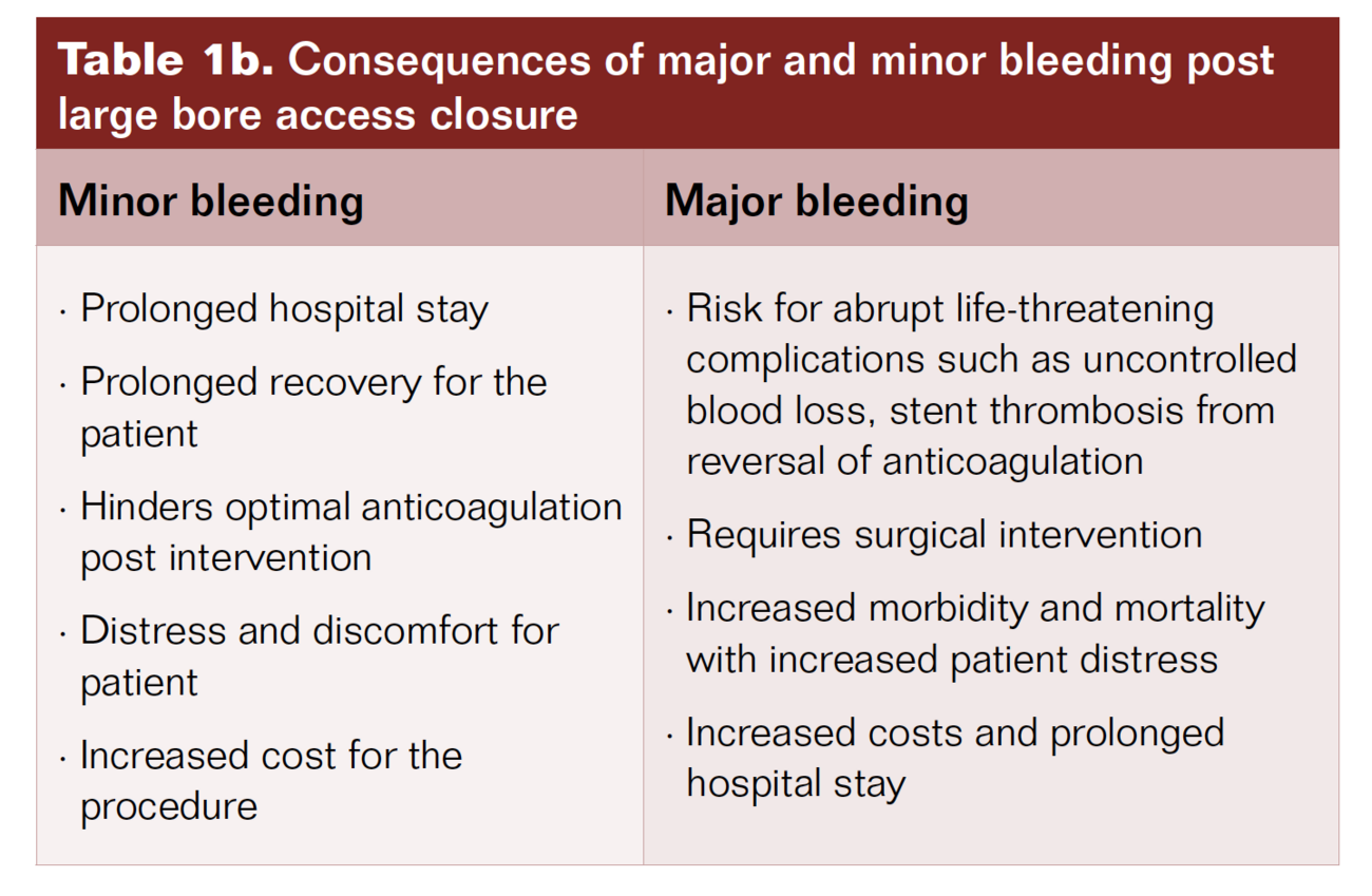
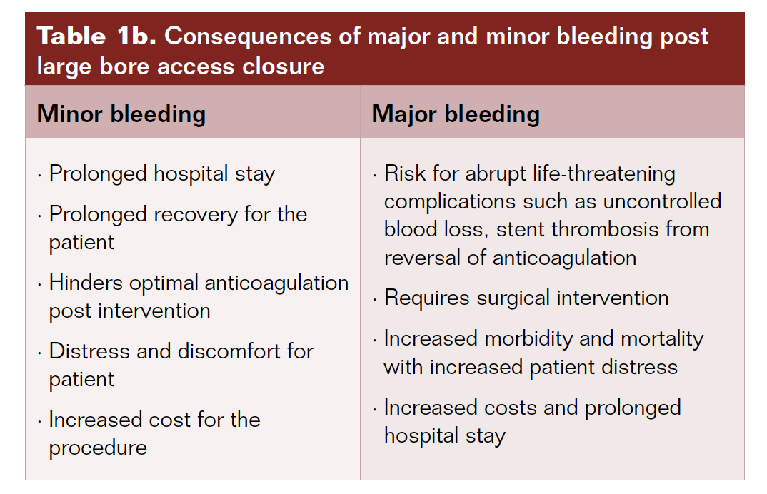
Complications with large bore access are depicted in Table 1a.14 Of these complications, bleeding is the most common. Vascular bleeding complications can be categorized as major and minor.2 Several consequences of minor or major bleeding are listed in Table 1b.
Comorbidities that predispose patients to major bleeding and access closure complications include:
1. Age >70 years;
2. Diabetes mellitus II with systemic vasculopathy;
3. Women with high bifurcation of the common femoral artery;
4. Obesity;
5. Peripheral vascular disease;
6. Chronic renal disease requiring dialysis;
7. Advanced heart failure with reduced ejection fraction (HFReF) with low output state.
These are comorbidities commonly associated with cardiovascular diseases.
Methods
A high-risk cohort of patients who underwent large bore access procedures at our institution were followed for immediate complications, early ambulation, length of hospital stay, and recovery. The sample size consisted of 124 subjects. The population has a variety of vascular comorbidities, including patient age >70 years old, female gender, peripheral arterial disease, diabetes mellitus, hyperlipidemia, chronic kidney disease, advanced heart failure patients with low output state leading to poor peripheral perfusion, and chronic stasis. The study period was from September 2014 until January 2021. As noted above, we have prior experience with step-down technique in successful hemostasis after brachial access in anticoagulated patients. Our experience was presented at the TCT conference in 1998 and published in the American Journal of Cardiology.1 Brachial access closure in anticoagulated patients is plagued with high risk for breakthrough bleeding due to the location and difficulty applying optimal manual pressure after sheath removal. Hence the Sones brachial technique, although it was the original approach for coronary angiography, was given up. Using step-down technique for brachial access closure, taking advantage of inherent vascular recoil, we achieved optimal hemostasis post brachial access in anticoagulated patients.
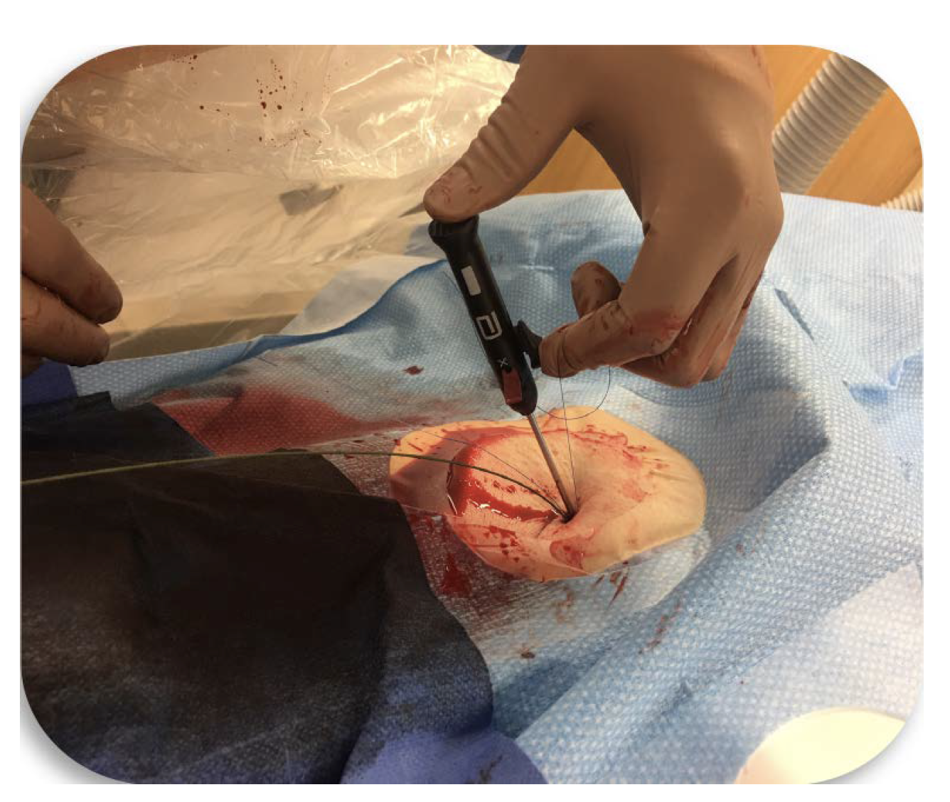
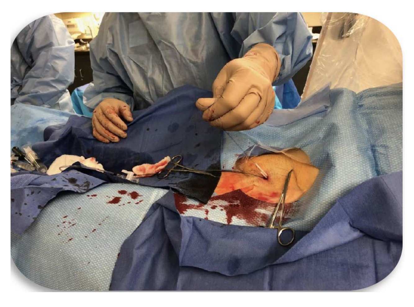
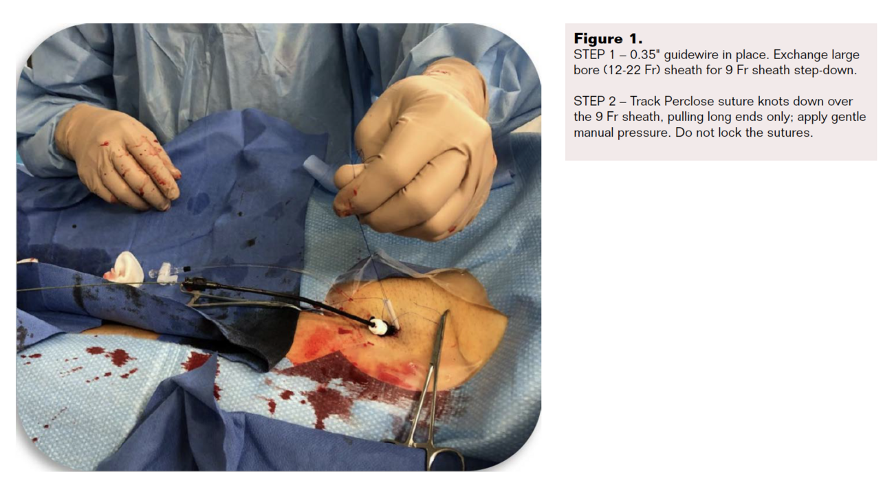
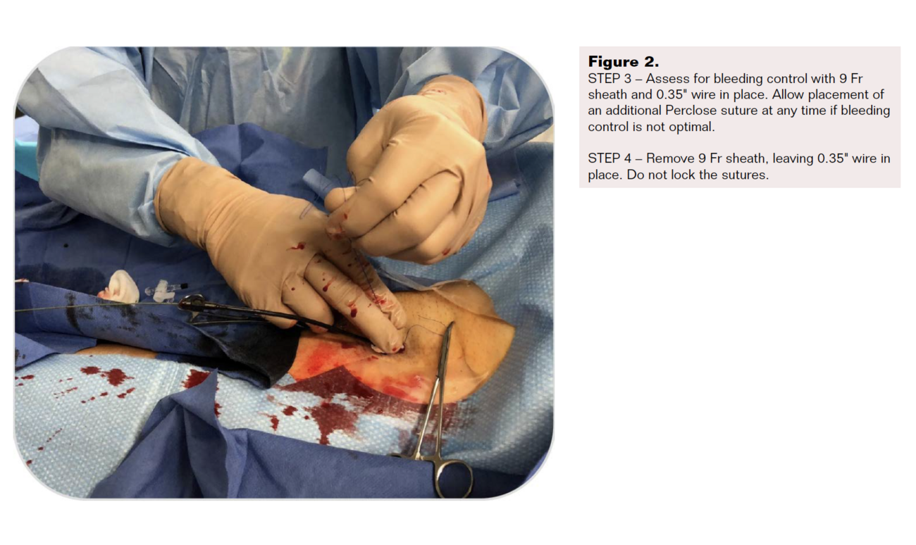
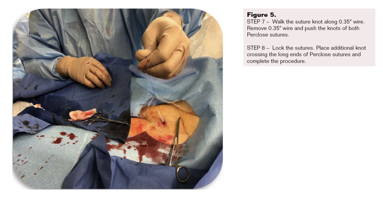
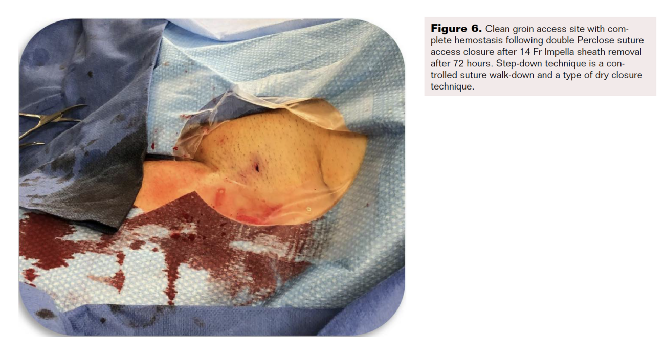
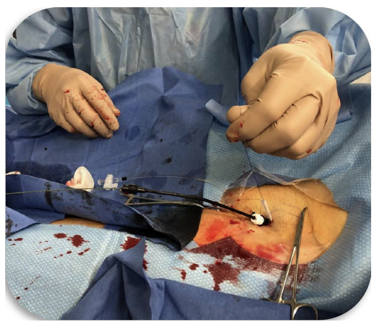
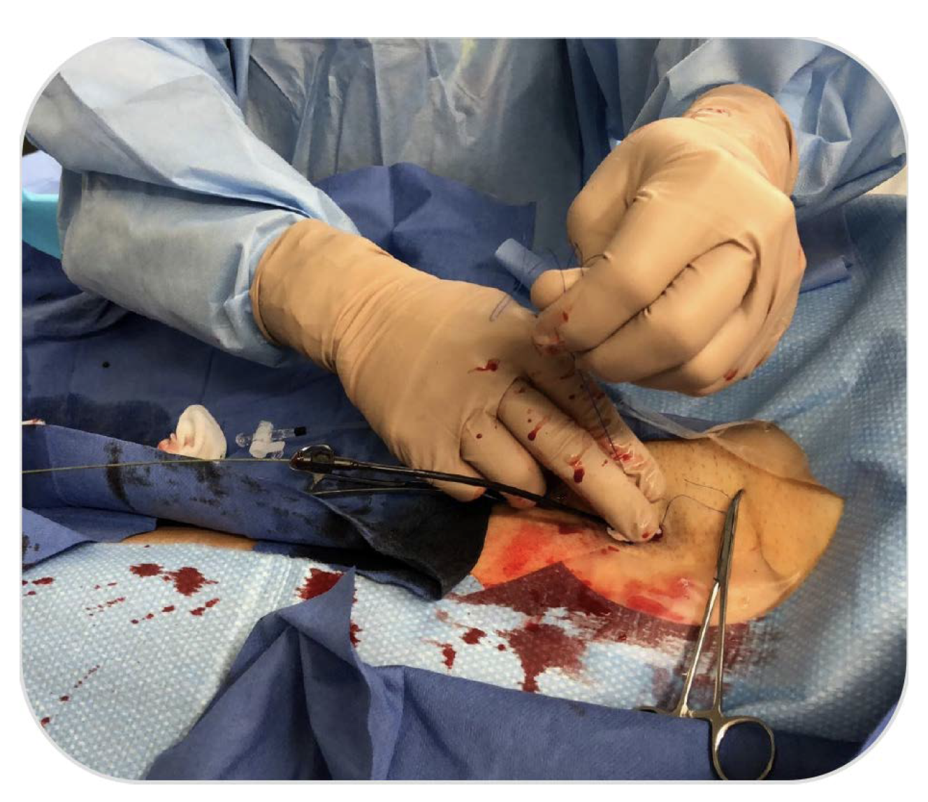
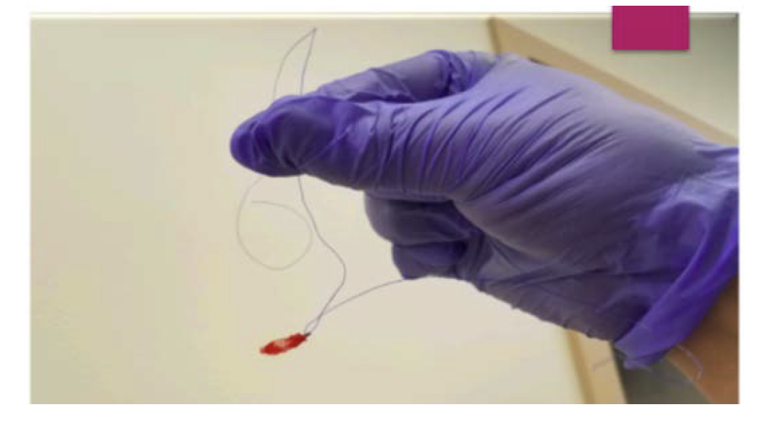
We developed a 0.35" guidewire-controlled step-down technique for larger bore access closure based on our brachial step-down experience. Our series is a community hospital-based real-world experience. Guidewire-controlled step-down technique was employed to achieve optimal large bore access closure with hemostasis as depicted in Figures 1 through 6.
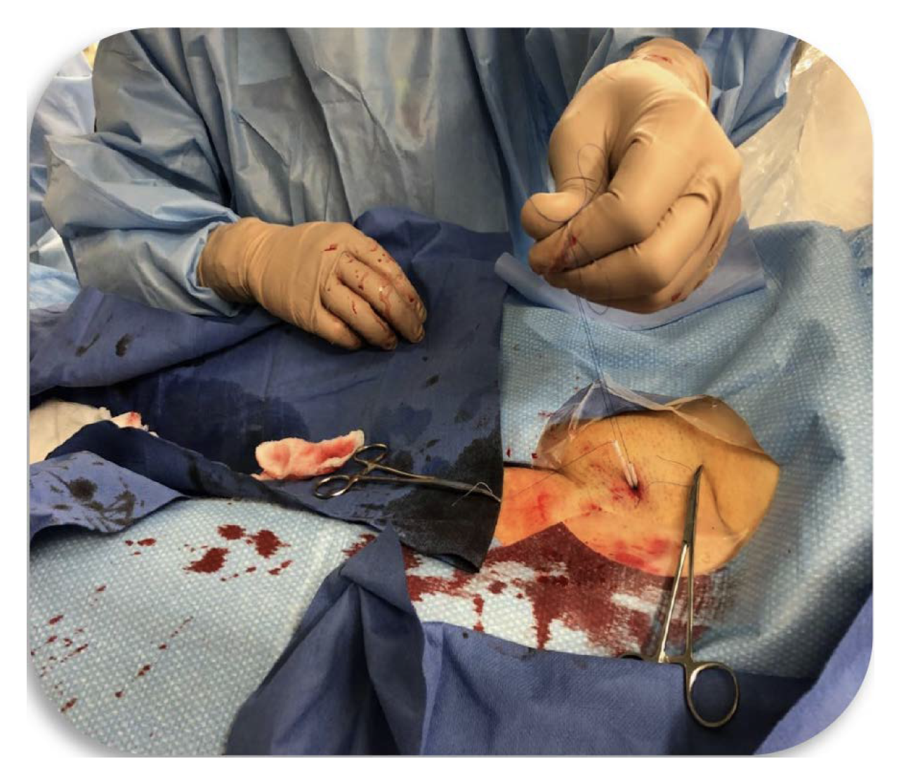
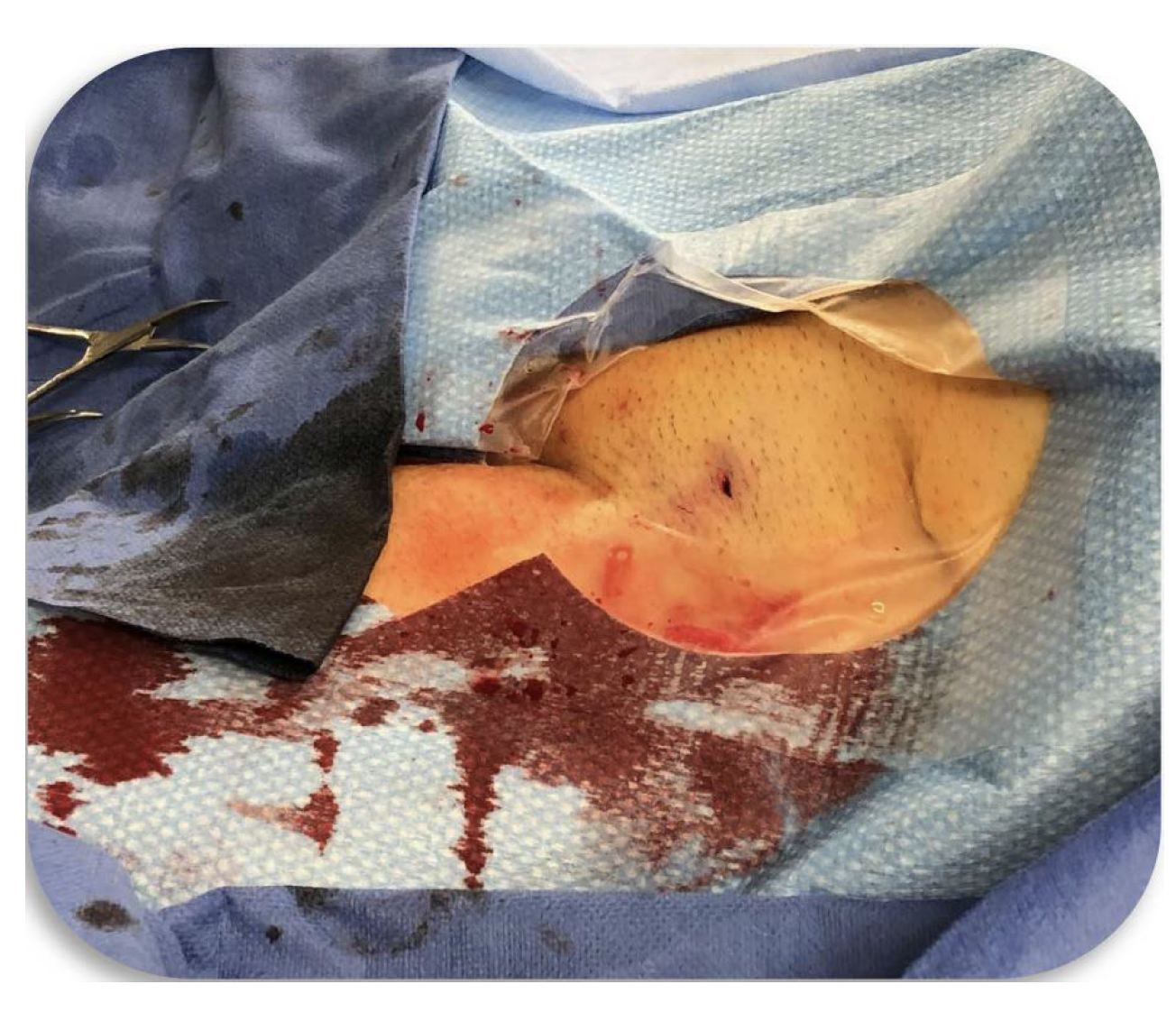
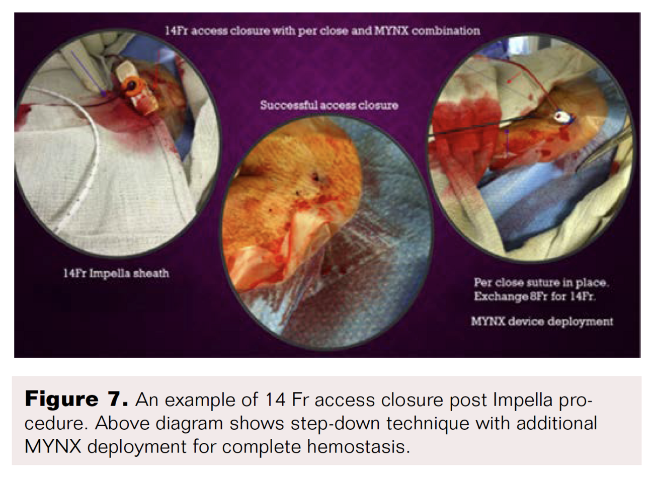
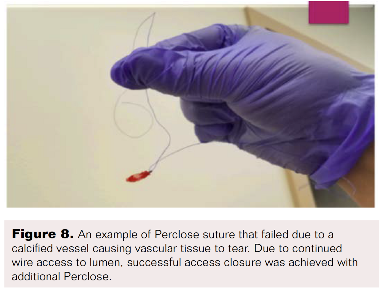
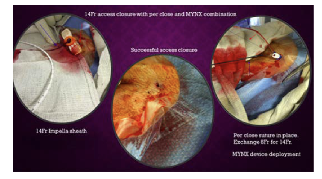
In the hospital, major and minor access complications were monitored in all patients. Time to ambulation post procedure, duration of hospital stay, and early readmissions were also monitored. Due to a controlled wire supported step-down technique, we were able to achieve access closure with a single Perclose suture in some cases where fibro-calcific femoral disease limited Perclose deliverability. Additional MYNX extravascular plugs can be used for optimizing hemostasis in single Perclose cases as shown in Figure 7. This is particularly useful in fibrocalcific ASVD patients where placing Perclose sutures is challenging. Figure 8 is an example of one such case where we saw tissue dehiscence with Perclose. Because of continued wire access to vessel lumen, we were able to reintroduce a second Perclose suture and complete successful access closure.
Discussion
Using wire-guided step-down technique for vascular closure, we were able to optimize closure and minimize overall bleeding and complications post large bore access. The principle behind this technique is to allow natural vascular recoil to aid in access closure. Vasospasm is a natural arterial vascular response to irritation or instrumentation that is mediated by vasa nervosa. This can be exacerbated in diseased vessels, diabetics, and stressful situations. A classic example of vasospasm is demonstrated during radial access, which can occur in 10% of cases.15 Step-down access closure technique takes advantage of vascular recoil to obtain optimal closure. This method becomes especially useful when utilized for access closure of large bore cannulation for Impella during complex high-risk coronary interventions, structural heart procedures, and percutaneous stent grafting.15 All these subgroups of patients have complvascular disease and are prone to access site complications.
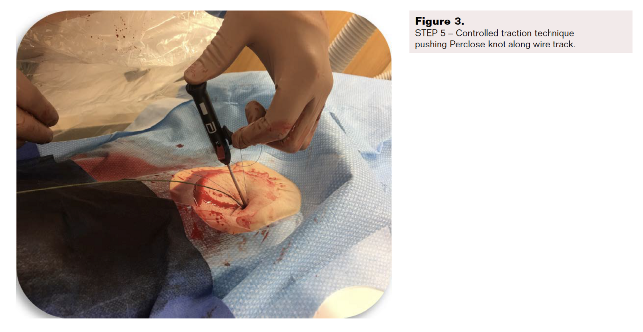
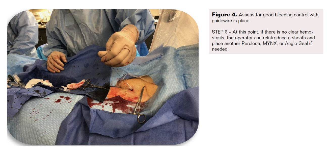
We are referencing our brachial closure experience as an example of prior step-down technique experience. Step-down technique was modified for large bore access adding a 0.35" guidewire support in this prospective study. We used this method to test the validity of step-down approach in reducing complications status post large bore access for AAA stent grafting and Impella placement for mechanical circulatory support, which require large bore access. Upon completion of Impella support, the device was removed along with the large bore sheath while maintaining vascular lumen access with the 0.35" guidewire. The Impella sheath was exchanged with a 9 Fr sheath over the 0.35" guidewire. While maintaining access to vascular lumen, control of hemostasis was accomplished by tracking sutures over the 9 Fr sheath. This minimizes blood loss during the closure and maintains a dry field. Maintaining lumen access and controlled closure allows for safety and confidence for the operator. Local pressure is applied during step-down process to activate vascular recoil and close the lumen gradually. After some time, the lumen size has now shrunk from the larger sheath size to the smaller sheath size. The next step is to remove the 9 Fr sheath while still maintaining access with the guidewire and optimally deliver a Perclose suture knot to the arteriotomy by sliding the knot down along the guidewire, “walking the suture down” toward the arteriotomy. Keeping access to the vascular lumen with the 0.35" wire allows for additional deployment of a suture or plug for persistent oozing and also securing optimal suture knot delivery to the arteriotomy. Controlled delivery of the purse-string suture also reduces long-term stricture and iatrogenic access site stenosis. Furthermore, step-down approach can be an optimal technique for post axillary Impella placement. Complicated approaches with intraluminal femoral access wiring and balloon tamponade (dry closure) have been used for access closure following axillary Impella implantation. Our technique is simple and more effective for optimal axillary access closure.
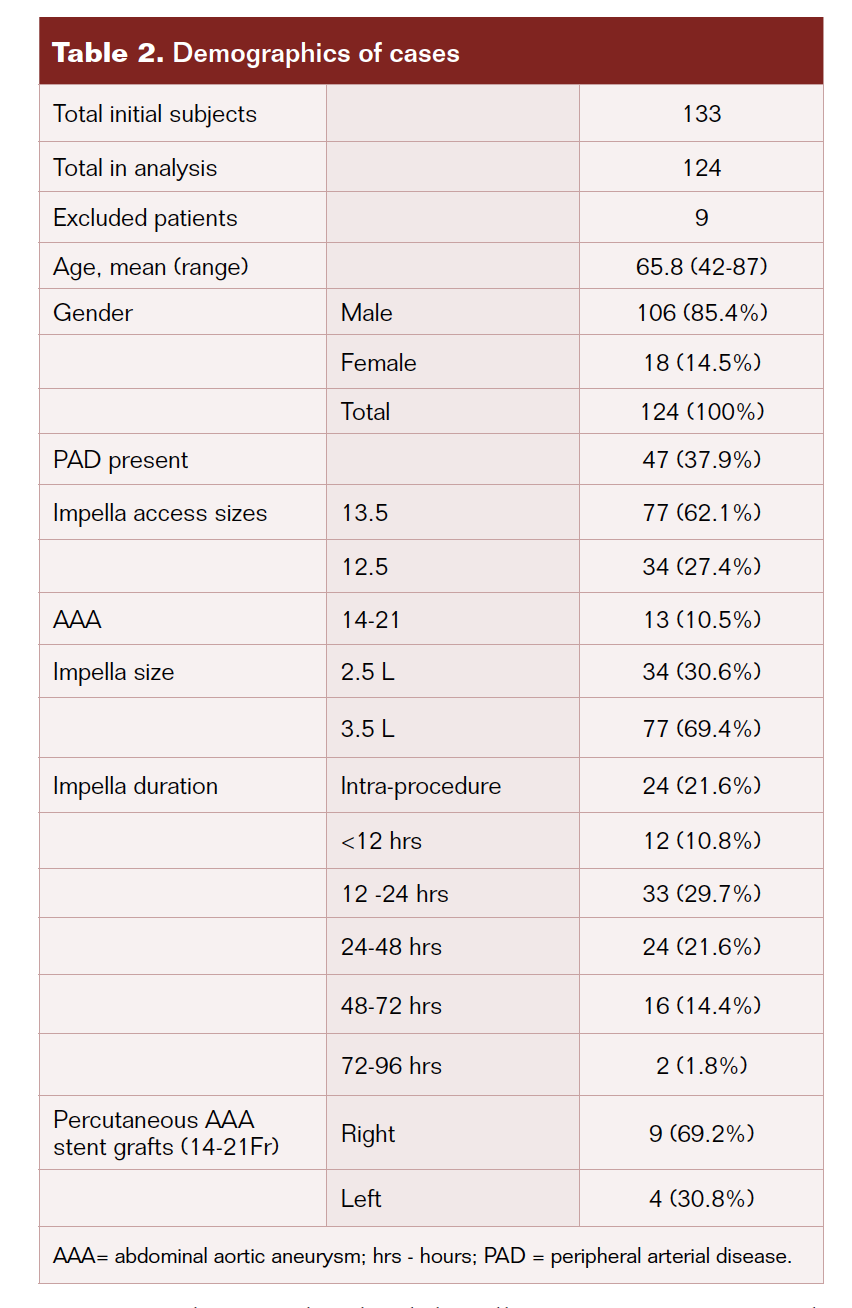
It is worth noting that more than one-third of the patients who underwent Impella insertion had a history of PAD (37.9%). Along with PAD, many had other vascular risk factors such as diabetes and chronic inflammatory disease (Table 2). Risk factors like these can make catheter insertion and removal difficult, as vascular integrity is compromised at baseline before any vascular intervention.

As mentioned above, the rate of bleeding complications plays a role in operator approach to left ventricular assist devices and access closure given its high complication rates. In 2008, Impella bleeding rates reported in FDA studies were 14.6% based on the PROTECT II RCT data. That number decreased to about 6.0% in 2018 through the STEMI DTU FDA pilot study.17-19 These complication rates have dropped over the course of 10 years and they do compare to our access closure technique. Of the 124 subjects who had successful removal of the Impella, no major complications were observed; only 7 subjects (5.6%) had minor closure complications, which consisted of hematoma and minor bleeding as met by the criteria in Table 3. There were no events that met the criteria for major bleeding. There were 5 subjects (7.0%) that required triple Perclose suture closure for persistent oozing. In some subjects with persistent oozing, we were able to control it by additional MYNX deployment, as depicted in Figure 7.
Conclusion
Controlled suture access closure with wire-supported step-down technique allows natural vascular recoil to aid in large bore access site closure. This method allows for optimal hemostatic control of the vascular site and promotes early ambulation while maintaining dry field during deployment and minimizing bleeding complications. We demonstrated these benefits, especially in high-risk patient groups including elderly population, women, diabetics, and patients with PAD in the setting of cardiogenic shock requiring large bore access for Impella use. All of our patients in this study were complex vascular patients and a high-risk group. Access failure complications in this subgroup across the data is in the range of 12%-15% while our group had no major complications, and minor complications were at 5.6%. This translates to significant cost effectiveness and reduced morbidity.
Using controlled step-down closure technique, interventionalists can be less hesitant to use large bore access procedures such as Impella and structural heart procedures for fear of complications because of large bore access.
Advantages of step-down technique access site closure:
(1) Minimizes blood loss and bleeding complications. Maintains dry field.
(2) Even though somewhat cumbersome, it is a controlled approach and improves operator confidence.
(3) Fewer vascular complications such as abrupt vessel occlusion and delayed stricture.
(4) Improved patient safety and operator comfort with large bore access.
(5) Early ambulation and more access to complex coronary and structural heart procedures.
(6) Less morbidity, reduced hospital stay, and more cost-effective solutions.
(7) In complex PAD, patients can achieve large bore access closure with a single Perclose.
(8) Optimal access closure following axillary Impella implantation.
(9) Guidewire-supported tracking of suture knots avoids failure by premature locking, suture snag, and premature breaking of suture with Perclose.
Limitations of our study:
(1) Single center experience with relatively small sample size.
(2) Less representation of women in this study.
(3) Somewhat cumbersome due to multiple steps to the technique.
(4) Reproducibility of the technique across multiple operators not tested.
(5) Not a multicenter randomized trial.
(6) Due to continued wire access, may have slight increase in thrombosis and distal embolization.
Patient selection for step-down closure:
(1) PAD patients.
(2) Obese patients.
(3) Patients requiring extended Impella duration.
(4) Diabetic women.
(5) Elderly patients.
Disclosure: The authors have completed and returned the ICMJE Form for Disclosure of Potential Conflicts of Interest. The authors report no conflicts of interest regarding the content herein.
Manuscript accepted July 15, 2021.
Address for correspondence: Ramesh K. Adiraju, MD; RENU-CA Research Institute, Lower Bucks Hospital, 501 Bath Road, Bristol, PA 19007. Email: renu-ca@comcast.net
REFERENCES
1. Adiraj R. Percutaneous brachial approach with step-down technique for vascular intervention (TCT abstract, #297; Am J Cardiol. 1998)
2. Adiraju RK. Access closure innovations. J Indian Coll Cardiol. 1 July 2017. http://www.sciencedirect.com/science/article/abs/pii/S1561881117301128.
3. Nabel EG, Braunwald E. A tale of coronary artery disease and myocardial infarction. N Eng J Med. 2012;366:54-63.
4. Burzotta F, Russo G, Previ L, Bruno P, Aurigemma C,Trani C. Impella pumps overview and access site management. Minerva Cardioangiol. 2018;66(5):606-611.
5. Goldsweig AM, Secemsky EA. Vascular access and closure for peripheral arterial intervention. Interv Cardiol Clin. 2020; 9:117-124.
6. Noori VJ, Eldrup-Jørgensen J. A systematic review of vascular closure devices for femoral artery puncture sites. J Vasc Surg. 2018;68:887-899.
7. van Wiechen MP, Ligthart JM, Van Mieghem NM. Large-bore vascular closure: new devices and techniques. Interv Cardiol. 2019;14:17-21.
8. Moriyama N, Lindström L, Laine M. Propensity-matched comparison of vascular closure devices after transcatheter aortic valve replacement using MANTA versus ProGlide. EuroIntervention. 2019;14(15):e1558-e1565.
9. Closure devices. Endovascular Today. 2021. Accessed March 12, 2021. https://evtoday.com/device-guide/us/closure-devices.
10. Saleem T, Baril DT. Vascular access closure devices. 2021. In: StatPearls [Internet]. Treasure Island, FL: StatPearls Publishing; 2021.
11. Sesana M, Vaghetti M, Corvaja N, et al. Effectiveness and complications of vascular access closure devices after interventional procedures. J Invasive Cardiol. 2000;12:395-399.
12. Yeni H, Axel M, Örnek A, Butz T, Maagh P, Plehn G. Clinical and subclinical femoral vascular complications after deployment of two different vascular closure devices or manual compression in the setting of coronary intervention. Int J Med Sci. 2016;13:255-259.
13. Kaki A,Blank N, Alaries MC, et al. Access and closure management of large bore femoral arterial access. J Interv Cardiol. 2018;31:969-977.
14. Adiraju RK. Optimal vascular access site hemostasis. Poster presented at: Society of Cardiac Angiography and Interventions; 2011.
15. Artery spasm. ScienceDirect.com. Accessed March 12, 2021. https://www.sciencedirect.com/topics/medicine-and-dentistry/artery-spasm.
16. Sandoval Y, Burke MN, Lobo AS, et al. Contemporary arterial access in the cardiac catheterization laboratory. JACC Cardiovasc Interv. 2017;10:2233-2241.
17. HeartRecovery.com. 2021. How do bleeding rates with Impella® compare to bleeding rates associated with other forms of mechanical circulatory support? Accessed March 12, 2021. https://www.heartrecovery.com/education/education-library/faq-how-do-bleeding-rates-with-Impella-compare-to-bleeding-rates-associated-with-other-forms-of-mechanical-circulatory-support.
18. O'Neill WW, Kleiman NS, Moses J. A prospective, randomized clinical trial of hemodynamic support with Impella 2.5 versus intra-aortic balloon pump in patients undergoing high-risk percutaneous coronary intervention: the PROTECT II study. Circulation. 2012;126:1717-1727.
19. Kapur NK, Alkhouli MA, DeMartini TJ, et al. Unloading the left ventricle before reperfusion in patients with anterior ST-segment elevation myocardial infarction. Circulation. 2019;139:337-346.
