


Traumatic Avulsion of Upper Eyelid Skin Following Surgery in a Patient With Multiple Myeloma and Amyloid Light-chain Amyloidosis
Abstract
Multiple myeloma (MM) is a common hematologic malignancy. Primary systemic amyloidosis or amyloid light-chain (AL) amyloidosis is a rare disease. PURPOSE: This article presents the case of a patient with MM and AL amyloidosis who experienced a severe case of medical adhesive-related skin injury. CASE STUDY: A 64-year-old man with MM, AL amyloidosis, and diabetes presented with a necrotic wound on his left heel that required surgical debridement. The patient experienced a traumatic avulsion of the right upper eyelid skin during the removal of the corneal abrasion preventive tape as well as traumatic avulsion of the left upper eyelid skin while the patient’s face was being cleansed. The avulsed right upper eyelid skin above the tarsus was repaired with a full-thickness skin graft. The partly avulsed left upper eyelid skin was repositioned, and an excisional biopsy was taken. Both upper eyelids healed uneventfully. The biopsy specimen revealed increased amyloid deposition in the dermis, subcutaneous tissue, and areas surrounding the veins and sweat glands. CONCLUSION: This case illustrates the increased risk of medical adhesive-related skin injury and other skin damage in patients with MM and AL amyloidosis. In these patients, the use of tape should be avoided to prevent intraoperative corneal abrasion.
Introduction
Multiple myeloma (MM) is characterized by malignant proliferation of plasma cells that accumulate in the bone marrow, where they interfere with the production of normal blood cells.1,2 Amyloid light-chain (AL) amyloidosis is characterized by the extracellular deposition of amyloid fibrils in tissues and organs. Twenty-five percent (25%) of patients with AL amyloidosis have skin involvement such as petechiae, purpuras, ecchymoses, bullous lesions, papules, and plaques.3 Amyloid deposition in vital organs such as the heart, kidney, and liver can lead to progressive organ dysfunction and death.4 Therefore, early diagnosis through skin findings of AL amyloidosis is crucial so that treatment can begin as soon as possible. A male patient who had avulsions on the eyelids following minor trauma due to increased amyloid infiltration of the skin in the late period of his illness is presented in this case report.
Case Report
A 63-year-old man with a 20-year history of diabetes mellitus was admitted to the plastic surgery clinic with a necrotic wound on his left heel that had been present for 2 months. His detailed medical history showed that he had undergone follow-up by the hematology clinic with the diagnosis of MM for 3 years. A relapse of MM was noted during routine follow-up in January 2015. During the same period, he was referred to the dermatology clinic because of periorbital ecchymosis. Dermatologic examination revealed no skin findings except periorbital ecchymosis. A punch biopsy of these lesions was performed. Histopathologic examination revealed subcorneal hemorrhage and spongiosis in the epidermis and upper dermis. Amyloid deposition was demonstrated with a Congo red stain, in which amyloid fibers showed dark red under the light microscope and showed birefringence under a polarized light microscope. These histopathologic findings led to a diagnosis of AL amyloidosis. Two (2) cycles of chemotherapy (cyclophosphamide, bortezomib, and dexamethasone) were administered, and autologous stem cell transplantation was performed.5
Physical examination findings and radiographic images revealed a necrotic wound and osteomyelitis on the patient’s left heel that was thought to be unrelated to MM or AL amyloidosis. Due to these findings, the patient had an elective indication for debridement. Written informed consent for publication of the clinical details and use of photographs was obtained from the patient.
During the procedure, necrotic wound and sequestered bone segments were debrided under general anesthesia. At the end of the surgery and while removing the eye protection tapes in the postextubation period, it was observed that the portion of the right upper eyelid skin above the tarsus was avulsed. Because it was not possible to remove the eyelid skin from the eye protection tape, the patient’s right eyelid was closed with an oily dressing (Figure 1A). Two (2) days later, the defect on the right upper eyelid was repaired with a full-thickness skin graft taken from the right posterior auricular region, and a “tie-over” dressing was used to secure the graft (Figure 1B). At the end of the procedure and while the patient’s face was being cleansed, an unintentional partial avulsion of the left upper eyelid skin occurred (Figure 1C). The skin was repositioned to the left upper eyelid, an excisional biopsy was taken, and the oily dressing was applied. Two (2) days later when the oily dressing was removed, the left upper eyelid skin had adhered smoothly to the bed. Five (5) days later when the tie-over dressing was removed, the skin graft that was applied to the right upper eyelid had adhered to the recipient bed (Figure 1D). The postoperative period was uneventful.
Histopathologic findings of the excisional biopsy showed that there was dense eosinophilic amorphous material in perivascular and interstitial areas of the dermis. After applying Congo red stain, the material was examined under a polarized light microscope, and the findings were consistent with amyloidosis.
Discussion
MM may manifest with kidney failure, anemia, hypercalcemia, lytic bone lesions, pathologic fractures, immunodeficiency, and hyperviscosity. MM can also affect the adrenal glands, lungs, liver, spleen, pancreas, lymph nodes, and skin.3,4,6–8
Cutaneous plasmacytomas are extremely rare, specific cutaneous findings of MM. They occur late in the course of the disease and present as erythematous nodules and plaques. They can present initially with common and nonspecific cutaneous lesions such as leukocytoclastic vasculitis, amyloidosis, pyoderma gangrenosum, or vesiculobullous disorder; it is difficult to suspect MM with the initial presentation of skin lesions mentioned above.1,9,10
AL amyloidosis may occur as an idiopathic condition or be associated with MM or lymphoma.2 Skin infiltration with amyloid leads to the formation of papules, nodules, or plaques.6 Hemorrhagic skin findings (petechia, purpura, and ecchymosis) are seen frequently on the eyelids, neck, axillae, and anogenital areas due to the amyloid infiltration of the vessel walls following a minor trauma such as amyloid purpura or pinch purpura.6,7 During coughing, the Valsalva maneuver, or proctoscopy, the signs and symptoms listed above can occur and are seen most commonly on the flexural regions, such as the eyelids and inframammary regions.6 There are some studies on severe skin or internal bleeding due to coagulation factor inhibitory circulating paraprotein, hyperfibrinolysis, platelet dysfunction, or isolated acquired factor X deficiency in amyloidosis; however, to the authors’ knowledge, skin avulsion due to minor traumas has not been reported in the literature.11,12
Corneal abrasions (CAs) are the most common corneal complications during general anesthesia, and their incidence varies between 0% and 44% depending on the method used for eye protection.13 For CA prophylaxis in the perioperative period, it is recommended to ensure eyelid closure by securing the eyelids with tape; applying lubricating eye ointment is recommended when this is not feasible.13–15 In the authors’ hospital, anesthesiologists prefer to use the tape method in the majority of patients to prevent perioperative CAs, as some studies report negative side effects of lubrication, such as blurred vision and ocular edema.16–18 In the current case, during the removal of protective tape the authors encountered skin avulsion of the right eyelid skin. No reports of this complication could be found in the authors’ review of the literature.
Medical adhesives are an integral part of health care delivery and are a component of a variety of products such as tapes and dressings. Medical adhesive-related skin injury (MARSI)19 has a significant, negative impact on patient safety as seen in the current case. The skin injury encountered on the right eyelid can be considered a serious example of MARSI.
The diagnosis of AL amyloidosis in MM is made by both clinical findings and histopathologic study. On histopathologic examination, lesions reveal eosinophilic amorphous material accumulation in the dermis and subcutaneous tissue as well as in the areas surrounding veins and sweat glands. When biopsy material is stained with Congo red, the finding of green birefringence with a polarized light microscope confirms amyloidosis.8 Histologic evaluation of the current case revealed that AL amyloidosis developed during the course of MM recurrence.
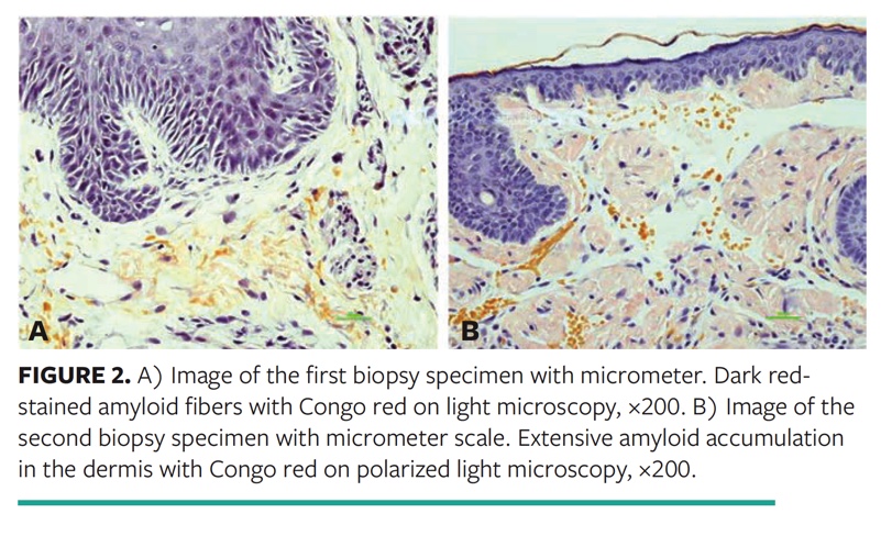
The second biopsy specimen taken from the patient was compared with the first biopsy specimen by using a high-resolution DS-Ri1 color camera and NIS-Elements imaging software (Nikon, Tokyo, Japan). Although amyloid deposition was diffusely seen in a great part of the tissue in the second biopsy specimen, the deposition was noted only in a limited area in the first (Figure 2A and Figure 2B). Increased amyloid deposition over time explains the clinical symptoms that have evolved from minor bruising to skin damage by minor traumatic events, as in the example the authors encountered on the current patient’s left eyelid.
Conclusion
The current case report describes a patient who experienced avulsions on the eyelids with trauma from tape removal due to increased amyloid infiltration of the skin. The probability of encountering patients with MM and AL amyloidosis has increased because of the increased prevalence of AL amyloidosis and the high incidence of MM in the growing elderly population. Achieving a promising treatment modality or total control of these diseases in this population is still problematic; hence, the possibility of experiencing a similar problem to what was seen in the current case report is also increasing. The risk for MARSI should be considered in these patients.
Affiliations
Dr. Ergün is a professor, Department of Plastic, Reconstructive and Aesthetic Surgery, Bezmialem Medical School, Bezmialem Vakif University, Istanbul, Turkey; Dr. Zengin is an assistant professor, Department of Anesthesiology and Reanimation, Marmara University Training and Research Hospital, Istanbul, Turkey; Dr. Ünal is a resident, Department of Plastic, Reconstructive and Aesthetic Surgery, Bezmialem Medical School, Bezmialem Vakif University, Istanbul, Turkey; and Dr. Yildiz is an assistant professor, Department of Pathology, Bezmialem Medical School, Bezmialem Vakif University, Istanbul, Turkey. Address all correspondence to: Selma Sönmez Ergün, MD, Estonşehir 3. Mahalle Ilgın Sokak CD Villa 7/2, Bahçeşehir/Başakşehir/İstanbul/Turkey; tel: + 90 212 691 52 56; fax: + 90 212 621 75 80; email: selmasonmezergun@yahoo.com
References
1. Harati A, Brockmeyer NH, Altmeyer P, Kreuter A. Skin disorders in association with monoclonal gammopathies. Eur J Med Res. 2005;10(3):93–104.
2. Behera B, Pattnaik M, Sahu B, Mohanty P, Jena S, Mohapatra L. Cutaneous manifestations of multiple myeloma. Indian J Dermatol. 2016;61(6):668–671. doi:10.4103/0019-5154.193682
3. Gertz MA, Lacy MQ, Dispenzieri A, Hayman SR. Amyloidosis. Best Pract Res Clin Haematol. 2005;18:709–727. doi:10.1016/j.beha.2005.01.030
4. Comenzo RL. Amyloidosis. Curr Treat Options Oncol. 2006;7(3):225–236. doi:10.1007/s11864-006-0015-8
5. Emiroglu N, Su O, Cengiz FP, Tosuner Z, Demirkesen C, Onsun N. Raccoon eyes in amyloidosis. G Ital Dermatol Venereol. 2017;152(1):80–82. doi:10.23736/S0392-0488.16.05160-9
6. Black MM, Upjohn E, Albert S. Amyloidoses. In: Bolognia JL, Jorizzo JL, Rapini RP, eds. Dermatology. 2nd ed. Mosby Elsevier; 2008:623–631.
7. Köse M, Buraniqi E, Akpinar TS, Kayacan SM, Tükek T. Relapse of multiple myeloma presenting as extramedullary plasmacytomas in multiple organs. Case Rep Hematol. 2015; 2015:452305. doi:10.1155/2015/452305
8. Lachmann HJ, Hawkins PN. Amyloidosis of the skin. In: Wolff K, Goldsmith LA, Katz SI, Gilchrest BA, Paller AS, Leffell DJ, eds. Fitzpatrick’s Dermatology in General Medicine. 7th ed. McGraw-Hill, 2008:1256–1265.
9. Bayer-Garner IB, Smoller BR. The spectrum of cutaneous disease in multiple myeloma. J Am Acad Dermatol. 2003;48(4):497–507. doi:10.1067/mjd.2003.180
10. Gül U, Kiliç A, Gönül M, Cakmak SK, Heper AO. A case of multiple myeloma presenting as a bullous dermatosis. Indian J Dermatol. 2008;53(2):83–84. doi:10.4103/0019-5154.41653
11. Kumar S, Sengupta RS, Kakkar N, Sharma A, Singh S, Varma S. Skin involvement in primary systemic amyloidosis. Mediterr J Hematol Infect Dis. 2013;5(1):e2013005. doi:10.4084/MJHID.2013.005
12. Colucci G, Alberio L, Demarmels Biasiutti F, Lämmle B. Bilateral periorbital ecchymoses. An often missed sign of amyloid purpura. Hamostaseologie. 2014;34(3):249–252. doi:10.5482/HAMO-14-03-0018
13. Bøggild-Madsen NB, Bundgarrd-Nielsen P, Hammer U, Jakobsen B. Comparison of eye protection with methylcellulose and paraffin ointments during general anesthesia. Can Anaesth Soc J. 1981;28(6):575–578. doi:10.1007/BF03007155
14. Harvey RR. Anesthesia for ophthalmic procedures. In: Nagelhout JJ, Plaus KL, eds. Nurse Anesthesia. 5th ed. Saunders; 2014:976–998.
15. Morris A , Bonanno L, Bennett M. Effectiveness of corneal abrasion prevention interventions for adults undergoing general anesthesia for more than one hour: a systematic review protocol. JBI Database System Rev Implement Rep. 2018;16(9):1785–1790. doi:10.11124/JBISRIR-2017-003670
16. Segal KL, Fleischut PM, Kim C, et al. Evaluation and treatment of perioperative corneal abrasions. J Ophthalmol. 2014;2014:901901. doi:10.1155/2014/901901
17. Carniciu AL, Fazzari MJ, Tabibian P, et al. Corneal abrasion following anaesthesia for non-ocular surgical procedures: a case-controlled study. J Perioper Pract. 2017;27(11):247–253. doi:10.1177/175045891702701102
18. Ganidagli S, Cengi M, Becerik C, Oguz H, Kilic A. Eye protection during general anaesthesia: comparison of four different methods. Eur J Anaesthesiol. 2004;21(8):665–667. doi:10.1017/s0265021504228137
19. McNichol L, Lund C, Rosen T, Gray M. Medical adhesives and patient safety: state of the science: consensus statements for the assessment, prevention, and treatment of adhesive-related skin injuries. Orthop Nurs. 2013;32(5):267–281. doi:10.1097/NOR.0b013e3182a39caf
