Kirschner Wires Combined With Elastic Tape for Multilayer Tension-Reducing Repair of a Large Stage 4 Pressure Injury of the Greater Trochanter: A Case Report
© 2025 HMP Global. All Rights Reserved.
Any views and opinions expressed are those of the author(s) and/or participants and do not necessarily reflect the views, policy, or position of Wounds or HMP Global, their employees, and affiliates.
Abstract
Background. Pressure injuries are common, difficult to manage, and carry a high economic burden. They are challenging to physicians and a burden to society. Case Report. An 89-year-old male, who had previously undergone internal fixation with screws and rods for a right intertrochanteric fracture, developed a deep circular open ulcer measuring 11 cm × 7.5 cm on his right hip after being bedridden for over 20 consecutive days because of symptoms of coughing and fatigue. Upon examination, the ulcer exhibited extensive necrotic tissue, purulent discharge, and a foul odor at its base. Following multiple debridement procedures, a multilayer tension reduction and repair surgery using the Kirschner wire technique in conjunction with elastic tape was performed on the 31st day of hospitalization. The wound achieved complete healing, and the patient was discharged on the 102nd day of admission. During the 6-month postoperative follow-up period, the appearance and sensory function of the wound skin were observed to have recovered satisfactorily. Conclusion. In this case, the use of Kirschner wires combined with elastic tape for multilayer tension reduction resulted in the repair of a large stage 4 pressure injury at the greater trochanter. The mechanism of action is discussed and analyzed to provide clinical evidence for such treatment.
Introduction
Pressure injury is a common issue in clinical practice, particularly among patients who are confined to bed for extended periods of time or who have limited mobility.1 Once pressure injuries have occurred, they can spread quickly and impede the healing process. The incidence of pressure injuries in hospitals ranges between 2.7% and 29.5%, and their prevalence ranges between 3.5% and 69%.2
Pressure injuries place a significant financial burden on the health care system, with a 2019 study reporting treatment expenditures for hospital-acquired pressure injury averaging approximately $10708 per patient in the United States in 2016.3 According to that study, 59% of an estimated $26.8 billion annual expenditure for treating hospital-acquired pressure injury in the United States was attributed to treating stage 3 and 4 pressure injuries.3 Traditional procedures for treating severe pressure injuries can be complex and risky, causing agony for patients and perhaps leading to subsequent injuries. The efficacy of current therapeutic procedures in treating pressure injuries is limited; thus, the search for novel treatment modalities has become critical.
Several authors of the present case report effectively incorporated the use of elastic therapeutic tape for the purpose of skin stretching to alleviate stress and enhance wound healing in a different case.4 The current report describes the use of Kirschner wires and a multilayer tension reduction technique using elastic tape to effectively treat severe pressure injury on the greater trochanter, resulting in favorable therapeutic outcomes. This case report demonstrates a novel and effective treatment procedure for greater trochanter pressure injury, detailing the patient’s medical history, therapy options, and follow-up results.
Case Report
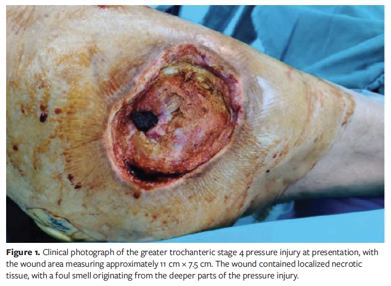
An 89-year-old male, with a history of internal fixation for a fracture in the right intertrochanteric region 2 years prior, presented to the Shenzhen Hospital (Futian) of Guangzhou University of Chinese Medicine after being bedridden for 20 consecutive days at home because of symptoms of cough and fatigue. He presented with a circular, significant, exposed pressure injury located on the right greater trochanter, measuring 11 cm × 7.5 cm. A pressure injury is characterized by notable full-thickness skin and tissue deficits, which include visible or palpable exposure of fascia, muscle, tendon, ligament, and bone. Additionally, there may be localized necrotic tissue and an unpleasant odor emanating from the deeper regions of the pressure injury (Figure 1). Informed consent was obtained from the patient for the publication of this case and the associated images.
Upon admission, laboratory tests showed the following results: a white blood cell count of 9.14 × 10⁹/L; a neutrophil percentage of 81.30%; a hemoglobin level of 118 g/L; a fast C-reactive protein level of 247.70 mg/L; an erythrocyte sedimentation rate of 101 mm/h; and an interleukin 6 concentration of 104.30 pg/mL. Pressure injury exacerbated by infection is a concern, and a culture of wound exudate revealed the presence of Staphylococcus aureus. Consequently, anti-infective therapy was initiated with cefoperazone sodium and sulbactam sodium injection, guided by the results of antimicrobial susceptibility testing. The patient underwent debridement and negative pressure wound therapy (NPWT) for the pressure injury on the right greater trochanter on the third and seventh days following admission to the general surgery department. On the 15th day following admission, he was transferred from the general surgery department to the orthopedics department. Further debridement and NPWT procedures were performed on the 17th and 24th days (Figure 2A).
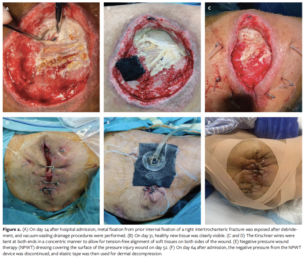
On the 31st day post-admission, the NPWT dressing was removed, at which time it was noted that a considerable quantity of necrotic tissue at the base of the pressure injury had been effectively excised, with healthy new tissue clearly visible (Figure 2B). Surgical debridement of the pressure injury was performed, using tension-reducing sutures with Kirschner wires and NPWT. Kirschner wires with a diameter of 1.5 mm were inserted vertically through the skin, approximately 2 cm to 3 cm from 1 border of the incision. The wires traversed the subcutaneous tissue, deep fascia, and muscular layers, and were placed under the deep fascia in a path perpendicular to the wound surface. The wires penetrated into the soft tissue on the other side and protruded approximately 2 cm to 3 cm from the wound border on that side. The Kirschner wires were bent at both ends in a concentric fashion to facilitate tension-free alignment of soft tissues on either side of the incision (Figure 2C, 2D). The exact degree of bending was determined with precision. Subsequently, both ends were contoured into an inverted “Ω” structure and cut at the points of curvature, preserving approximately 1 cm of the ends beyond these junctions. A total of 5 Kirschner wires were used. Two wires were positioned 2 cm apart because of the excessive size of the lesion, which precluded primary closure intraoperatively. The bending points at both ends were originally secured with No. 10 silk thread before being covered with NPWT dressing across the wound surface.
Post-admission evaluations on the 38th, 45th, 52nd (Figure 2E), and 59th days indicated little local exudation as the healing process progressed towards closure. On the 64th day post-admission, NPWT was terminated, and elastic tape was then used for dermal decompression (Figure 2F).
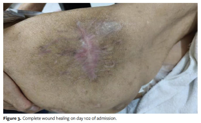
As wound healing progressed, the Kirschner wires used for stabilization were progressively extracted, culminating in their complete removal on the 97th day post-admission. The patient was released on the 102nd day, and a satisfactory recovery was observed during a 6-month postoperative follow-up period, which confirmed the restoration of appearance and sensory function in the affected skin (Figure 3).
Discussion
Pressure injuries can arise in individuals who are confined to bed for prolonged periods.1 These lesions may significantly diminish quality of life, particularly in patients with underlying medical conditions,5 and the lesions pose a considerable danger of infection.2 Therefore, it is crucial to implement proactive preventative methods and effective care procedures for pressure injuries. The current primary treatment options include routine surgical excision of necrotic tissue,6 flap reconstruction, NPWT, synthetic skin replacements, and topical growth agents.7 Nonetheless, there are occasions when these remedies are unsuccessful, leading to suboptimal healing results or extended recuperation duration. Drawing from their clinical knowledge, the authors of the current report have used Kirschner wires with elastic tape to enable multilayer tension reduction as a novel approach to address these issues.
Prior approaches and devices for skin expansion use the viscoelastic properties of the skin to facilitate closure via the mechanisms of mechanical creep and stress relaxation.8 Moreover, the subcutaneous tissue and deep fascia have considerable elasticity.9 In the current case, Kirschner wires were meticulously inserted into the epidermis, subcutaneous tissue, and deep fascia on both sides of the incision edge. Simultaneously, elastic tape was used to leverage the inherent elasticity, mechanical extensibility, and biological stretch capability of the adjacent skin, subcutaneous tissue, and deep fascia. This technique enabled the targeted closure of the incision in a concentrated fashion, entirely alleviating tension without generating voids beneath the skin. This innovative methodology exhibits clear advantages over traditional methods. It substantially alleviates the tissue tearing and impairment of wound edge perfusion caused by conventional suture traction, therefore facilitating a large reduction in suture line tension. The modifiable traction force applied to surrounding tissues facilitates a more uniform distribution of tension throughout the wound bed. The progressive traction-closure technique enables gradual wound approximation, potentially eliminating the necessity for extensive skin grafting or flap-transfer procedures. This innovation mitigates iatrogenic injury to donor sites for graft or flap harvesting. Moreover, it avoids numerous difficulties associated with conventional reconstruction techniques, such as graft or flap viability issues, extended healing times for donor sites, intensive postoperative scar care, recurrent ulceration of scar tissue, and the need for subsequent revision procedures. This method clinically reduces the local tissue healing duration by optimizing the biomechanical distribution of tension and minimizing shear stress.10
The findings of the current case demonstrate that the use of Kirschner wires in conjunction with elastic tape for the management of severe pressure injuries is not only straightforward but also facilitates wound closure without exerting stress during treatment, resulting in expedited healing and reduced complications. This procedure may serve as a useful clinical approach for addressing soft tissue anomalies associated with pressure injuries.
Limitations
This report has limitations. It presents a single patient case, and not every patient may have had such a positive reaction to this treatment approach. Further research is needed to assess the efficacy of using Kirschner wires together with elastic tape in the particular scenarios outlined in this article. Such a study should encompass larger patient cohorts and consider various wound dimensions and sites.
Conclusion
The prior clinical experience of the authors of the current report indicates that the exclusive use of elastic tape is helpful for managing small chronic wounds on the lower back, provided there is sufficient healthy skin surrounding the lesion for adequate application of the tape. The simultaneous use of elastic tape and Kirschner wires is a viable method for addressing extensive pressure injuries. Because of their stiffness, Kirschner wires should be avoided for treating wounds on the posterior portion of the body, as they may disrupt repose or perhaps worsen the lesion.
The incorporation of Kirschner wires and elastic tape in a multilayer tension-reduction technique is an effective approach for repairing extensive pressure injuries, systematically targeting various anatomical layers, including the skin, subcutaneous tissue, and deep fascia, while promoting wound healing—especially in cases involving substantial muscle attachment. This case report seeks to offer thorough information on the clinical care of extensive pressure injuries, along with specific recommendations for surgeons addressing these intricate cases. Further studies clarifying clinical experiences are needed to provide robust data concerning the effectiveness of this combination method in treating extensive pressure injuries.
Author and Publication Information
Authors: Xu Qiliang, MD1; Liang XiaoHua, MD2; Chen Haoxiong, MD1; Zhao Liang, MD1; Tan Jingchao, MD3; Zhang Linlin, MD1; and Yang Junxing, MD1
Affiliations: 1Shenzhen Hospital (Futian) of Guangzhou University of Chinese Medicine, Shenzhen, China; 2Guangxi University of Chinese Medicine, Nanning, Guangxi Province, China; 3Guangzhou Orthopedic Hospital, Guangzhou, China
Acknowledgments: Xu Qiliang, MD, and Liang XiaoHua, MD, are considered as joint first authors.
Disclosure: The authors disclose no financial or other conflicts of interest.
Ethical Approval: Informed consent was obtained from the patient for the publication of this case and the associated images.
Correspondence: Yang Junxing, MD; Department of Orthopaedics, Shenzhen Hospital (Futian) of Guangzhou University of Chinese Medicine, No.6001, Beihuan Avenue, Futian District, Shenzhen, China; dryang@gzucm.edu.cn
Manuscript Accepted: June 27, 2025
References
1. Mervis JS, Phillips TJ. Pressure ulcers: pathophysiology, epidemiology, risk factors, and presentation. J Am Acad Dermatol. 2019;81(4):881-890. doi:10.1016/j.jaad.2018.12.069
2. Fogerty M, Guy J, Barbul A, Nanney LB, Abumrad NN. African Americans show increased risk for pressure ulcers: a retrospective analysis of acute care hospitals in America. Wound Repair Regen. 2009;17(5):678-684. doi:10.1111/j.1524-475X.2009.00522.x
3. Padula WV, Delarmente BA. The national cost of hospital-acquired pressure injuries in the United States. Int Wound J. 2019;16(3):634-640. doi:10.1111/iwj.13071
4. Fuming G, Qiliang X, Haoxiong C, Xuecheng H, Junxing Y. Therapeutic taping to offload wound margin strain and as an adjunct to wound closure: a case report. Wounds. 2023;35(4):E146-E148. doi:10.25270/wnds/21074
5. Gorecki C, Nixon J, Madill A, Firth J, Brown JM. What influences the impact of pressure ulcers on health-related quality of life? A qualitative patient-focused exploration of contributory
factors. J Tissue Viability. 2012;21(1):3-12. doi:10.1016/j.jtv.2011.11.001
6. Alam W, Hasson J, Reed M. Clinical approach to chronic wound management in older adults. J Am Geriatr Soc. 2021;69(8):2327-2334. doi:10.1111/jgs.17177
7. Eriksson E, Liu PY, Schultz GS, et al. Chronic wounds: treatment consensus [published correction appears in Wound Repair Regen. 2022;30(4):536. doi:10.1111/wrr.13035]. Wound Repair Regen. 2022;30(2):156-171. doi:10.1111/wrr.12994
8. Cheng LF, Lee JT, Hsu H, Wu MS. Simple skin-stretching device in assisted tension-free wound closure. Ann Plast Surg. 2017;78(3 Suppl 2):S52-S57. doi:10.1097/SAP.0000000000001006
9. Joodaki H, Panzer MB. Skin mechanical
properties and modeling: a review. Proc Inst Mech Eng H. 2018;232(4):323-343. doi:10.1177/0954411918759801
10. Topaz M, Carmel NN, Silberman A, Li MS, Li YZ. The TopClosure® 3S System, for skin stretching and a secure wound closure. Eur J Plast Surg. 2012;35(7):533-543. doi:10.1007/s00238-011-0671-1